1 Programa de Pós-graduação em Ciências da Reabilitação, Universidade Nove de Julho (UNINOVE), São Paulo, SP, Brazil 2 Programa de Pós-graduação em Biofotônica Aplicada às Ciências da Saúde, UNINOVE, São Paulo, SP, Brazil
3 Departamento de Ciências Exatas, UNINOVE, São Paulo, SP, Brazil
4 Departamento de Estomatologia, Faculdade de Odontologia, Universidade de São Paulo (USP), São Paulo, SP, Brazil
Received: 06/21/2013 Revised: 10/21/2013 Accepted: 02/17/2014 a r t i c l e
Effect of low-level laser therapy on the modulation of the
mitochondrial activity of macrophages
Nadhia H. C. Souza1, Raquel A. M. Ferrari1,2, Daniela F. T. Silva3,
Fabio D. Nunes4, Sandra K. Bussadori2, Kristianne P. S. Fernandes2
ABSTRACT | Background: Macrophages play a major role among the inlammatory cells that invade muscle tissue following an injury. Low-level laser therapy (LLLT) has long been used in clinical practice to accelerate the muscle repair process. However, little is known regarding its effect on macrophages. Objective: This study evaluated the effect of LLLT on the mitochondrial activity (MA) of macrophages. Method: J774 macrophages were treated with lipopolysaccharide (LPS) and interferon – gamma (IFN-γ) (activation) for 24 h to simulate an inlammatory process, then irradiated with LLLT using two sets of parameters (780 nm; 70 mW; 3 J/cm2 and 660 nm; 15 mW; 7.5 J/cm2).
Non-activated/non-irradiated cells composed the control group. MA was evaluated by the cell mitochondrial activity (MTT) assay (after 1, 3 and 5 days) in three independent experiments. The data were analyzed statistically. Results: After 1 day of culture, activated and 780 nm irradiated macrophages showed lower MA than activated macrophages, but activated and 660 nm irradiated macrophages showed MA similar to activated cells. After 3 days, activated and irradiated (660 nm and 780 nm) macrophages showed greater MA than activated macrophages, and after 5 days, the activated and irradiated (660 nm and 780 nm) macrophages showed similar MA to the activated macrophages. Conclusions: These results show that 660 nm and 780 nm LLLT can modulate the cellular activation status of macrophages in inlammation, highlighting the importance of this resource and of the correct determination of its parameters in the repair process of skeletal muscle.
Keywords: macrophages; low-level laser therapy; muscle repair; rehabilitation.
HOW TO CITE THIS ARTICLE
Souza NHC, Ferrari RAM, Silva DFT, Nunes FD, Bussadori SK, Fernandes KPS. Effect of low-level laser therapy on the modulation of the mitochondrial activity of macrophages. Braz J Phys Ther. 2014 July-Aug; 18(4):308-314. http://dx.doi.org/10.1590/ bjpt-rbf.2014.0046
Introduction
Although the pattern of gene expression in muscle regeneration parallels the pattern corresponding to embryonic muscle development, the microenvironments in which the two processes occur are dramatically different1.
This difference is due to the abundance of inflammatory cells in regenerative muscle, the concentration of which might exceed 100,000 cells/ mm3[1,2]. These inflammatory cells are activated
cells able to release numerous soluble molecules, particularly cytokines, which can affect the viability, differentiation and transcriptional activities of regenerative muscle cells1.
Skeletal muscle initially responds to injury through a Th1-driven inlammatory response, which mostly involves neutrophils and macrophages
with the M1 phenotype. M1 macrophages release cytokines (tumor necrosis factor-alpha - TNF-α – and interleukin 6 – IL6) and proinlammatory enzymes (cyclooxygenase 2 – COX-2) and produce nitric oxide (NO); all these factors contribute to further tissue damage1,3-5.
Forty-eight hours after injury, the muscle tissue exhibits M2 macrophages, which reduce the population of M1 macrophages through the release of anti-inlammatory cytokines, including IL-101. The
number of M2 macrophages reaches its peak four days later and remains high for many days1.
M2 macrophages are primarily activated by cytokines IL-4, IL-10 and IL-136 and express
cytokines such as IL-101.
The complexity and the antagonism of the macrophages phenotypes involved in the inlammatory process triggered by muscle injury point to the need to consider such cells as targets for therapeutic interventions1.
Among the therapeutic interventions applied to accelerate skeletal muscle repair following different types of injuries, low-level laser therapy (LLLT) stands out7-12.
Notwithstanding, few studies have assessed the isolated effect of LLLT on macrophages, particularly on mitochondrial activity (MA) in these cells13.
Based on the information above, it seems safe to assume that much research is still needed to understand the effects of laser therapy on the macrophages involved in muscle repair and to establish ideal dosimetry parameters to modulate and accelerate the process.
This study sought to contribute to illing that gap by assessing the effect of LLLT on the mitochondrial activity of (M1) macrophages activated to simulate inlammation.
Method
Cell culture
The macrophage J774 cell line was grown in Dulbecco’s Modified Eagle Medium (DMEM, Vitrocell, Campinas, SP, Brazil) supplemented with 10% fetal bovine serum (FBS) and 2 mM L-glutamine (Vitrocell, Campinas, SP, Brazil). Cultures were kept in an incubator (HEPA class 3110, Thermo Electron
Corporation, Marietta, OH, USA) at 37°C and in
a wet environment with 5% CO2. Cell growth was assessed every 24 hours using an inverted phase microscope (Eclipse TE 2000U, Nikon, Melville, NY, USA).
Inflammation simulation
Macrophages were treated with 1 μg/mL Escherichia coli (E coli) O26:B6 lipopolysaccharide (LPS) (Sigma, St. Louis, MO) and 0.2 μg/mL interferon-gamma (IFN-γ) (Sigma, St. Louis, MO, EUA) to simulate phenotype M1. To simulate inflammation and cell suffering, macrophages cells were grown in DMEM with 5% FBS14-17. The
procedure to culture cells for the control groups was the same, but without the addition of LPS and IFN-γ. After 24 hours, the plates were washed three
times with buffered saline solution. The cells were then detached using a cell scraper and transferred to 50-mL Falcon tubes (Techno Plastic Products [TPP],
Trasadingen, Switzerland).
Low-level laser therapy (LLLT)
The 50-mL tubes containing cell suspensions were centrifuged (1,200 rpm at 10°C for ive minutes using a Centrifuge Excelsa 4-280R, Fanem, São Paulo, SP, Brazil). Then, the lower end of the tubes was subjected to irradiation from underneath, allowing the laser beam to strike the cell pellet without
passing through the culture medium17. Irradiation
was performed in continuous mode using a
Twin-laser (MM Optics, São Carlos, SP, Brazil) in a
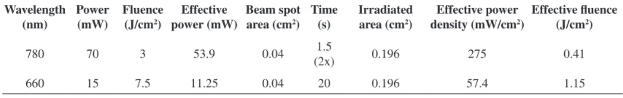
partially darkened room to avoid interference from external light sources. The cells in the control group were subjected to the same procedures but were not irradiated. The irradiation parameters (described in Table 1) were selected based on previous studies18-20.
The device output power was assessed using a power meter (Laser Check, MM Optics, São Carlos, SP,
Brazil). Table 1 describes the output values as well as the effective values considering the passage of light through the polypropylene tubes with the cell precipitates, as previously described21.
Experimental groups
Group 1
Control (non-activated, non-irradiated macrophages); Group 2: macrophages activated by means of LPS and IFN-γ; Group 3: macrophages irradiated with 660-nm laser; Group 4: macrophages activated by means of LPS and IFN- γ and irradiated with 660-nm laser; Group 5: macrophages irradiated with 780-nm laser; Group 6: macrophages activated by means of LPS and IFN-γ and irradiated with 780-nm laser.
Cell mitochondrial activity assay – MTT
The MTT assay is a colorimetric assay able to assess the ability of mitochondrial enzyme succinate dehydrogenase in viable cells to cleave MTT [3-(4,5-dimethylthiazol-2-yl)-2,5-diphnyltetrazolium bromide] tetrazolium rings, resulting in dark-blue formazan crystals. As the cell membrane is impermeable to formazan crystals, they are retained within viable cells and released following cell lysis. Macrophages (1 x 10³/well) were incubated in
then washed with 100 μL of phosphate-buffered saline (PBS); MTT (0.5 μg/mL) was added; and the plates were incubated three hours in a CO2 incubator at 37°C. Next, 100 μL of isopropanol was added, and an absorbance reading was performed at 620 nm using a plate reader (2020, Anthos, Eugendorf,
Austria).
Statistical analysis
All the tests were independently performed three times, and eight replicates were prepared from each sample. Data analysis included the calculation of the mean and standard deviation and analysis of variance (ANOVA), for which purpose the software
GraphPad InStat-3 was used. Statistical signiicance
was assessed by means of Tukey’s test and was considered acceptable when p≤0.05.
Results
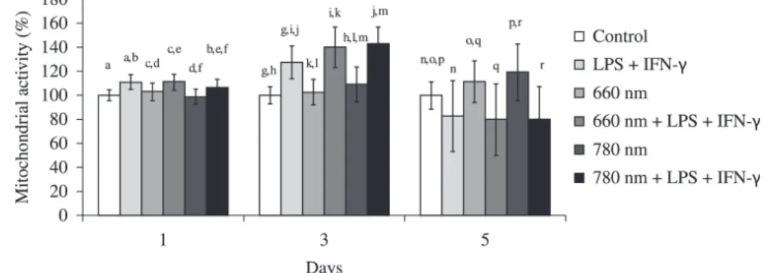
Activated cells (simulated inflammation) After one day of culture, the MA of activated macrophages was similar to the MA of activated macrophages irradiated at 660 nm and higher than the MA of activated macrophages irradiated at 780 nm (p<0.05). The MA of activated macrophages irradiated at 660 nm was higher (p<0.05) than the MA of activated macrophages irradiated at 780 nm. The MA of activated macrophages was higher (p<0.001) than the MA of the control group. The MA of activated and irradiated (660 nm and 780 nm) macrophages was higher (p<0.001) than the MA of the non-activated cells irradiated with the corresponding energy parameters (Figure 1). After three days of culture, the MA of the activated macrophages irradiated at 660 nm or 780 nm laser was higher (p<0.01 and p<0.001, respectively) than the MA of the activated macrophages. The MA of the activated macrophages irradiated by the 660 nm laser was similar to the MA of the activated cells irradiated at 780 nm. The MA of the activated macrophages was not only higher (p<0.001) than the control
group (non-activated, non-irradiated cells), but the difference was more patent (Figure 1).
After ive days of culture, the MA of the activated macrophages was similar to the MA of activated macrophages irradiated at 660 nm or 780 nm laser. The MA of the activated macrophages irradiated at 660 nm laser was similar to the MA of the activated cells irradiated by the 780 nm laser. The MA of the activated macrophages was lower (p<0.01) than the MA of the control group (Figure 1). Upon comparison of activated and irradiated cultures (using 660- and 780-nm lasers) with irradiated non-activated cultures, an inversion of the behavior found on days one and three was observed, as the MA of the activated and irradiated cells was lower than the MA of the non-activated macrophages irradiated with the corresponding energy parameters (p<0.001).
Non-activated cells
After one day of culture, there was no difference between the MA of the control group and the groups of irradiated cells. The MA of the cells irradiated at 660 nm was higher (p<0.01) than the MA of the cells irradiated at 780 nm (Figure 1). After three days of culture, irradiation at 660 nm did not induce changes in MA compared to non-irradiated cells, but irradiation at 780 nm induced an increase in MA compared to the control group (p<0.001). The MA of the cells irradiated at 660 nm was lower (p<0.05) than the MA of the cells irradiated at 780 nm (Figure 1). After ive days of culture, irradiation at 660 nm (p<0.05) and particularly at 780 nm (p<0.001) increased the MA of the irradiated cells compared to the non-irradiated cells. The MA of cells irradiated at 660 nm was similar to the MA of cells irradiated at 780 nm (Figure 1).
Discussion
The modulation of the various stages of skeletal muscle repair is mainly accomplished through changes in the activation proile of macrophages, resulting in changes in the phenotype and function
Table 1. Low-level laser therapy (LLLT) parameters.
Wavelength (nm)
Power (mW)
Fluence (J/cm2)
Effective power (mW)
Beam spot area (cm2)
Time (s)
Irradiated area (cm2)
Effective power density (mW/cm2)
Effective fluence (J/cm2)
780 70 3 53.9 0.04 (2x)1.5 0.196 275 0.41
of such cells1. For that reason, macrophages are
considered to be targets for therapeutic intervention1.
Several experimental and clinical studies conducted within the context of muscle injury rehabilitation have shown that LLLT can modulate the process of muscle repair22-28. However, no study
has yet assessed whether laser therapy can change the state of activation of macrophages.
In this study, we assessed the effect of LLLT applied with two different energy parameters on the mitochondrial activity of J774 macrophages one, three and ive days after irradiation. The cells were cultured under conditions of nutrient deiciency and treated with LPS and IFN-γ to simulate inlammation and induce the appearance of macrophage phenotype M1.
Previous studies that assessed the effects of LLLT and LED (light emitting diode) therapy on
macrophages or their precursors (monocytes) did not evaluate their mitochondrial activity but did evaluate various functions of such cells13,14,18-20,29-31.
Mitochondria exert a crucial modulatory effect on the pathway of activation of inflammatory macrophages that leads to the production of cytokines, i.e., the MAPK (Mitogen Activated Protein
Kinases) and NF-κb (Nuclear Factor-KappaB)
pathways32. When an inlammatory stimulus (e.g., LPS + IFNγ) triggers macrophage activation, the mitochondria amplify the MAPK pathway, resulting in increases of cytokines and other inlammatory mediators production33. The MTT assay, which was
used in this study, assesses mitochondrial activity and directly relects the status of cell activation32,33.
After one and three days of culture, the MA of the macrophages treated with IFNγ and LPS increased compared to the non-activated cells, showing that the
activation model used in this study was effective. The indings after ive days of culture were the opposite of the earlier ones, as the MA of the activated cells was lower than in the control group. The reason might be that by 5 days, the cell activation and/or viability had decreased as a function of the intense stimulation to which they had been subjected on the previous days and/or the action of the products they secreted.
The MA of the activated cells irradiated at 780 nm decreased after one day of culture. After three days of culture, irradiation at 660 nm and 780 nm exerted a positive modulation of the MA of macrophages, which might denote an increase in cell activation. After ive days of culture, irradiation with either laser no longer modulated the mitochondrial activity of the activated cells.
Relative to the non-activated cells, irradiation at 780 nm exerted a positive modulation of the MA after three days of culture. The same effect was found after ive days of culture with both laser energy parameters (660 and 780 nm).
Only the study by Young et al.13 assessed viability
and proliferation in an irradiated monocyte line, though with an 820 nm pulsed laser (15 mW; 2.4 J/cm2; 0.3 J).
After 36 hours of culture, the trypan blue exclusion
test showed an increase in the number of viable cells compared to the non-irradiated group. It is dificult, however, to compare those results with ours because the dosimetry parameters, methods and outcome are all different. In addition, Young et al.13 used
monocytes, whereas we assessed a macrophage cell line.
In fact, many studies have shown that LLLT has effects on several cell types, mostly through the activation of the mitochondrial respiratory chain, resulting in increased ATP production and the
induction of transcription factors34,35. Our indings,
therefore, might indicate that the energy applied to cells by means of laser irradiation was able to stimulate the above mentioned mechanisms, increasing the activation of non-activated macrophages (780-nm laser on day three and 660- and 780-nm lasers on day ive) and amplifying the effects in the activated macrophages.
Our results showed that the MA of activated cells irradiated at 780 nm was reduced after one day of culture. This inding corroborates the reports by Sousa et al.18, who observed a reduction in
TNF-α production 24 hours after the irradiation of activated M1 macrophages using the same dosimetry parameters and methods that we used.
Although in vitro studies afford standardized,
highly reproducible models and allow cell and molecular assessment, the results of such studies cannot be correlated with eventual clinical outcomes. Nevertheless, previous knowledge of the effect of LLLT and other therapeutic resources on the various cell types that compose muscle tissue is of paramount importance for the formulation of in vivo protocols that can exert more effective modulation of the muscle repair process.
In addition, accurate knowledge of the optical properties of the cells/tissues to be irradiated as well as of the barriers through which the light will pass is crucial. In the experimental model used in this study, the lasers had to pass through the test tube bottom to reach the macrophages. As a consequence, a part of the output energy was lost due to relection, dispersion and absorption by the polypropylene that composed the test tubes21. For that reason, the
effective (remaining) power values were included in the calculation of the power density and the energy density, which was performed according Silva et al.21.
For dosimetry parameters to be transferred from one experimental model to another, the behavior of light relative to the various barriers through which it passed before reaching the target should be accurately known. The absorption coeficient of the targeted tissue should also be known, as the therapeutic effects of light are fully dependent on the amount of absorbed energy.
Once the appropriate experimental data at the cell level and the data from animal and human studies become available, the clinical use of therapeutic resources will be based on scientiic evidence rather than on mere empiricism.
Conclusion
LLLT at 660 nm (15 mW, 7.5 J/cm2) and 780
nm (70 mW, 3 J/cm2) might modulate the activation
of J774 macrophages in a inlammatory condition simulation. Further studies are needed to elucidate the mechanisms that underlie such modulation as well as to assess the effects of irradiation on other relevant functions of macrophages.
Acknowledgements
To the Fundação de Amparo à Pesquisa do Estado de São Paulo (FAPESP grant 2011/14474-9), Brazil, to the Conselho Nacional de Desenvolvimento Científico e Tecnológico (CNPq grant no. 303662/2012-3), Brazil, and to the Coordenação de Aperfeiçoamento de Pessoal de Nível Superior (CAPES/PROSUP) for their inancial support. To Carlos Pelleschi Taborda (ICB – USP), for providing the J774 macrophage cell line.
References
1. Tidball JG, Villalta SA. Regulatory interactions between muscle and the immune system during muscle regeneration. Am J Physiol Regul Integr Comp Physiol. 2010;298(5):R1173-87. PMid:20219869 PMCid:PMC2867520. http://dx.doi.org/10.1152/ ajpregu.00735.2009
2. Wehling M, Spencer MJ, Tidball JG. A nitric oxide synthase transgene ameliorates muscular dystrophy in mdx mice. J Cell Biol. 2001;155(1):123-31. PMid:11581289 PMCid:PMC2150800. http://dx.doi.org/10.1083/ jcb.200105110
3. Villalta SA, Nguyen HX, Deng B, Gotoh T, Tidball JG. Shifts in macrophage phenotypes and macrophage competition for arginine metabolism affect the severity of muscle pathology in muscular dystrophy. Hum Mol Genet. 2009;18(3):482-96. PMid:18996917 PMCid:PMC2638796. http://dx.doi.org/10.1093/hmg/ ddn376
4. Schwab N, Waschbisch A, Wrobel B, Lochmüller H, Sommer C, Wiendl H. Human myoblasts modulate the function of antigen-presenting cells. J Neuroimmunol. 2008;200(1-2):62-70. PMid:18644633. http://dx.doi. org/10.1016/j.jneuroim.2008.06.012
5. Nguyen HX, Tidball JG. Interactions between neutrophils and macrophages promote macrophage killing of rat muscle cells in vitro. J Physiol. 2003;547(Pt 1):125-32. PMid:12562965 PMCid:PMC2342622. http://dx.doi. org/10.1113/jphysiol.2002.031450
6. Gordon S. Alternative activation of macrophages. Nat Rev Immunol. 2003;3(1):23-35. PMid:12511873. http://dx.doi. org/10.1038/nri978
low-energy laser irradiation. Anat Rec. 1993;235(3):374-80. PMid:8430907. http://dx.doi.org/10.1002/ ar.1092350306
8. Bibikova A, Oron U. Regeneration in denervated toad (Bufo viridis) gastrocnemius muscle and the promotion of the process by low energy laser irradiation. Anat Rec. 1995;241(1):123-8. PMid:7879917. http://dx.doi. org/10.1002/ar.1092410116
9. Oliveira NM, Parizzotto NA, Salvini TF. GaAs (904-nm) laser radiation does not affect muscle regeneration in mouse skeletal muscle. Lasers Surg Med. 1999;25(1):13-21. http://dx.doi.org/10.1002/ (SICI)1096-9101(1999)25:1<13::AID-LSM3>3.0.CO;2-7 10. Weiss N, Oron U. Enhancement of muscle regeneration
in the rat gastrocnemius muscle by low energy laser irradiation. Anat Embryol (Berl). 1992;186(5):497-503. http://dx.doi.org/10.1007/BF00185463
11. Lopes-Martins RA, Marcos RL, Leonardo PS, Prianti AC Jr, Muscará MN, Aimbire F, et al. Effect of low-level laser (Ga-Al-As 655 nm)on skeletal muscle fatigue induced by electrical stimulation in rats. J Appl Physiol. 2006;101(1):283-8. PMid:16627677. http://dx.doi. org/10.1152/japplphysiol.01318.2005
12. De Almeida P, Lopes-Martins RÁ, Tomazoni SS, Silva JA Jr, De Carvalho PT, Bjordal JM, et al. Low-level laser therapy improves skeletal muscle performance, decreases skeletal muscle damage and modulates mRNA expression of COX-1 and COX-2 in a dose-dependent manner. Photochem Photobiol. 2011;87(5):1159-63. PMid:21749398. http:// dx.doi.org/10.1111/j.1751-1097.2011.00968.x
13. Young S, Bolton P, Dyson M, Harvey W, Diamantopoulos C. Macrophage responsiveness to light therapy. Lasers Surg Med. 1989;9(5):497-505. PMid:2811573. http:// dx.doi.org/10.1002/lsm.1900090513
14. Gavish L, Perez LS, Reissman P, Gertz SD. Irradiation with 780 nm diode laser attenuates inlammatory cytokines but upregulates nitric oxide in lipopolysaccharide-stimulated macrophages: implications for the prevention of aneurysm progression. Lasers Surg Med. 2008;40(5):371-8. PMid:18563774. http://dx.doi.org/10.1002/lsm.20635 15. Mesquita-Ferrari RA, Ribeiro R, Souza NHC, Silva CAA,
Martins MD, Bussadori SK, et al. No effect of low-level lasers on in vitro myoblast culture. Indian J Exp Biol. 2011;49(6):423-8. PMid:21702221.
16. Da Silva TD, Mesquita-Ferrari RA, Souza NHC, Silva CAA, Martins MD, Bussadori SK, et al. Efeito da laserterapia de baixa potencia sobre a proliferação de mioblastos C2C12. Fisioter Bras. 2010;11(3):216-20. 17. Fujihara NA, Hiraki KR, Marques MM. Irradiation
at 780 nm increases proliferation rate of osteoblasts independently of dexamethasone presence. Lasers Surg Med. 2006;38(4):332-6. PMid:16526043. http://dx.doi. org/10.1002/lsm.20298
18. Sousa LR, Cavalcanti BN, Marques MM. Effect of laser phototherapy on the release of TNF-alpha and MMP-1 by endodontic sealer-stimulated macrophages. Photomed Laser Surg. 2009;27(1):37-42. PMid:19182976. http:// dx.doi.org/10.1089/pho.2007.2220
19. Bolton PA, Young S, Dyson M. Macrophage responsiveness to light therapy- a dose response study.
Tissue repair research Unit Division of anatomy. 1990;2(3):101-6.
20. Bolton P, Young S, Dyson M. Macrophage responsiveness to light therapy with varying Power and energy densities. Laser Ther. 1991;3:105-11.
21. Silva DF, Mesquita-Ferrari RA, Fernandes KP, Raele MP, Wetter NU, Deana AM. Effective transmission of light for media culture, plates and tubes. Photochem Photobiol. 2012;88(5):1211-6. PMid:22540924. http:// dx.doi.org/10.1111/j.1751-1097.2012.01166.x
22. Dourado DM, Favero S, Baranauskas V, Da Cruz-Holing MA. Effects of the Ga-As laser irradiation on myonecrosis caused by Bothrops Moojeni snake venom. Lasers Surg Med. 2003;33(5):352-7. PMid:14677163. http://dx.doi. org/10.1002/lsm.10237
23. Barbosa AM, Villaverde AB, Guimaraes-Souza L, Ribeiro W, Cogo JC, Zamuner SR. Effect of low-level laser therapy in the inflammatory response induced by Bothrops jararacussu snake venom. Toxicon. 2008;51(7):1236-44. PMid:18439641. http://dx.doi. org/10.1016/j.toxicon.2008.02.007
24. Barbosa AM, Villaverde AB, Sousa LG, Munin E, Fernandez CM, Cogo JC, et al. Effect of low-level laser therapy in the myonecrosis induced by Bothrops jararacussu snake venom. Photomed Laser Surg. 2009;27(4):591-7. PMid:19530909. http://dx.doi. org/10.1089/pho.2008.2296
25. Mesquita-Ferrari RA, Martins MD, Silva JA Jr, Da Silva TD, Piovesan RF, Pavesi VC, et al. Effects of low-level laser therapy on expression of TNF-α and TGF-b in skeletal muscle during the repair. Lasers Med Sci. 2011;26(3):335-40. PMid:21053039. http://dx.doi. org/10.1007/s10103-010-0850-5
26. De Souza TO, Mesquita DA, Ferrari RA, Dos Santos Pinto D Jr, Correa L, Bussadori SK, et al. Phototherapy with low-level laser affects the remodeling of types I and III collagen in skeletal muscle repair. Lasers Med Sci. 2011;26(6):803-14. PMid:21761120. http://dx.doi. org/10.1007/s10103-011-0951-9
27. Baptista J, Martins MD, Pavesi VC, Bussadori SK, Fernandes KP, Dos Santos Pinto D Jr, et al. Inluence of laser photobiomodulation on collagen IV during skeletal muscle tissue remodeling after injury in rats. Photomed Laser Surg. 2011;29(1):11-7. PMid:20701543. http:// dx.doi.org/10.1089/pho.2009.2737
28. Fernandes KP, Alves AN, Nunes FD, Souza NH, Silva JA Jr, Bussadori SK, et al. Effect of photobiomodulation on expression of IL-1b in skeletal muscle following acute injury. Lasers Med Sci. 2013;28(3):1043-6. P M i d : 2 3 1 7 9 3 0 8 . h t t p : / / d x . d o i . o r g / 1 0 . 1 0 0 7 / s10103-012-1233-x
29. Mehrsai A, Afsharpad M, Afsharpad M, Mohydin M, Ansari B, Pourmand G, et al. The effect of low-level helium-neon (HeNe) laser radiation on the secretion of cytokines that promote chronic graft rejection – An in vitro study. Med Laser App. 2009;24(3):194-200. http:// dx.doi.org/10.1016/j.mla.2009.03.001
inflammatory protein-2 (MIP-2) mRNA expression and generation of intracellular reactive oxygen species in alveolar macrophages. Photomed Laser Surg. 2010;28(6):763-71. PMid:21142721. http://dx.doi. org/10.1089/pho.2009.2638
31. Dube A, Bansal H, Gupta PK. Modulation of macrophage structure and function by low level He-Ne laser irradiation. Photochem Photobiol Sci. 2003;2(8):851-5. PMid:14521221. http://dx.doi.org/10.1039/b301233f 32. Emre Y, Nübel T. Uncoupling protein UCP2: when
mitochondrial activity meets immunity. FEBS Lett. 2010;584(8):1437-42. PMid:20227410. http://dx.doi. org/10.1016/j.febslet.2010.03.014
33. Gerlier D, Thomasset N. Use of MTT colorimetric assay to measure cell activation. J Immunol M e t h o d s . 1 9 8 6 ; 9 4 ( 1 - 2 ) : 5 7 - 6 3 . h t t p : / / d x . d o i . org/10.1016/0022-1759(86)90215-2
34. Chung H, Dai T, Sharma SK, Huang YY, Carroll JD, Hamblin MR. The nuts and bolts of low-level laser
(light) therapy. Ann Biomed Eng. 2012;40(2):516-33. PMid:22045511 PMCid:PMC3288797. http://dx.doi. org/10.1007/s10439-011-0454-7
35. Gao X, Xing D. Molecular mechanisms of cell proliferation induced by low power laser irradiation. J Biomed Sci. 2009;16:4. PMid:19272168 PMCid:PMC2644974. http:// dx.doi.org/10.1186/1423-0127-16-4
Correspondence