Regulatory T and B cells in asthmatic women: variations from pregnancy to postpartum
Treg and Breg: pregnancy to postpartum
Martins C, PhD
Corresponding address: Immunology, NOVA Medical School, Campo dos Mártires da Pátria, 1169-056 Lisbon, Portugal
Mailing address: catarina.martins@nms.unl.pt
Affiliation: 1 CEDOC, Chronic Diseases Research Center, NOVA Medical School|FCM; Universidade Nova de Lisboa, Lisbon, Portugal
Lima J, MD
Affiliation: 2 Ginecologia e Obstetrícia, Hospital CUF Descobertas, Lisbon, Portugal; 1 CEDOC, Chronic Diseases Research Center, NOVA Medical School|FCM, Universidade Nova de Lisboa, Lisbon, Portugal Nunes G, BSc
Affiliation: 1 CEDOC, Chronic Diseases Research Center, NOVA Medical School|FCM; Universidade Nova de Lisboa, Lisbon, Portugal
Borrego LM, MD, PhD
Affiliation: 1 CEDOC, Chronic Diseases Research Center, NOVA Medical School|FCM; Universidade Nova de Lisboa, Lisbon, Portugal; 4 Imunoalergologia, Hospital CUF Descobertas, Lisbon, Portugal
This article has been accepted for publication and undergone full peer review but has
not been through the copyediting, typesetting, pagination and proofreading process,
which may lead to differences between this version and the Version of Record. Please
Abstract
Background: Allergic asthma and rhinitis are common in pregnancy. The immune mechanisms
underlying the effects of pregnancy in asthma and vice-versa are not completely understood.
Objectives: This work aimed to study the evolution of regulatory T and B cells in asthmatic pregnant
women, from late pregnancy till postpartum.
Methods: Four groups of women were enrolled for this study: third trimester pregnant women,
asthmatic (n=24) and healthy (n=43), and non-pregnant women, asthmatic (n=33) and healthy (n=35).
Pregnant women were also evaluated postpartum (>6 weeks after delivery). Blood samples were taken
from each woman and flow cytometry was used to characterize circulating regulatory T and B cells.
Foxp3 expression was assessed within CD4DimCD25Hi regulatory T cells.
Results: In asthmatic and healthy pregnant women, regulatory T cells did not oscillate significantly from
pregnancy to postpartum, but CD24HiCD38Hi regulatory B cells, decreased in pregnancy, rose significantly
postpartum. Foxp3 expression in regulatory T cells was also impaired during pregnancy in asthmatic and
healthy pregnant women, recovering postpartum. Nevertheless, asthmatic pregnant women presented
higher Foxp3 expression than healthy pregnant women (p=0.007), probably due to the use of control
medication.
Conclusions: Women with controlled asthma present variations in regulatory cell subsets during
pregnancy and postpartum. The similar pattern observed for Foxp3 expression and CD24HiCD38Hi
regulatory B cells during this period corroborates the interaction established between regulatory T and
B cells in immune responses. Considering the immunomodulatory potential of these immune mediators,
more studies are needed to evaluate their relation with asthma and rhinitis complications in pregnancy.
Resumen
Antecedentes: El asma y la rinitis alérgica son enfermedades comunes durante el
embarazo. A pesar de ello, no están completamente esclarecidos los mecanismos
inmunológicos del embarazo implicados en el asma y viceversa.
Objetivos: Este trabajo tuvo como objetivo el estudiar la evolución de los linfocitos T y
B reguladores en mujeres asmáticas embarazadas, desde fases tardías del embarazo
hasta después del parto.
Métodos: Se incluyeron cuatro grupos de mujeres para este estudio: mujeres
embarazadas en su tercer trimestre, asmáticas (n = 24) y sanas (n = 43), y mujeres no
embarazadas, asmáticas (n = 33) y sanas (n = 35). Las mujeres embarazadas también
fueron evaluadas después del parto (> 6 semanas después del parto). Se tomaron
muestras de sangre de cada mujer y se realizó citometría de flujo para caracterizar los
linfocitos T y B reguladores circulantes. La expresión de Foxp3 se evaluó en los
linfocitos T reguladores CD4
DimCD25
Hi.
Resultados: En las mujeres embarazadas, tanto sanas como asmáticas, los linfocitos T
reguladores no oscilaron de manera significativa desde el embarazo hasta después del
parto. Sin embargo, en los linfocitos B reguladores CD24
HiCD38
Hi, se observó una
disminución durante el embarazo que aumentó significativamente después del parto.
La expresión de Foxp3 en los linfocitos T reguladores también se vio alterada durante
el embarazo tanto en las mujeres embarazadas asmáticas como en las sanas,
normalizándose en el posparto. No obstante, las mujeres asmáticas embarazadas
presentaron niveles de expresión de Foxp3 superiores a los de las mujeres
embarazadas sanas (p = 0,007), probablemente debido a la utilización de medicación
de control.
Conclusiones: Las mujeres con asma controlada, durante el embarazo y después del
parto, presentan variaciones en los diferentes subtipos linfocitos reguladores. El similar
comportamiento que se observa para la expresión de Foxp3 y los linfocitos B
reguladores CD24
HiCD38
Hiapoya la interacción que se establece en la respuesta
inmunitaria, entre los linfocitos T y B reguladores, durante este período. Teniendo en
cuenta el potencial inmunomodulador de estos mecanismos, se necesitan más
estudios para evaluar su relación con las complicaciones del asma y la rinitis durante
el embarazo.
Introduction
For women in fertile age, allergic asthma and rhinitis can present risks for pregnancy [1]. Disease course
may improve, remain unchanged, or worsen during pregnancy, and asthma exacerbations may have a
significant impact in fetal development and birth weight, besides being related with preterm delivery
[1]. Also, maternal asthma is a risk factor for the development of asthma in asthmatic women´s progeny
[2].
Pregnancy challenges the immune system, as it must assume tolerance towards the semi-allogenic
fetus, without compromising mother defenses. Th2 cells, hallmark of atopic diseases, are typically
associated to gestation and appear to contribute to the maintenance of pregnancy [3]. Nonetheless,
more than biased Th2 immune response, pregnancy presents rather strict regulatory mechanisms that
balance cytokine production [4].
Regulatory T cells (Tregs) are known to be expanded during early pregnancy [5], and seem to be
decreased and/or functionally impaired in asthmatics [6, 7]. Yet, Tregs and IL10, an anti-inflammatory
cytokine secreted by these cells, have been implicated in asthma´s amelioration [8]. Recently, B cells
have been considered to have an important role in Th2 responses and airway inflammation [9]. As for
regulatory B cells (Bregs), its impaired regulatory activity was also reported in allergic asthma [10], and
Bregs have a close relation to Tregs [11].
Modifications in T and B cell subsets in healthy [12-14] and asthmatic [15, 16] pregnant women have
been reported. However, not much is known about B cells neither about the immune profile in
postpartum, when an immune reactivation is believed to happen [17].
Our aim was to monitor pregnant women with asthma, from late pregnancy till postpartum in order to
characterize Tregs, Bregs and IL10 expression, and the impact of pregnancy on these immune
Methods
Study Subjects
Sequential pregnant women with atopic asthma (AP), healthy pregnant women (HP), as well as
non-pregnant women with atopic asthma (ANP) and healthy non-non-pregnant women (HNP) of reproductive
age followed at CUF Descobertas Hospital were enrolled for this study. All women were informed about
the nature of the study and provided written informed consent at recruitment. Blood samples were
collected once for non-pregnant women and twice for pregnant: the pregnancy time point occurred in
the third trimester of gestation ( between weeks 31 and 36 of gestation) and the postpartum time point
at least 6 weeks after delivery. All women completed questionnaires for demographic and clinical data
in each time point. Diabetes; hypertension; autoimmune diseases; any active infectious disease
including hepatitis and HIV; any other allergic (except for atopic dermatitis and allergic rhinitis) or
respiratory disease; and smoking for the last 6 months before sample collection, represented exclusion
criteria for all groups. Multiple gestation, pregnancy complications and prenatal use of any medication
(other than vitamins, folic acid, and iron supplements) were exclusion criteria for HP. The same criteria
were applied to AP, except for therapeutics.
AP and ANP groups included women with both atopic asthma and rhinitis, physician diagnosed
according to current international guidelines [18-20], with documented sensitization to aeroallergens
(by skin prick tests and/or specific IgE quantification). Immunotherapy, performed in the past or
present, was considered an exclusion criteria for AP and ANP. Asthma and rhinitis were treated
according to the current guidelines [19, 20]. All AP and ANP were under inhaled corticosteroid (ICS)
therapy (low-median dose, 200-400 ug beclomethasone /daily), and/or long-acting Beta-agonists (LABA)
and antileukotrienes. At recruitment, AP and ANP presented no asthma exacerbation for at least 6
weeks before sample collection. Asthma symptoms were evaluated according to GINA guidelines [19,
20], using daytime symptoms, night waking due to asthma, reliever needed for symptoms and activity
limitation due to asthma in the assessment of disease control. Table 1 resumes demographic and
The study was approved by CUF Descobertas Hospital and NOVA Medical School Ethics Committees. All
investigations were conducted according to the Declaration of Helsinki.
Regulatory T and B cells
Peripheral blood samples were collected into EDTA-containing tubes using standard aseptic
venipuncture techniques. Cells were processed less than 24 hours after collection.
For Tregs phenotyping, the Human FoxP3 Buffer Set (BD Pharmingen, San Jose, CA) was used, according
to the manufacturer’s instructions. First, cells were lysed with BD FACS Lysing (BD Biosciences), and
incubated with anti-CD25 PE (clone BC96, Biolegend, San Diego, CA) and anti-CD4 PerCP Cy5.5 (clone
SK3, Biolegend). After being fixed and permeabilized with the reagents supplied in the kit set, cells were
stained with Anti-Foxp3 Alexa Fluor 488 (clone 259D/C7, BD Pharmingen). At least 10,000 CD4+T cells
were acquired.
For the evaluation of Bregs, the panel of mAbs included anti-CD19 PerCP Cy5.5 (clone HIB19, Biolegend),
anti-CD24 PE (clone ML5, Biolegend), anti-CD27 FITC (clone O323, Biolegend) and anti-CD38 APC (clone
HIT2, Biolegend). A lyse-wash protocol was used. Briefly, cells were incubated for 15 minutes with
monoclonal antibodies (mAbs), and then lysed with BD FACS Lysing (BD Biosciences). Acquisition was
performed after a wash step with PBS. At least 2,000 CD19+ B cells were acquired.
Cell culture for intracellular cytokine stimulation
Heparin whole blood was used to evaluate IL10 expression in T and B cells. Cells were stimulated with
PMA (50ng/mL; Sigma Aldrich, St. Louis, MO), calcium ionophore (1µg/mL, Sigma Aldrich) and LPS
(10µg/mL, Sigma Aldrich) [21], and incubated for 5h, at 37ºC in a 5% CO2 atmosphere, in the presence of
Brefeldin A (1.0μg/ml, BD Pharmingen). For each sample, unstimulated tubes were incubated in parallel,
and were further used as stimulation and staining controls. The protocol was performed according to
the instructions of Cytofix-Cytoperm kit (BD Pharmingen). Briefly, the initial surface staining step
included an incubation with anti-CD3-FITC (clone SK7, BDBiosciences), anti-CD8 APC (clone SK1,
Biolegend) and anti-CD19 PerCP Cy5.5 (clone HIB19, Biolegend). After fixing and permeabilizing, cells
were marked with anti-IL10 PE (clone JES3-19F1, Biolegend). A minimum of 2,000 B cells (CD19+) or
Multicolor Flow Cytometry and Data Analysis
Flow cytometry was performed in a 4-color BD FACSCalibur (BD Biosciences, San Jose, CA), equipped
with a 488nm blue laser and a 647nm red laser. Equipment setup, calibration and quality control
protocols were performed to assure trough time stability in measurements. Figure 1 presents the gating
strategy for Treg assessment and Foxp3 expression (assessed in geometric mean values of Mean
Fluorescence Intensity (MFI) units). To reduce the impact of day-to-day variation on this evaluation, a
ratio between the MFI of Foxp3 in CD4DimCD25Hi Tregs and the MFI of Foxp3 in CD4- Lymphocytes was
performed. The gating strategy for Bregs subsets and IL10 secretion are presented in Figures 2 and 3.
All evaluations were performed directly in whole blood.
Statistical Analysis
Absolute frequencies were used in categorical variables, expressed also as percentages, and analyzed by
Fisher’s exact test or Chi-square. D'Agostino & Pearson test was carried out to assess normality of
distributions. Data normally distributed were presented as mean±standard deviation, otherwise as
median and interquartile range. Multiple group evaluation was performed with One-way ANOVA
(ANOVA I) or Kruskal-Wallis test, followed by Tukey’s or Dunn’s multiple comparison tests, respectively.
Regarding paired groups, data were analyzed with paired Student’s t-test or Wilcoxon test. Unpaired
Student’s t-test or Mann–Whitney test were used to compare each 2 independent groups.
Statistical significance was defined by a p-value <0.05. All data were analyzed using GraphPad Prism
software, version 6.01 for Windows (GraphPad Software, La Jolla, California, USA,
http://www.graphpad.com). Tukey box-and-whiskers graphs, also obtained with GraphPad Prism, were
used to present results.
Results
Demographic and anthropometric data
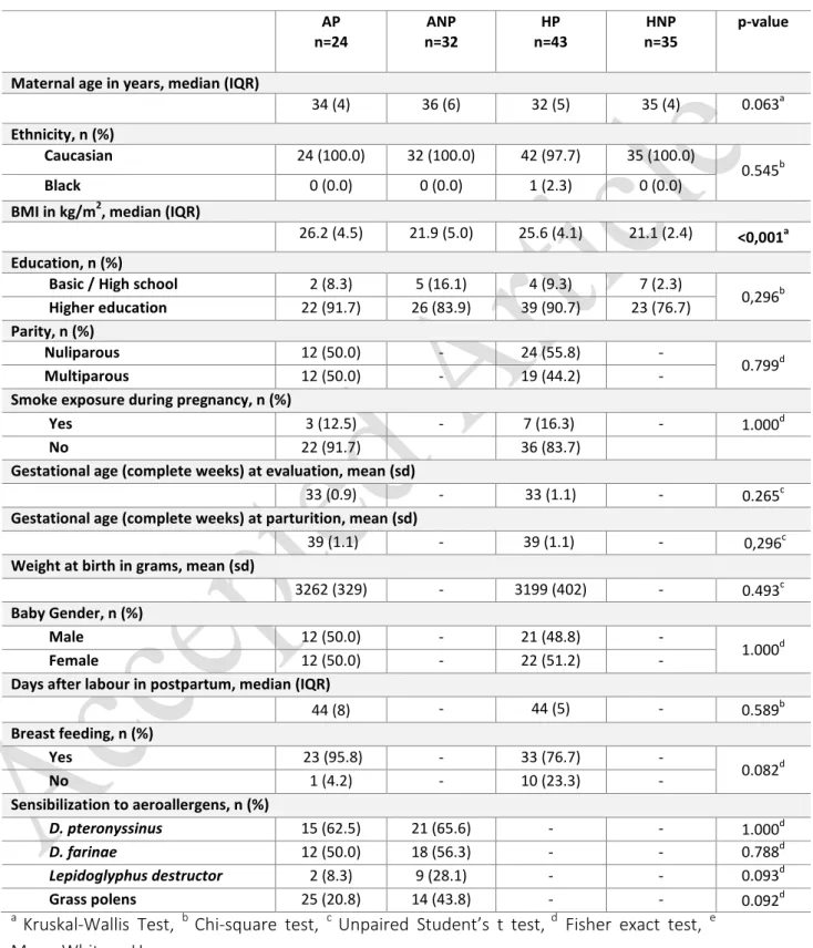
Demographic and anthropometric evaluations are summarized in Table 1. Analyzing all groups of
women (AP, n=24; ANP, n=32; HP, n=43; HNP, n=35) for demographic data, statistical significance was
significant p-value, with the subsequent Dunn's multiple comparisons reporting significantly higher BMI
values in pregnant women (p<0.001 for HP vs HNP and p=0.001 for AP vs ANP). Regarding parity, AP and
HP groups were comparable.
Asthma and/or rhinitis complications were reported as illustrated in Table 1: 4 (16.7%) AP presented
asthma exacerbations and 2 (8.3%) presented rhinitis (nasal symptoms) in the 3rd trimester of
pregnancy. Postpartum, 1 AP (4.2%) reported asthma exacerbation and 2 AP (8.3%) reported rhinitis
(nasal symptoms). None of these complications required hospitalization or treatment at emergency
department. Though medicated during pregnancy, postpartum only 13 AP (54.2%) remained under
therapeutics (ICS therapy and/or LABA and antileukotrienes).
Final postpartum measurements were carried out on median 44 (8) days after delivery.
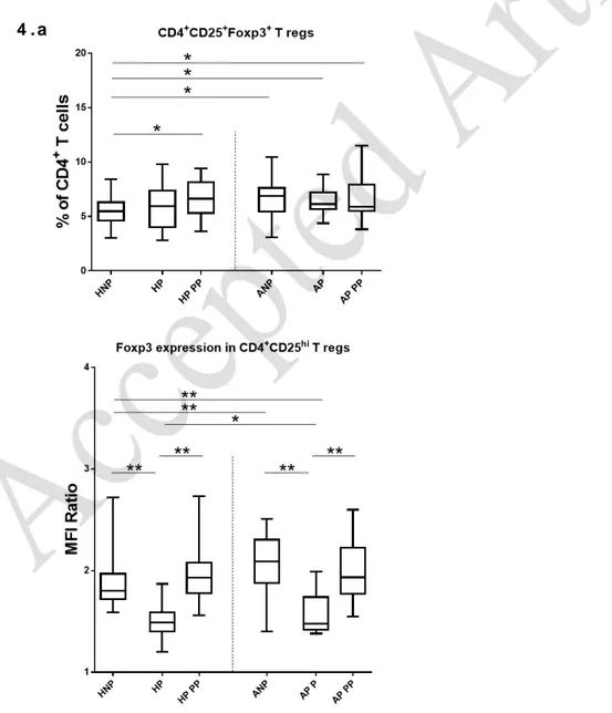
Increase of CD4+CD25HiFoxp3+ Tregs in ANP and in AP postpartum
In the comparison of non-pregnant asthmatic and non-asthmatic patients, significant differences were
observed in CD4+CD25HiFoxp3+ Tregs (p=0.002), increased in ANP (Figure 4.a).
Following the evolution of pregnant women from 3rd trimester of pregnancy to postpartum, no
differences were reported either within AP or HP in the paired evaluation of time points.. Comparing
Tregs of 3rd trimester pregnant women vs non-pregnant women, no significant differences were found
for the comparisons of AP vs ANPand HP vs HNP. Nevertheless, in postpartum, Tregs were increased in
both AP and HP compared to HNP (AP vs HNP, p=0.041; HP vs HNP, p=0.005), which seems to point
towards an increase of Tregs after pregnancy.
Foxp3 expression is increased in asthmatic women, but oscillates from pregnancy to postpartum
We also studied the levels of Foxp3 expression in CD4DimCD25Hi Tregs (Figure 4.a). In ANP, Foxp3
expression was augmented (p<0.001) compared to HNP.
During pregnancy, and at least until delivery (data not shown), decreased Foxp3 expression within
CD4DimCD25Hi Tregs was observed in both AP and HP. This decrease was more pronounced in HP, with
poorer expression levels then the ones observed in AP (p=0.007). Both groups of women significantly
down regulated Foxp3 expression in the third trimester of pregnancy, compared to non-pregnant
women (vs ANP and vs HNP; p<0.001). Postpartum,Foxp3 expression levels increased significantly in AP
expression levels similar to the ones observed in ANP; HP recovered their Foxp3 expression levels as
well, reaching values similar to HNP.
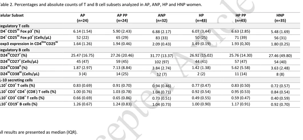
CD24HiCD38Hi regulatory B cells are augmented postpartum in AP and HP
As displayed in table 2, CD24HiCD27+ Bregs showed similar levels in all groups of women studied, and in
all time points. Thus, no differences were observed either in their monitoring from pregnancy to
postpartum in AP and HP (Figure 4.b). CD24HiCD38Hi Bregs, a transitional subset of B cells, displayed
mostly pregnancy-associated changes. First, considering ANP vs HNP, comparable values were
obtained. No differences were reported regarding comparisons of AP vs HP, in the time points analyzed.
Along pregnancy, CD24HiCD38Hi Bregs fluctuate with the same pattern observed for Foxp3 expression:
reduced proportions of circulating CD24HiCD38Hi Bregs during pregnancy in AP and HP (compared to
non-pregnant controls; p≤0.004). Postpartum, both AP and HP raised significantly the proportions of
CD24HiCD38Hi Bregs, compared to the levels observed in the 3rd trimester of pregnancy (p<0.001), but
also compared to ANP and HNP (p≤0.003).
In 3rd trimester, a positive correlation was found between CD24HiCD38Hi Bregs and the levels of Foxp3
expression in HP (p=0.002, r=0.457; Spearman correlation test), but significance is lost postpartum.
In ANP, the percentages of IL10-secreting T cells (CD3, CD4 and CD8) were increased compared to HNP
(p=0.024 for CD3; p=0.034 for CD4; p=0.009 for CD8). However, 3rd trimester AP presented only
significantly increased percentages of IL10-secreting B cells, but not T cells, compared to both HP
(p=0.022) and HNP (p=0.008). Postpartum, IL10-secreting T cells (CD3 and CD8) were again augmented
in AP compared to HNP (p=0.046 for CD3; p=0.016 for CD8), but also IL10-secreting B cells (p=0.031).
Curiously, in postpartum, HP presented higher percentages of IL10-secreting B cells compared to HNP
(p=0.019), possibly sustaining an impaired IL10 production by B cells in healthy pregnancy. Within the
pregnant women groups (AP and HP), paired analyses revealed no significant changes from pregnancy
to postpartum in AP, but a significant increase of IL10-secreting B cells in HP in the later evaluation
Discussion
Regulatory cells have an important role in immune tolerance and inflammation. To our knowledge this is
the first study to report variations of regulatory T and B cell subsets in women with asthma, from the
third trimester of pregnancy till postpartum, addressing also IL10 secretion profiles. It seems that
though Tregs do not oscillate expressively along this period, CD24HiCD38Hi Bregs decrease in the third
trimester of pregnancy, and present an important increase postpartum, in both asthmatic and healthy
women. Moreover, the reduction of Foxp3 expression in Tregs in late pregnancy (and at least until
delivery), was also overcome 6 weeks after delivery. Considering IL10 production, women with asthma
presented distinguishing features from healthy, with more IL10 producing T cells than HNP. In the 3rd
trimester of pregnancy, IL10-secreting B cells were augmented in AP, compared to HP, but no
differences were reported in postpartum.
Tregs have been involved in several human pathologies . Their number increases during early pregnancy
and declines from mid-gestation onwards [5]. As observed in our study, other authors reported similar
Tregs percentages in third trimester pregnant women and non-pregnant controls [5, 22]. Wegienka et
al. [23], reported a progressive augment of Tregs percentages from the prenatal period until the first 12
months postpartum, in both atopic and non-atopic patients, similarly to our findings. Despite our
monitoring timings are not the same, we also found that at 6 weeks after delivery, both AP and HP
presented higher percentages of Tregs compared to healthy non-pregnant controls. Different strategies
for cell identification, distinct gestational periods or confrontation with animal models are also possible
additional biases.
Our results shown that pregnant and non-pregnant women, with controlled asthma, present higher
percentages of Tregs than HNP, which are maintained in postpartum. Some studies have already
reported an increased Tregs population in children with asthma under ICS treatment, compared to
non-asthmatic age-matched controls [24]. Nevertheless, Bohacs et al. [15] reported comparable Tregs in
healthy non-pregnant and asthmatic (pregnant and non-pregnant) women, although asthmatics
presented higher values. Similar values for Tregs were reported in asthmatic patients taking ICS,
independently of the regularity on its intake. We also observed comparable values in treated and
untreated AP in postpartum (data not shown). Taking into consideration that all AP had been under ICS
may suggest that the impact of ICS in circulating Tregs frequency may be sustained even after the
treatment is stopped. Another effect of ICS is the upregulation of Foxp3 expression. Karagiannidis et al.
[25] reported that ICS-treated asthmatic patients presented significantly higher values of Foxp3
expression than healthy controls. By contrast, Provoost et al. [26] only reported augmented expression
in ICS treated asthmatics compared to untreated patients, but not compared to healthy controls.
Methodological differences can justify these opposite results; nevertheless, our data seem to be in
accordance with Karagianiddis [25]. Not only ANP presented higher Foxp3 expression than HNP, but
also, during pregnancy, AP showed higher levels compared to HP. Yet, an important decrease in Foxp3
expression in pregnancy was observed in both AP and HP. Recently, progesterone receptors have been
identified in Tregs [27], supporting the immunomodulatory potential of pregnancy hormones. Without
compromising the regulatory potential of Tregs during pregnancy, and even promoting their
proliferation [28] and proportions [29], progesterone and 17β-estradiol are thought to reduce Foxp3
expression on Tregs in second trimester pregnant women [30]. Our data support these findings,
extending this decrease to the third trimester of gestation. Furthermore, we have shown that
ICS-treated AP also have this pregnancy-derived decrease of Foxp3 expression, though maintaining higher
values than HP. Recognizing that Foxp3 expression can be induced in activated T cells, and that our
study did not assess the functional status of Tregs, we can only speculate a functional normality for
these cells in AP. Considering the protective role of Tregs towards asthma manifestations [31], AP
presenting controlled asthma at recruitment is in favor of a normal function of regulatory cells in these
patients. Nevertheless, further studies are needed to complete and confirm our observations.
In postpartum, Foxp3 expression levels normalized in AP and HP, reaching non-pregnant controls,
reinforcing that pregnancy induces modulation of Foxp3 expression.
Bregs evaluation in asthmatic pregnant women constitutes the innovation of our study. The regulatory
capacities of B cells have been recently reported [32], though there´s a lack of consensus on their
phenotype [33]. Both CD24HiCD27+ and CD24HiCD38Hi B cell subsets have been considered to have
regulatory functions, namely by the secretion of IL10 [10, 34]. Patients with asthma and/or AR have
shown decreased frequencies of CD24HiCD27+ Bregs [10, 35], but increased CD24HiCD38Hi Bregs has also
been spotted in asthmatic patients without medication [10]. In our study, Bregs subsets presented no
were under medication. As defended for Tregs [24], our data suggest that the beneficial effects of
therapeutics (such as ICS) in the course of atopic diseases can also be related to the normalization of
Bregs frequencies in treated patients.
Interestingly, CD24HiCD38Hi Bregs are modulated by pregnancy. Pregnancy is associated with B cell
lymphopenia and B cell lymphopoiesis arrestment [13], which probably decreases circulating transitional
B cell subsets such as CD24HiCD38Hi Bregs. These modifications were importantly overcome in
postpartum, with CD24HiCD38Hi Bregs reaching levels significantly higher than those observed in
non-pregnant controls, traducing maternal immune system’s recovery after gestation. Interestingly, in mice,
transitional B cells express high levels of prolactin receptors and hyperprolactinemia has been shown to
increase transitional but not mature B cells [36]. A prolactin-mediated response can thus explain the
accumulation of transitional B cells postpartum, when the hormone levels raise up to 30 times,
compared to pre-pregnancy levels [37].
It is believed that the promotion of Tregs differentiation is one of the mechanisms by which
CD24HiCD38Hi Bregs operate in healthy individuals [34]. Thus, the elevation of CD4+CD25HiFoxp3+ Tregs
postpartum in HP and AP, also reported by other authors [23], may be mediated by CD24HiCD38Hi Bregs.
Recent experimental data also concluded that CD24HiCD38Hi Bregs promote Foxp3 expression in
co-cultured CD4 T cells [34]. In our study, Foxp3 expression and CD24HiCD38Hi Bregs evolved similarly from
pregnancy to postpartum, and there was a positive correlation between these parameters in HP,
reinforcing the idea of Bregs modulating Tregs.
The profile of CD24HiCD38Hi Bregs and Foxp3 expression is comparable in asthmatic and non-asthmatic
women, sustaining a pregnancy-derived pattern, independently of either therapeutics or asthma and
rhinitis.
Studies approaching IL10 production by PBMCs during pregnancy described similar levels before
pregnancy and in late pregnancy [38]. In line with these reports, our data showed that IL10-secreting T
and B cells were similar in 3rd trimester HP women, compared to HNP. However, in postpartum, HP
presented increased frequencies of IL10+ B cells after LPS stimulation. The increase of circulating
CD24HiCD38Hi Bregs in postpartum (the subset in which IL10 secretion is mainly described in B cells) [32,
34], could be a possible explanation. Other studies addressed the production of IL10 in asthmatic and
differences in PBMC production were identified comparing asthmatic and non-asthmatic women [39],
IL10 serum levels were increased in allergic mothers (at delivery) and their children [40]. As far as we
know, we report for the first time the distinct profiles of IL10-secreting T and B cells in asthmatic
pregnant and non-pregnant women. We stress out that ANP presented increased IL10-secreting T cells,
but during pregnancy IL10-secreting B cells were increased in asthmatic women, compared to healthy
ones. Nonetheless, Tregs can secrete IL10, as well as other subsets of T cells, such as Th2 cells [4]. Th2
cells, associated to both pregnancy and allergic diseases, may support the similar levels observed
amongst AP, ANP and HP. However, during pregnancy, AP have a distinct capacity of B cells to secrete
IL10, compared to HP and HNP, presenting similar (vs HP) or even lower (vs HNP) frequencies of
CD24HiCD38Hi Bregs. Corroborating what was observed in ANP, in postpartum, AP also presented
increased IL10-secreting B and T cells, compared to HNP.
At follow-up, we realized that several AP women stopped therapeutics during postpartum. Considering
the distinguishing features of immune profiles in postpartum, we compared AP with and without
therapeutics, and concluded that studied parameters were similar in both subgroups, being
independent from therapeutics.
Monitoring AP pre-pregnancy and throughout more pregnancy time points would have been ideal,
thought difficult to accomplish from a financial and practical point of view. Nevertheless, important
immune events are known to occur during the 3rd trimester of pregnancy, recovering in the postpartum,
as we were also able to identify in our study.
Overall, we conclude that pregnant women with controlled asthma present a similar profile towards
healthy pregnant women, though with few distinctive features regarding mostly Foxp3 expression. The
changes observed in postpartum reinforce the idea of being pregnancy-dependent, probably hormone
driven. More studies will clarify if these parameters can be used to assess complications during
pregnancy in women with asthma and eventually influence the immune profile of the asthmatic
Acknowledgements
The authors would like to thank Tiago Domingues for technical support on statistical analysis.
Conflicts of interests
The authors declare no conflicts of interests. The authors alone are responsible for the content and writing of the paper. The authors have no financial sources to declare for this work.
Conflicts of interests
The authors declare no conflicts of interests. The authors alone are responsible for the content and
References
1.
Namazy J, Schatz M. The Treatment of Allergic Respiratory Disease During Pregnancy. J
Investig Allergol Clin Immunol. 2016;26(1):1-7; quiz 2p following 7.
2.
Xu R, DeMauro SB, Feng R. The impact of parental history on children's risk of asthma:
a study based on the National Health and Nutrition Examination Survey-III. J Asthma Allergy.
2015;8:51-61.
3.
Piccinni MP, Lombardelli L, Logiodice F, Kullolli O, Romagnani S, Le Bouteiller P. T
helper cell mediated-tolerance towards fetal allograft in successful pregnancy. Clin Mol
Allergy. 2015;13(1):9.
4.
Halonen M, Lohman IC, Stern DA, Spangenberg A, Anderson D, Mobley S, Ciano K, Peck
M, Wright AL. Th1/Th2 patterns and balance in cytokine production in the parents and infants
of a large birth cohort. J Immunol. 2009;182(5):3285-93.
5.
Zhao JX, Zeng YY, Liu Y. Fetal alloantigen is responsible for the expansion of the
CD4(+)CD25(+) regulatory T cell pool during pregnancy. J Reprod Immunol. 2007;75(2):71-81.
6.
Hartl D, Koller B, Mehlhorn AT, Reinhardt D, Nicolai T, Schendel DJ, Griese M,
Krauss-Etschmann S. Quantitative and functional impairment of pulmonary CD4+CD25hi regulatory T
cells in pediatric asthma. J Allergy Clin Immunol. 2007;119(5):1258-66.
7.
Lin YL, Shieh CC, Wang JY. The functional insufficiency of human CD4+CD25 high
T-regulatory cells in allergic asthma is subjected to TNF-alpha modulation. Allergy.
2008;63(1):67-74.
8.
Bohm L, Maxeiner J, Meyer-Martin H, Reuter S, Finotto S, Klein M, Schild H, Schmitt E,
Bopp T, Taube C. IL-10 and regulatory T cells cooperate in allergen-specific immunotherapy to
ameliorate allergic asthma. J Immunol. 2015;194(3):887-97.
9.
Drake LY, Iijima K, Hara K, Kobayashi T, Kephart GM, Kita H. B cells play key roles in
th2-type airway immune responses in mice exposed to natural airborne allergens. PLoS One.
2015;10(3):e0121660.
10.
van der Vlugt LE, Mlejnek E, Ozir-Fazalalikhan A, Janssen Bonas M, Dijksman TR,
Labuda LA, Schot R, Guigas B, Moller GM, Hiemstra PS, Yazdanbakhsh M, Smits HH.
CD24(hi)CD27(+) B cells from patients with allergic asthma have impaired regulatory activity in
response to lipopolysaccharide. Clin Exp Allergy. 2014;44(4):517-28.
11.
Amu S, Saunders SP, Kronenberg M, Mangan NE, Atzberger A, Fallon PG. Regulatory B
cells prevent and reverse allergic airway inflammation via FoxP3-positive T regulatory cells in a
murine model. J Allergy Clin Immunol. 2010;125(5):1114-24 e8.
12.
Tilburgs T, Roelen DL, van der Mast BJ, van Schip JJ, Kleijburg C, de Groot-Swings GM,
Kanhai HH, Claas FH, Scherjon SA. Differential distribution of CD4(+)CD25(bright) and
CD8(+)CD28(-) T-cells in decidua and maternal blood during human pregnancy. Placenta.
2006;27 Suppl A:S47-53.
13.
Muzzio DO, Soldati R, Ehrhardt J, Utpatel K, Evert M, Zenclussen AC, Zygmunt M,
Jensen F. B cell development undergoes profound modifications and adaptations during
pregnancy in mice. Biol Reprod. 2014;91(5):115.
14.
Rolle L, Memarzadeh Tehran M, Morell-Garcia A, Raeva Y, Schumacher A, Hartig R,
Costa SD, Jensen F, Zenclussen AC. Cutting edge: IL-10-producing regulatory B cells in early
human pregnancy. Am J Reprod Immunol. 2013;70(6):448-53.
15.
Bohacs A, Cseh A, Stenczer B, Muller V, Galffy G, Molvarec A, Rigo J, Jr., Losonczy G,
Vasarhelyi B, Tamasi L. Effector and regulatory lymphocytes in asthmatic pregnant women. Am
J Reprod Immunol. 2010;64(6):393-401.
16.
Toldi G, Molvarec A, Stenczer B, Muller V, Eszes N, Bohacs A, Bikov A, Rigo J, Jr.,
17.
Ostensen M, Sicher P, Forger F, Villiger PM. Activation markers of peripheral blood
mononuclear cells in late pregnancy and after delivery: a pilot study. Ann Rheum Dis.
2005;64(2):318-20.
18.
Bousquet J, Khaltaev N, Cruz AA, Denburg J, Fokkens WJ, Togias A, Zuberbier T,
Baena-Cagnani CE, Canonica GW, Van Weel C, Agache I, Aït-Khaled N, Bachert C, Blaiss MS, Bonini S,
Boulet LP, Bousquet PJ, Camargos P, Carlsen KH, Chen Y, Custovic A, Dahl R, Demoly P, Douagui
H, Durham SR, Van Wijk RG, Kalayci O, Kaliner MA, Kim YY, Kowalski ML, Kuna P, Le LTT,
Lemiere C, Li J, Lockey RF, Mavale-Manuel S, Meltzer EO, Mohammad Y, Mullol J, Naclerio R,
O’Hehir RE, Ohta K, Ouedraogo S, Palkonen S, Papadopoulos N, Passalacqua G, Pawankar R,
Popov TA, Rabe KF, Rosado-Pinto J, Scadding GK, Simons FER, Toskala E, Valovirta E, Van
Cauwenberge P, Wang DY, Wickman M, Yawn BP, Yorgancioglu A, Yusuf OM, Zar H,
Annesi-Maesano I, Bateman ED, Kheder AB, Boakye DA, Bouchard J, Burney P, Busse WW, Chan-Yeung
M, Chavannes NH, Chuchalin A, Dolen WK, Emuzyte R, Grouse L, Humbert M, Jackson C,
Johnston SL, Keith PK, Kemp JP, Klossek JM, Larenas-Linnemann D, Lipworth B, Malo JL,
Marshall GD, Naspitz C, Nekam K, Niggemann B, Nizankowska-Mogilnicka E, Okamoto Y, Orru
MP, Potter P, Price D, Stoloff SW, Vandenplas O, Viegi G, Williams D. Allergic Rhinitis and its
Impact on Asthma (ARIA) 2008*. Allergy. 2008;63:8-160.
19.
From the Global Strategy for Asthma Management and Prevention, Global Initiative for
Asthma (GINA) 2014. Available from: http://www.ginasthma.org/. Assessed on 30/09/2015.
2014.
20.
From the Global Strategy for Asthma Management and Prevention, Global Initiative for
Asthma (GINA) 2015. Available from: http://www.ginasthma.org/. Assessed on 30/09/2015.
2015.
21.
Yanaba K, Bouaziz JD, Haas KM, Poe JC, Fujimoto M, Tedder TF. A regulatory B cell
subset with a unique CD1dhiCD5+ phenotype controls T cell-dependent inflammatory
responses. Immunity. 2008;28(5):639-50.
22.
Loewendorf AI, Nguyen TA, Yesayan MN, Kahn DA. Normal human pregnancy results in
maternal immune activation in the periphery and at the uteroplacental interface. PLoS One.
2014;9(5):e96723.
23.
Wegienka G, Havstad S, Bobbitt KR, Woodcroft KJ, Zoratti EM, Ownby DR, Cole Johnson
C. Within-woman change in regulatory T cells from pregnancy to the postpartum period. J
Reprod Immunol. 2011;88(1):58-65.
24.
Singh AM, Dahlberg P, Burmeister K, Evans MD, Gangnon R, Roberg KA, Tisler C, Dasilva
D, Pappas T, Salazar L, Lemanske RF, Jr., Gern JE, Seroogy CM. Inhaled corticosteroid use is
associated with increased circulating T regulatory cells in children with asthma. Clin Mol
Allergy. 2013;11(1):1.
25.
Karagiannidis C, Akdis M, Holopainen P, Woolley NJ, Hense G, Ruckert B, Mantel PY,
Menz G, Akdis CA, Blaser K, Schmidt-Weber CB. Glucocorticoids upregulate FOXP3 expression
and regulatory T cells in asthma. J Allergy Clin Immunol. 2004;114(6):1425-33.
26.
Provoost S, Maes T, van Durme YM, Gevaert P, Bachert C, Schmidt-Weber CB, Brusselle
GG, Joos GF, Tournoy KG. Decreased FOXP3 protein expression in patients with asthma.
Allergy. 2009;64(10):1539-46.
27.
Areia A, Vale-Pereira S, Alves V, Rodrigues-Santos P, Moura P, Mota-Pinto A.
Membrane progesterone receptors in human regulatory T cells: a reality in pregnancy. BJOG.
2015;122(11):1544-50.
28.
Prieto GA, Rosenstein Y. Oestradiol potentiates the suppressive function of human CD4
CD25 regulatory T cells by promoting their proliferation. Immunology. 2006;118(1):58-65.
29.
Mao G, Wang J, Kang Y, Tai P, Wen J, Zou Q, Li G, Ouyang H, Xia G, Wang B.
Progesterone increases systemic and local uterine proportions of CD4+CD25+ Treg cells during
midterm pregnancy in mice. Endocrinology. 2010;151(11):5477-88.
30.
Mjosberg J, Svensson J, Johansson E, Hellstrom L, Casas R, Jenmalm MC, Boij R,
CD4dimCD25highFoxp3+ Tregs in human second trimester pregnancy is induced by
progesterone and 17beta-estradiol. J Immunol. 2009;183(1):759-69.
31.
Thorburn AN, Hansbro PM. Harnessing regulatory T cells to suppress asthma: from
potential to therapy. Am J Respir Cell Mol Biol. 2010;43(5):511-9.
32.
Blair PA, Norena LY, Flores-Borja F, Rawlings DJ, Isenberg DA, Ehrenstein MR, Mauri C.
CD19(+)CD24(hi)CD38(hi) B cells exhibit regulatory capacity in healthy individuals but are
functionally impaired in systemic Lupus Erythematosus patients. Immunity. 2010;32(1):129-40.
33.
Braza F, Chesne J, Castagnet S, Magnan A, Brouard S. Regulatory functions of B cells in
allergic diseases. Allergy. 2014;69(11):1454-63.
34.
Flores-Borja F, Bosma A, Ng D, Reddy V, Ehrenstein MR, Isenberg DA, Mauri C.
CD19+CD24hiCD38hi B cells maintain regulatory T cells while limiting TH1 and TH17
differentiation. Sci Transl Med. 2013;5(173):173ra23.
35.
Kamekura R, Shigehara K, Miyajima S, Jitsukawa S, Kawata K, Yamashita K, Nagaya T,
Kumagai A, Sato A, Matsumiya H, Ogasawara N, Seki N, Takano K, Kokai Y, Takahashi H, Himi T,
Ichimiya S. Alteration of circulating type 2 follicular helper T cells and regulatory B cells
underlies the comorbid association of allergic rhinitis with bronchial asthma. Clin Immunol.
2015;158(2):204-11.
36.
Ledesma-Soto Y, Blanco-Favela F, Fuentes-Panana EM, Tesoro-Cruz E,
Hernandez-Gonzalez R, Arriaga-Pizano L, Legorreta-Haquet MV, Montoya-Diaz E, Chavez-Sanchez L,
Castro-Mussot ME, Chavez-Rueda AK. Increased levels of prolactin receptor expression
correlate with the early onset of lupus symptoms and increased numbers of transitional-1 B
cells after prolactin treatment. BMC Immunol. 2012;13:11.
37.
Zen M, Ghirardello A, Iaccarino L, Tonon M, Campana C, Arienti S, Rampudda M,
Canova M, Doria A. Hormones, immune response, and pregnancy in healthy women and SLE
patients. Swiss Med Wkly. 2010;140(13-14):187-201.
38.
Hanna N, Hanna I, Hleb M, Wagner E, Dougherty J, Balkundi D, Padbury J, Sharma S.
Gestational age-dependent expression of IL-10 and its receptor in human placental tissues and
isolated cytotrophoblasts. J Immunol. 2000;164(11):5721-8.
39.
Vanders RL, Gibson PG, Wark PA, Murphy VE. Alterations in inflammatory, antiviral and
regulatory cytokine responses in peripheral blood mononuclear cells from pregnant women
with asthma. Respirology. 2013;18(5):827-33.
40.
Prokesova L, Lodinova-Zadnikova R, Zizka J, Kocourkova I, Novotna O, Petraskova P,
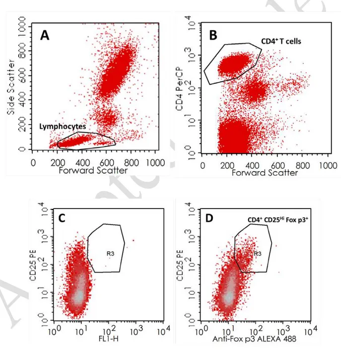
Figure 1. Gating strategies for the identification of distinct regulatory T cells subsets.
A-B. The first step of the analysis of Tregs was the identification of CD4
+T cells, recognized as
the CD4
+cells within the lymphocyte gate. C-D. Identification of CD4
+CD25
HiFoxp3
+regulatory T
cells with dot plots of FMO (C) and Foxp3 (D) tubes. E. CD4 vs CD25 dot plot, showing the
identification of CD4
DimCD25
Hiregulatory T cells. F. Histogram with Foxp3 expression within
CD4
DimCD25
Hiregulatory T cells (grey line), overlaid on Foxp3 expression within CD4
-Lymphocytes (black line). Geo MFI means were further in the ratio MFI of Foxp3 in
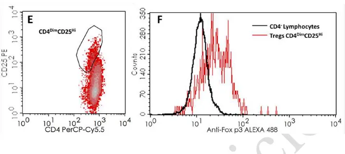
Figure 2. Gating strategies for the identification of distinct regulatory B cells subsets.
B cells were identified as the CD19
+population within the lymphocyte gate, as displayed in dot
plots A and B. C and D dotplots present the identification of regulatory B cells subsets
according to their expression of CD24, CD27 and CD38 (CD24
HiCD27
+Bregs and CD24
HiCD38
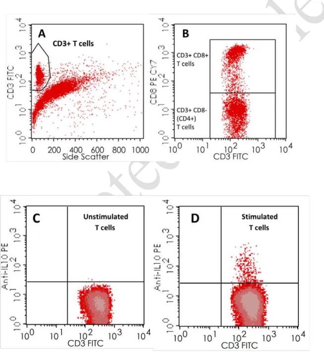
HiFigure 3. Gating strategies for the identification IL10 secretion in T and B cells.
A-B. T cells were identified according the their positive expression of CD3 as shown in the
CD3/SSC (Side Scatter) dot plot, and were further gated in CD8 negative T cells (CD4) and CD8
positive T cells (CD8), according to their expression of CD8. C-D. CD19
+B cells (gated as shown
in Figure 2) and CD4
+and CD8
+T cells (gated as shown in Figure 3 – A and B) were analyzed for
Figure 4. Immune parameters in HNP, HP, ANP and AP
a) Representative Tukey box-and-whiskers of CD4
+CD25
HiFoxp3
+Regulatory T cells subsets
frequencies and Foxp3 expression within CD4
DimCD25
HiTregs in HNP, HP, ANP and AP,
including the postpartum evaluation of pregnant women.
b) Representative Tukey box-and-whiskers of circulating Regulatory B cells subsets frequencies
in HNP, HP, ANP and AP, including the postpartum evaluation of pregnant women.
c) Representative Tukey box-and-whiskers of IL-10 secreting T and B cells in HNP, HP, ANP and
AP, including the postpartum evaluation of pregnant women.
All comparisons performed with Mann-Whitney U, except paired groups comparisons (AP vs
AP PP; and HP vs HP PP), which were performed with Wilcoxon test.
Central line: median; box: interquartile range; whiskers: range; dots: outliers.
* p<0.05; ** p<0.001. AP – Asthmatic pregnant women; ANP – Asthmatic non-pregnant
women; HP – Healthy pregnant women; HNP – Healthy non-pregnant women; PP –
postpartum.
4 . a
Table 1. Demographic and anthropometric comparisons between the groups of women
recruited.
a
Kruskal-Wallis Test,
bChi-square test,
cUnpaired Student’s t test,
dFisher exact test,
eMann-Whitney U.
AP – asthmatic pregnant women, ANP – asthmatic non-pregnant women, HP – healthy
pregnant women, HNP – healthy non-pregnant, IQR – interquartile range, sd – standard
deviation.
AP n=24 ANP n=32 HP n=43 HNP n=35 p-valueMaternal age in years, median (IQR)
34 (4) 36 (6) 32 (5) 35 (4) 0.063a Ethnicity, n (%)
Caucasian 24 (100.0) 32 (100.0) 42 (97.7) 35 (100.0)
0.545b
Black 0 (0.0) 0 (0.0) 1 (2.3) 0 (0.0)
BMI in kg/m2, median (IQR)
26.2 (4.5) 21.9 (5.0) 25.6 (4.1) 21.1 (2.4) <0,001a Education, n (%)
Basic / High school 2 (8.3) 5 (16.1) 4 (9.3) 7 (2.3)
0,296b Higher education 22 (91.7) 26 (83.9) 39 (90.7) 23 (76.7)
Parity, n (%)
Nuliparous 12 (50.0) - 24 (55.8) -
0.799d
Multiparous 12 (50.0) - 19 (44.2) -
Smoke exposure during pregnancy, n (%)
Yes 3 (12.5) - 7 (16.3) - 1.000d
No 22 (91.7) 36 (83.7)
Gestational age (complete weeks) at evaluation, mean (sd)
33 (0.9) - 33 (1.1) - 0.265c
Gestational age (complete weeks) at parturition, mean (sd)
39 (1.1) - 39 (1.1) - 0,296c
Weight at birth in grams, mean (sd)
3262 (329) - 3199 (402) - 0.493c
Baby Gender, n (%)
Male 12 (50.0) - 21 (48.8) -
1.000d
Female 12 (50.0) - 22 (51.2) -
Days after labour in postpartum, median (IQR)
44 (8) - 44 (5) - 0.589b
Breast feeding, n (%)
Yes 23 (95.8) - 33 (76.7) -
0.082d
No 1 (4.2) - 10 (23.3) -
Sensibilization to aeroallergens, n (%)
D. pteronyssinus 15 (62.5) 21 (65.6) - - 1.000d
D. farinae 12 (50.0) 18 (56.3) - - 0.788d
Lepidoglyphus destructor 2 (8.3) 9 (28.1) - - 0.093d
Table 2. Percentages and absolute counts of T and B cell subsets analyzed in AP, ANP, HP and HNP women.
Celular Subset AP
(n=24)
AP PP (n=24)
ANP (n=32)
HP (n=43)
HP PP (n=43)
HNP (n=35) Regulatory T cells
CD4+ CD25Hi Fox p3+ (%) 6.14 (1.54) 5.90 (2.43) 6.88 (2.17) 6.07 (3.44) 6.63 (2.85) 5.48 (1.69)
CD4+ CD25Hi Fox p3+ (Cells/µL) 52 (22) 65 (29) 83 (33) 50 (25) 71 (39) 56 (31)
Foxp3 expression in CD4DimCD25Hi 1.64 (1.26) 1.94 (0.46) 2.09 (0.43) 1.49 (0.19) 1.93 (0,30) 1.80 (0.25) Regulatory B cells
CD24HiCD27+ (%) 25.47 (16.75) 27.26 (20.46) 31.77 (13.37) 26.92 (15.02) 25.76 (14.30) 27.46 (49.80)
CD24HiCD27+ (Cells/µL) 45 (47) 59 (45) 102 (97) 44 (41) 57 (47) 54 (40)
CD24HiCD38H (%) 1.87 (2.97) 7.13 (8.84) 3.84 (2.74) 1.42 (1.38) 5.62 (5.58) 3.63 (2.48)
CD24HiCD38Hi (Cells/µL) 3 (4) 14 (25) 12 (7) 2 (2) 11 (14) 8 (8)
IL-10 secreting cells
IL10+ CD3+ T cells (%) 0.83 (0.69) 0.91 (0.70) 0.94 (0.46) 0.77 (0.47) 0.83 (0.50) 0.72 (0.57) IL10+ CD3+ CD4+ (CD8-) T cells (%) 1.00 (0.76) 1.03 (0.78) 1.04 (0.71) 0.92 (0.54) 0.95 (0.53) 0.84 (0.54) IL10+ CD3+ CD8+ T cells (%) 0.66 (0.69) 0.65 (0.86) 0.73 (0.51) 0.49 (0.55) 0.59 (0.47) 0.40 (0.59) IL10+ CD19+ B cells (%) 1.26 (0.67) 1.24 (0.83) 1.04 (0.73) 1.00 (0.90) 1.17 (0.91) 0.92 (0.70)