Braz J Otorhinolaryngol. 2014;80(2):152-155
www.bjorl.org
Brazilian Journal of
OTORHINOLARYNGOLOGY
1808-8694/$ - see front matter © 2014 Associação Brasileira de Otorrinolaringologia e Cirurgia Cérvico-Facial. Published by Elsevier Editora Ltda. All rights reserved.
DOI: 10.5935/1808-8694.20140031
ORIGINAL ARTICLE
Please cite this article as: Huang X, Chen Y, Zhang F, Yang Q, Zhang G. Peripheral Th17/Treg cell-mediated immunity imbalance in allergic rhinitis patients. Braz J Otorhinolaryngol. 2014;80:152-5
Study conducted at Third Hospital, Sun Yat-sen University, Guangdong, China. * Corresponding author.
E-mail: [email protected] (Q. Yang).
Peripheral Th17/Treg cell-mediated immunity imbalance in
allergic rhinitis patients
,
Xuekun Huang
a, Yulian Chen
a, Fucheng Zhang
b, Qintai Yang
a,*, Gehua Zhang
aa Department of Otolaryngology, Third Hospital, Sun Yat-sen University, Guangdong, China b Central Laboratory, Third Hospital of Sun Yat-sen University, Guangdong, China
Received 8 July 2013; accepted 10 November 2013
KEYWORDS Rhinitis allergic, seasonal;
Th17 cells; T-lymphocytes, regulatory
PALAVRAS-CHAVE Rinite alérgica, sazonal; Células Th17;
Linfócitos T reguladores
Abstract
Introduction: Allergic rhinitis (AR) is an IgE-mediated non-infectious disease of the nasal muco-sa following contact with allergens.
Objective: To investigate the peripheral Th17 cells and CD4 + CD25 + Foxp3 + regulatory T (Treg) cells and the expression of cytokines in the serum of AR patients.
Methods: The peripheral blood of 14 patients with AR (AR group) and six healthy subjects (con-trol group) was collected from March to May of 2012. Flow cytometry was performed to detect the Th17 cells and Treg cells, and enzyme-linked immunosorbent assay (ELISA) to measure the serum levels of IL-17 and TGF-β1.
Results: The proportion of Th17 cells in the AR group was markedly higher than that in the control group (p < 0.01). The proportion of Treg cells in the AR group was also dramatically reduced when compared with the control group (p < 0.01). In the AR group, serum IL-17 levels were markedly higher than those in the control group (p < 0.01). In the AR group, serum TGF-β1.
levels were signiicantly lower than those in the control group (p < 0.01).
Conclusion: The imbalance of peripheral Th17/Treg cells plays an important role in the patho-genesis of AR.
© 2014 Associação Brasileira de Otorrinolaringologia e Cirurgia Cérvico-Facial. Published by Elsevier Editora Ltda. All rights reserved.
Desequilíbrio imunológico periférico mediado por células Th17/Treg em pacientes com rinite alérgica
Resumo
Introdução: A rinite alérgica (RA) é uma doença não infecciosa da mucosa nasal mediada por IgE após o contato com alérgenos.
Objetivo: Investigar as células Th17 periféricas e CD4 + CD25 + Foxp3 + células T reguladoras (Treg) e a expressão sérica de citocinas em pacientes com RA.
Métodos: De março a maio de 2012, foi coletado o sangue periférico de 14 pacientes com RA (grupo RA) e seis indivíduos saudáveis (grupo controle). A detecção das células Th17 e células
Immunity imbalance in allergic rhinitis 153
Introduction
Allergic rhinitis (AR) is an IgE-mediated non-infectious dise-ase of the nasal mucosa following contact with allergens.1
Previous studies have demonstrated that imbalance of Th1/ Th2 cell-mediated immunity has an important role in the pathogenesis of AR, which is characterized by Th2
cell-me-diated inlammation.2 In recent years, there is evidence
showing that regulatory T (Treg) cells are another subset of T lymphocytes involved in allergic diseases and can regulate the pathological and physiological immune response, whi-ch may promote self-immune tolerance and maintenance of immune balance.3 CD4+CD25+ Treg cells are the major
type of Treg cells; they can secret some cytokines, including TGF-β1 and IL-10. Forkhead box p3 (Foxp3) gene is a critical regulatory gene in the development and functional mainte-nance of CD4+CD25+ Treg cells.4
Th17 cells are a subset of T lymphocytes that play
cru-cial roles in pro-inlammatory inlammation and autoimmu
-ne diseases. RORγt is a key nuclear transcription factor of
Th17 cells, and IL-17 is an important effector cytokine
se-creted by these cells. IL-17 can exert a pro-inlammatory
effect, promote the production of chemokines (such as IL-8 and monocyte chemoattractant protein-1, among others) in the local tissues, facilitate the proliferation of monocytes and neutrophils, and stimulate the generation of IL-6 and
prostaglandin E2, resulting in local inlammation.5,6 Some
researchers, using real-time polymerase chain reaction (RT--PCR) and enzyme-linked immunosorbent assay (ELISA) for the detection of associated transcription factors and cy-tokines of Th17 and Treg cells in the peripheral blood of AR
patients, observed that the expression of RORγt and IL-17
increased and the expression of Foxp3 (transcription factor of Treg cells) and TGF-β1 (associated cytokine) decreased.7
In the present study, low cytometry (FCM) and ELISA were
employed to detect the Th17/Treg cells and serum levels of IL-17 and TGF-β1, respectively, aiming to explore the role of Th17/Treg cells in the pathogenesis of AR.
Materials and methods
Subjects
From March to May of 2012, a total 14 patients with AR were
admitted into this department: ive males and nine fema -les, with a mean age of 29.76 years. The diagnosis of AR
was made according to its diagnostic criteria.1 Skin- prick
test results were positive for dust mite; the severity of AR was moderate or severe in all 14 patients. These patients had not been treated with glucocorticoids in the past month and had not received anti-histamine therapy and
allergen-s-peciic immunotherapy. Patients with sinusitis, asthma, and
intolerance to acetylsalicylic acid were excluded. Additio-nally, six healthy volunteers without symptoms of AR were recruited as controls: two males and four females, with a mean age of 30.83 years. All subjects signed an informed consent prior to their enrollment in the study.
Main reagents and instrument
Flow cytometer (FACSCalibur; BD - USA) was used for de-tection and Cellquest software (BD - USA) was employed for the acquisition of data and subsequent analysis. Microplate reader (ELX-800; Bio-Tek - USA), PMA, ionomycin calcium, BFA (MultiSciences), Human Regulatory T Cell Staining Kit (eBioscience - USA), APC-conjugated human CD8 mAb (BD - USA), PerCP-Cy5.5 conjugated human CD3 mAb, PE-con-jugated human IL-17 mAb and corresponding istype control
(eBioscience - USA), ixation solution and membrane rup -ture solution (Invitrogen - USA), anti-human IL-17 ELISA kit (eBioscience - USA), and TGF-β1 ELISA kit (RayBio - USA) were used in the present study.
Sample collection
In the morning, venous blood samples (4 mL) were collected from the ulnar vein and anti-coagulated with heparin. Then, 2 mL of blood were subjected to centrifugation at 3,000 rpm for 15; the serum was collected and stored at -20°C for the detection of IL-17 and TGF-β1. In addition, the
re-maining 2 mL of blood were used for low cytometry for the
detection of Th17 cells and Treg cells.
Detection of Th17 cells by FCM
In brief, 250 μL of peripheral blood were mixed with 50 μg/L PMA, 2.0 μmol/L monensin, and 750 μmoL/L ionomycin
calcium, followed by incubation at 37°C in an environment with 50 mL/L CO2 for 4 h. The cell suspension was trans-ferred into a 1.5 ml-PE tube followed by centrifugation at 2,500 rpm for 6 min. The supernatant was removed, and the cells were washed with PBS twice for FCM. The cells were
treated with 10 μL of PECy5-CD3Ab and 10 μL of FITC-CD8Ab
at room temperature, in a dark environment, for 30 min.
Resultados: A percentagem de células Th17 no grupo RA foi bem maior do que no grupo controle (p < 0,01). A proporção de células Treg no grupo RA também foi drasticamente menor quando
comparada ao grupo controle (p < 0,01). No grupo RA, o nível sérico de IL-17 foi signiicativa -mente maior do que no grupo controle (p < 0,01).
Conclusão: O desequilíbrio de células Th17/Treg periféricas desempenha um papel importante na patogênese da RA.
154 Huang X et al.
After washing in PBS twice, the cells were ixed in 300 μL of ixation solution in a dark environment at 4°C for 15 min. Af -ter centrifugation, the supernatant was removed. Following the addition of membrane rupture solution, centrifugation was performed at 3,000 rpm and the supernatant was remo-ved. After washing in PBS twice, cells were transferred into
two tubes and treated with 20 μL of PE-IL-17Ab and 10 μL of
isotype control PE-IgG1, respectively, in a dark environment at room temperature for 30 min. After washing in PBS twice, cells were re-suspended in 0.3 mL of PBS and subjected to FCM. CellQuest software was employed for data analysis.
Detection of Treg cells by FCM
Cells were transferred into sample tube and control tube. Cells in the sample tube were treated with CD4/CD25/ Foxp3, and those in the control group were treated with Foxp3 isotype control (CD4/CD25/mouse IgG). Then, 100
μL of anti-coagulated blood was added followed by incu -bation in a dark environment at room temperature for 20 min. Following the addition of 1 mL of hemolytic agent, incubation was performed at room temperature in a dark environment for 10 min followed by centrifugation at 1,000 rpm for 5 min. The supernatant was removed and cells were re-suspended in 1 mL of PBS followed by centrifugation at 1,000 rpm for 5 min. The supernatant was then removed,
and 0.5 mL of Foxp3 ixation solution were added, followed
by incubation in a dark environment at room temperature for 20 min. Cells were re-suspended in 1 mL of PBS, and centrifuged at 1,000 rpm for 5 min. The supernatant was removed, and 0.5 mL of Foxp3 membrane rupture solution was added to each tube, followed by centrifugation at 1,000 rpm for 5 min. These cells were re-suspended in 0.5 mL of Foxp3 membrane rupture solution, followed by incubation in a dark environment at room temperature for 15 min. Af-ter centrifugation at 1,000 rpm for 5 min, the supernatant
was removed. Then, 10 μL of PE Foxp3Ab were added to the sample tube and 10 μL of isotype control were added to the
control tube, followed by incubation at room temperature in a dark environment for 30 min. Cells were re-suspended in 1 mL of PBS and centrifuged at 1,000 rpm for 5 min. The supernatant was removed and the cells were re-suspended in 0.4 mL of PBS. FCM was performed and CellQuest softwa-re was employed for data analysis.
Detection of serum IL-17 and TGF-β1 by ELISA
The detection of serum contents of IL-17 and TGF-β1 was
performed according to the manufacturer’s instructions. The lower limit of detection was 0.5 pg/mL for IL-17 and 9 pg/mL for TGF-β1.
Statistical analysis
Statistical analysis was performed with SPSS version 16.0. Data with normal distribution were expressed as mean ± standard deviation. Student’s t-test for independent varia-bles was performed for comparisons between two groups.
A value of p < 0.05 was considered statistically signiicant.
Results
Proportion of Th17 cells in AR group and control
group
FCM demonstrated that the proportion of Th17 cells in the AR group was markedly higher than that of the control group (p < 0.01; Table 1).
Proportion of Treg cells in AR group and control
group
In the AR group, the proportion of Treg cells was markedly reduced when compared to the control group (p < 0.01; Ta-ble 1).
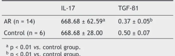
Serum contents of IL-17 and TGFβ1 in AR group and con-trol group
ELISA was performed to detect the serum contents of IL-17 and TGF-β1 in the AR group and control group. Data are shown in Table 2. Results demonstrated that the IL-17 le-vel in the AR group was markedly higher than that of the control group (p < 0.01). In the AR group, the TGF-β1 level was dramatically reduced when compared with the control group (p < 0.01).
Table 1 Proportion of Th17 cells and regulatory T (Treg) cells in the allergic rhinitis (AR) group and control group (mean ± standard deviation).
Group Th17 (%) Treg (%)
AR (n = 14) 1.69 ± 0.64a 1.82 ± 0.76b
Control (n = 6) 0.59 ± 0.19 5.19 ± 2.10
a p < 0.01 vs. control group. b p < 0.01 vs. control group.
Table 2 Serum levels of IL-17 and TGF-β1 in the allergic rhinitis (AR) group and control group (mean ± standard deviation; pg/mL)
IL-17 TGF-β1
AR (n = 14) 668.68 ± 62.59a 0.37 ± 0.05b
Control (n = 6) 668.68 ± 28.00 0.50 ± 0.07
a p < 0.01 vs. control group. b p < 0.01 vs. control group.
Discussion
Previous studies have demonstrated that the serum level of
IL-17 in AR patients was signiicantly increased when compa -red with controls,8 and that the proportion of IL-17 positive
Immunity imbalance in allergic rhinitis 155
higher than that in healthy subjects.9 Ciprandi et al.10,11
observed that the serum IL-17 level was positively related to the severity of symptoms of AR and positively associated with the number of peripheral eosinophils. The peripheral monocytes from AR patients who were treated with pollen allergen and FCM showed the proportion of Th17 cells was markedly higher than that in the control group. In the pre-sent study, the results indicated that the proportion of
pe-ripheral Th17 cells in AR patients was signiicantly higher
than that of the controls, accompanied by the increase in
serum level of IL-17. These indings suggest that Th17 cells
can secret a large amount of IL-17, which is involved in the pathogenesis of AR.
Additionally, the proportion of CD4 + CD25 + Treg cells
in peripheral blood of AR patients was signiicantly lower
than that of the controls, accompanied by the reduction of serum TGF-β1 level. This suggests that the reduction of peripheral Treg cell count leads to a compromised function of these cells. In healthy subjects, the proportion of CD4 + CD25 + Treg cells was approximately 5% to 10% in the peri-pheral blood. The CD4 + CD25 + Treg cells can exert inhi-bitory effect on the immune response via the intercellular interaction or secretion of inhibitory cytokines (such as IL-10 and TGF-β).4 The present results demonstrated that the
proportion of peripheral CD4 + CD25 + Treg cells and the serum level of TGF-β1 were reduced. Xu et al.12 conirmed
that the Foxp3 + positive lymphocytes and Foxp3 mRNA ex-pression in the nasal mucosa and peripheral monocytes of AR patients were markedly reduced when compared with healthy controls, and that Foxp3 and CD4 + CD25 + Treg cells in allergy patients were markedly reduced.13 This suggests
that the compromised inhibitory effect of Treg cells leads to the occurrence of allergic diseases.
Th17 cells and Treg cells are derived from naïve T cells.
Th17 cells mediate the inlammatory reaction, whereas Treg
cells mediate immune tolerance. There is antagonism in the function and differentiation of Th17 cells and Treg cells. Thus, the balance of Treg cells and Th17 cells is crucial for the maintenance of the immune status. In the present study, the results demonstrated that the proportion of Th17 cells in the peripheral blood of AR patients was markedly incre-ased and that of Treg cells was dramatically reduced. This suggests that Th17/Treg imbalance plays an important role in the pathogenesis of AR. Further research in the Th17/ Treg balance may provide a novel strategy for the treatment of AR.
Conlicts of interest
The authors declare no conlicts of interest.
References
1. Bousquet J, Khaltaev N, Cruz AA, Denburg J, Fokkens WJ, To-gias A, et al. Allergic rhinitis and its impact on asthma (ARIA) 2008 Update (in collaboration with the World Health Organiza-tion, GA2LEN and AllerGen). Allergy. 2008;63(Suppl 86):8-160.
2. Agrawal DK, Shao Z. Pathogenesis of allergic airway inlamma -tion. Curr Allergy Asthma Rep. 2010;10:39-48.
3. Ozdemir C, Akdis M, Akdis CA. T regulatory cells and their counterparts-masters of immune regulation. Clin Exp Allergy. 2009;39:626-39.
4. Pai SY, Truitt ML, Ting CN, Leiden JM, Glimcher LH, Ho IC. Cri-tical roles for transcription factorGATA-3 in thymocyte develo-pment. Immunity. 2003;19:863-75.
5. Harrington LE, Hatton RD, Mangan PR, Turner H, Murphy TL, Murphy KM, et al. Interleukin 17-producing CD4+ effector T cells develop via a lineage distinct from the T helper type 1 and 2 lineages. Nat Immunol. 2005;6:1123-32.
6. Ivanov II, McKenzie BS, Zhou L, Tadokoro CE, Lepelley A, Lafail-le JJ, et al. The orphan nucLafail-lear receptor RORgammat directs
the differentiation program of proinlammatory IL-17+ T helper
cells. Cell. 2006;126:1121-33.
7. Zhang C, Hong S, Hu G. The expression of Treg/Th17 cells rela-ted transcription factors and cytokines in PBMCs and plasma in patients with allergic rhinitis. Lin Chung Er Bi Yan Hou Tou Jing Wai Ke Za Zhi. 2012; 26:209-11.
8. Huang X, Yang Q, Chen Y, Zhang GH, Li Y. Expressions of IL-17, IL-21 and IL-23 in the serum of allergic rhinitis patients. J Med Biochem. 2011;30:323-7.
9. Ba L, Du J, Liu Y, Shang T, Yang F, Bian P. The expression and
signiicance of interleukin-17 and the iniltrating eosinophils
in nasal polyps. J Clin Otorhino Laryngol Head Neck Surg. 2010;24:53-6.
10. Ciprandi G, De Amici M, Murdaca G, Fenoglio D, Ricciardolo F, Marseglia G, et al. Serum interleukin-17 levels are related to clinical severity in allergic rhinitis. Allergy. 2009;64:1375-8. 11. Ciprandi G, Filaci G, Battaglia F, Fenoglio D. Peripheral Th- 17
cells in allergic rhinitis: New evidence. Int Immuno Pharmacol. 2010;10:226-9.
12. Xu G, Mou Z, Jiang H, Cheng L, Shi J, Xu R, et al. A possib-le ropossib-le of CD4+CD25+ T cells as well as transcription factor Foxp3 in the dysregulation of allergic rhinitis. Laryngoscope. 2007;117:876-80.