DOI: http://dx.doi.org/10.1590/2446-4740.0720
*e-mail: [email protected]
Received: 12 February 2015 / Accepted: 30 December 2015
Biomechanical performance of Bio Cross-Pin and EndoButton
for ACL reconstruction at femoral side: a porcine model
Ari Digiácomo Ocampo Moré, André Luiz Almeida Pizzolatti, Eduardo Alberto Fancello, Carlos Rodrigo de Mello Roesler*
Abstract Introduction: The method of graft ixation is critical in anterior cruciate ligament (ACL) reconstruction surgery. Success of surgery is totally dependent on the ability of the implant to secure the graft inside the bone tunnel until complete graft integration. The principle of EndoButton is based on the cortical suspension of the graft. The Cross-Pin is based on graft expansion. The aim of this study was to evaluate the biomechanical performance of EndoButton and Bio Cross-Pin to ix the hamstring graft at femoral side of porcine knee joints and evaluate whether they are able to support of loading applied on graft during immediate post-operative tasks. Methods: Fourteen ACL reconstructions were carried out in porcine femurs ixing supericial lexor tendons with Titanium EndoButton (n = 7) and with 6 × 50 mm HA/PLLA Bio Cross-Pin (n = 7). A cyclic loading test was applied with 50-250 N of tensile force at 1 Hz for 1000 cycles. The displacement was measured at 20, 100, 500 and 1000 load cycles to quantify the slippage of the graft during the test. Single-cycle load-to-failure test was performed at 50 N/mm to measure ixation strength. Results: The laxity during cyclic loading and the displacement to failure during single-cycle test were lower for the Bio Cross-Pin ixation (8.21 ± 1.72 mm) than the EndoButton (11.20 ± 2.00 mm). The Bio Cross-Pin (112.22 ± 21.20 N.mm–1) was signiicantly stiffer than the EndoButton ixation (60.50 ±10.38 N.mm–1). There was no signiicant difference between Bio Cross-Pin (failure loading: 758.29 ± 188.05 N; yield loading: 713.67 ± 192.56 N) and EndoButton strength (failure loading: 672.52 ± 66.56 N; yield loading: 599.91 ± 59.64 N). Both are able to support the immediate post-operative loading applied (445 N). Conclusion: The results obtained in this experiment indicate that the Bio Cross-Pin technique promote stiffer ixation during cyclic loading as compared with EndoButton. Both techniques are able to support the immediate post-operative loading applied.
Keywords: Biomechanics, ACL reconstruction, EndoButton, Bioabsorbable Cross-Pin.
Introduction
The anterior cruciate ligament (ACL) replacement with hamstring graft has been widely performed with positive results. This procedure, however, requires
great care at postoperative period. Although the four-stranded hamstrings have higher strength and
stiffness than patellar tendon (Hamner et al., 1999), the integration into the bone is delayed due to the lack of bone block (Blickenstaff et al., 1997; Rodeo et al.,
1993; Weiler et al., 2002a, b). This factor makes the early postoperative period a critical time for
successful surgery (Becker et al., 2001; Brown et al., 1996; Wilson et al., 1999). The ixation of graft is
totally dependent of implant performance during
this period. A poor ixation can lead to graft slippage and result in knee instability or failure of ixation (Fu et al., 1999; Giurea et al., 1999; Magen et al.,
1999; Shen et al., 2010). Furthermore, the slippage impairs the integration and ligamentization process
(Rodeo et al., 1993, 2006).
EndoButton and Cross-pin are techniques based on different mechanical principles. The graft is suspended inside the bone tunnel by both techniques.
The anchor point, however, is different. EndoButton is an extra-articular device made of metallic button and a polyurethane ribbon (Endotape). The button is supported by the external cortical portion of the bone Endotape links the graft to the not supported
central part of the metallic button. The Cross-Pin is
an intra-articular device that traverses the joint and is stabilized with one tip ixed to the cortical wall and the other ixed inside cancellous bone. The graft passes around this pin to be ixed. During the graft tension,
the button and the pin are submitted to bending forces
(Figure 1). The diameter of Cross-Pin is higher than
Endotape, increasing the volume when the tendon loops. In theory, this effect causes expansion and compression of the graft against the tunnel wall resulting in highest strength (Milano et al., 2006).
Previous studies have shown that the interference screws ixation on tibia is the weak point in ACL
reconstruction when Bio Cross-Pin or EndoButton are used at femoral site (Shen et al., 2010). However,
other methods of ixation on tibia such as Washer Loc
and Tandem Washer have higher strength compared
to Bio Cross-Pin or EndoButton (Kousa et al., 2003a; Kousa et al., 2003b; Magen et al., 1999). The femoral
site becomes the weak point in this case. Therefore, the aim of this study was to assess the mechanical
properties and the failure mode of Bio Cross-Pin and
EndoButton with respect to the ixation of Hamstring graft. Our hypothesis was that different mechanical
principle to secure the graft inside the bone tunnel results in different performances not being able to support post-operative loading. A porcine model
was used to compare the laxity, the strength, the linear stiffness and the energy associated with each
technique. Therefore, to evaluate the Cross-Pin and
EndoButton performance for femoral ixation we
apply the load directly on the graft.
Methods
Fourteen porcine knees of Landrace specimens with 2 years old and 400 kg weight were purchased
from a commercial slaughterhouse in the state ready
for consumption. They were harvested and stored at –20 °C. This method allowed for harvesting of the soft tissue. Each femur was dissected and the supericial lexor tendon with approximately 5 mm diameter was extracted and used as a double graft. The use of the autograft instead of an artiicial graft was preferred to
mimic the clinical practice of use a graft retrieved from
the patient. These grafts were then ixed to the femurs by the two ixation techniques: Titanium EndoButton linked with 30 mm polyurethane Endotape (n = 7) and by Bio Cross-Pin HA/PLLA 70 30 6 × 50 mm (n = 7) (Figure 2).
Fixation technique
The ixation procedure followed the same clinical
protocol established for ACL reconstruction at
femoral side of human knees. A 6 mm over-the-top guiding device was used to locate the anatomical ACL insertion. The tunnel was drilled to match the graft diameter. A 6 mm diameter bone tunnel was
drilled in inside-out technique to the EndoButton
ixation. After this, a 30 mm tunnel was drilled with a 9 mm diameter for the graft positioning. On the Bio Cross-Pin ixation a 9 mm tunnel diameter was
drilled for the graft positioning.
Mechanical testing
Immediately after the graft ixation, each femur was clamped to a custom device with bone cement (PMMA) and screws. This device was then placed in the servo-hydraulic testing machine (Brasvalvulas,
São Paulo, Brazil) to guarantee the alignment between the tunnel axis and loading direction. Therefore, the testing was conducted in a worst condition scenario. The free end of the graft was ixed in the load cell
leaving a gage length of 30 mm to mimic the human
intra articular ACL length (Figure 3). Each specimen
was then submitted to cyclic and single-cyclic
loading-to-failure test.
Figure 1. Mechanical methods of graft ixation. Left: Bio Cross-Pin (one cortical support point). Right: EndoButton (external cortical
support point).
Figure 2. Bio Cross-Pin HA/PLLA 70 30 6 × 50 mm and Titanium
The cyclic test started with a preconditioning static tensile load of 50 N for 2 min followed by 1000 load cycles at 1 Hz between 50 N and 250 N. The slippage of the graft-ixation device interface was measured
indirectly through the graft lengthening after 20, 100, 500 and 1000 load cycles. This measurement combined
the effect of the ixation device slippage and tendon stretch. The procedure was suficiently accurate for the purposes of this study, in accordance with clinical practice (Kousa et al., 2003a). Failure during cyclical
loading was assumed to occur in cases in which a complete slippage of the ixation device was observed. The specimens that did not fail were then submitted to a single-cycle load-to-failure test with a force rate
of 50 mm/min after the static preconditioning load. The values for the ultimate failure load, yield point load, displacement at ultimate failure load, displacement at
yield load, linear stiffness, and energy were obtained from the load-displacement curves (200 Hz sampling rate). The force x displacement curves was used to calculate these variables because we are not focusing
the graft strain but the mechanical behavior of the
whole system bone-graft-implant. The specimens were kept moistened by spraying with physiological solution (0.9% NaCl).
Statistical analysis
A two-way split-plot ANOVA was used (ixation technique – between factor; and displacement by cycles – within factor) to test laxity during cyclic loading. The t student test was used to compare the ultimate
failure load, the displacement at ultimate failure load, the yield load, the displacement at yield load,
and energy between the two graft ixation techniques during the single-cycle loading test. The Wilcoxon rank sum test was used to compare the linear stiffness variable. The probability level was set at 0.05.
Results
No ixation devices failed during the cyclic loading test. The Bio Cross-Pin displacement was lower than EndoButton (F = 6.92; P = 0.011) (Figure 4). The interaction between the ixation methods and cycles numbers, however, was not signiicant (F = 0.66; P = 0.61), showing that the numbers of cycles chosen
for the test did not change the pattern of displacement
between the two ixation methods.
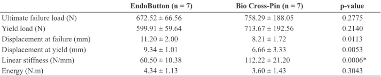
Table 1 shows the results of single-cyclic
load-to-failure test. No signiicant difference of ultimate failure load, yield load, and energy was observed between the Bio Cross-Pin and EndoButton ixation. The EndoButton revealed a smaller standard
deviation of ultimate failure load and yield load. The mean displacement at ultimate failure load and
displacement at yield load for Bio Cross-Pin was signiicantly lower than EndoButton. The Bio Cross-Pin technique showed signiicantly higher linear stiffness
than the EndoButton.
The Endotape rupture was the most common
failure mode. This failure mode occurred in 5 cases.
The EndoButton was pulled out of the bone tunnel with plastic deformation of the button and left intact the Endotape in 2 cases. The Bio Cross-Pin broke in
5 cases, and the tendon failed in 2 cases.
Discussion
Studies in animal models have been widely
realized to understand the biomechanical performance
of several devices to ix the hamstring tendons in the
femoral side in ACL reconstruction. A variety of testing
parameters were used in these studies, including the use of femur-graft-tibia complex, the direction of
pulling out the tendon, the rate of loading, and cyclic
test protocol. However, the interpretation of implant performance at femoral site testing the whole complex may be dificult since that the ixation on the tibia fail before that on femur fails (Shen et al., 2010).
Figure 3. Position of femur during biomechanical test.
Figure 4. Average and standard deviation displacement of ixations
Therefore, the best way to assess the Bio Cross-Pin
and EndoButton performance is to apply the loading
directly at the graft. The existence of different methods makes it dificult to compare results. Despite a lack of similar methods, however, the results obtained at present work were similar with others from literature (Ahmad et al., 2004; Milano et al., 2006; Miyata et al., 2000; Rodríguez et al., 2014; Shen et al., 2010).
Our data showed that the Bio Cross-Pin ixation had lower laxity during cyclic loading than EndoButton. This superior performance means that the Bio Cross-Pin is more secure to support the loads applied
during aggressive rehabilitation protocol. The laxity is related to loss of ixation by elongation of the graft,
graft slippage, and plastic deformation of device. The sum of these factors may increase the graft
micromovement and delay graft healing (Fu et al.,
1999). The elongation of polyurethane Endotape may
be responsible for the high displacement occurring
with the EndoButton (Höher et al., 2000). Previous studies have shown that the biomechanical performance
of the Bio Cross-Pin is superior that the EndoButton
during cyclic and failure loading test (Ahmad et al.,
2004; Rodríguez et al., 2014; Shen et al., 2010). Despite any difference in strength observed
during the failure test, Bio Cross-Pin was stiffer than
EndoButton. In theory, the stiffness is an important
variable to be considered. The ixation device should
promote stiffness near the native ACL to avoid an
excessive graft motion and knee laxity until graft integration, (To et al., 1999; Wu et al., 2009). A lower
stiffness can, therefore, increase the displacement
associated with anterior translation and may result in an unstable knee. There is consensus in the literature
that the EndoButton have less stiffness than the Bio
Cross-Pin (Milano et al., 2006; Rodríguez et al., 2014;
Trump et al., 2011). Considering the energy that the
femur-graft complex has absorbed up to the point of ixation failure, there was not differences between
the both techniques evaluated.
The yield load has been advocated as the most important variable to be evaluated in the performance
of ixation (Kousa et al., 2003a). In the present approach, the yield load is assumed to be the last
point of linear region of load-displacement curve.
Theoricaly, this point represents the ixation resistance before yielding. The irst signiicant slippage of the
graft occurs at this point. The stiffness decreases
and the ixation tends to fail. The mean yield load for the Bio Cross-Pin was signiicantly higher than those for the EndoButton. The Endobutton showed a yield point very close to the failure load when the endotape broke (Figure 5). It happened in 5 cases.
In two cases, the button pulled out the bone tunel with
visible plastic deformation. When the failure did not occur in the Endotape, the displacement-load curve
showed a different pattern. The yield point was located away from the failure load. The Bio Cross-Pin broke in all cases tested. This failure mode was similar to Rodríguez et al. (2014).
The yield loading displayed by both ixations was suficient to support the loading applied during daily tasks and accelerated rehabilitation programs. Morrison
(1970) reported 169 N as the ACL force during normal level walking. Noyes et al. (1984) estimated that the
maximum force on the graft ixation device occurs while descending stairs and is approximately 445 N. Previous studies have shown that ligament loads of this
magnitude can be generated during quadriceps muscle
contraction at full knee extension (Rupp et al., 1999).
All of the constructs, therefore, can be considered to be secure enough for their intended use.
It is worth noting that some methods used in this experiment should be highlighted. One is the use of
animal tissues. The porcine bone does not have the same bone mineral density as a healthy human bone. Previous studies have observed that the porcine bone did not resist the load applied. As a result, the button
pulled out from the cortical bone (Ahmad et al.,
2004; Rylander et al., 2014). Studies performed with
human bone have not described this failure mode
(Kousa et al., 2003a). Furthermore, the bone quality
(Brown et al., 2004) and the button position on the femur (Conner et al., 2010) may alter the failure mode device. The advantage of using animal bone
is its mineral density likeness and easy acquisition.
The second aspect to be noted is the direction of bone
tunnel axis during graft tension testing. The method
Table 1. Results of single-cycle failure-to-load test (Mean ± SD).
EndoButton (n = 7) Bio Cross-Pin (n = 7) p-value
Ultimate failure load (N) 672.52 ± 66.56 758.29 ± 188.05 0.2775
Yield load (N) 599.91 ± 59.64 713.67 ± 192.56 0.2140
Displacement at failure (mm) 11.20 ± 2.00 8.21 ± 1.72 0.0113
Displacement at yield (mm) 9.34 ± 1.01 6.66 ± 3.33 0.0053
Linear stiffness (N/mm) 60.50 ± 10.38 112.22 ± 21.20 0.0006*
Energy (N.m) 4.34 ± 1.13 3.60 ± 1.43 0.3043
used in our study did not represent the functional
loading applied to the graft during lexion-extension movement (Zhang et al., 2007). Therefore, the results do not warrant comparison to a clinical situation. These results, however, help us understand the biomechanical performance of the ixation, when submiting the device to a worst case condition.
The results obtained indicate that both types of
ixations analyzed in this study showed suficient
strength to resist the loads applied during functional
activities in the early intensive rehabilitation. However, the Bio Cross-Pin performance was the best. The laxity at cyclic test was lower, and the linear stiffness was
higher than those for EndoButton. The main failure
mode of EndoButton was endotape rupture while Bio Cross-Pin broke in all cases. Further research is
needed to determine the clinical relevance of these
indings relating the ACL replacement at femoral side.
Acknowledgements
The authors would like to thank FAPESC, FINEP and CNPq for the inancial support.
References
Ahmad CS, Gardner TR, Groh M, Arnouk J, Levine WN. Mechanical Properties of Soft Tissue Femoral Fixation Devices for Anterior Cruciate Ligament Reconstruction. The American Journal of Sports Medicine. 2004; 32(3):635-40. http://dx.doi.org/10.1177/0363546503261714. PMid:15090378. Becker R, Voigt D, Stärke C, Heymann M, Wilson GA, Nebelung W. Biomechanical properties of quadruple tendon and patellar tendon femoral fixation techniques. Knee Surgery, Sports Traumatology, Arthroscopy. 2001; 9(6):337-42. http://dx.doi.org/10.1007/s001670100223. PMid:11734869.
Blickenstaff KR, Grana WA, Egle D. Analysis of a semitendinosus autograft in a rabbit model. The American Journal of Sports Medicine. 1997; 25(4):554-9. http:// dx.doi.org/10.1177/036354659702500420. PMid:9240991. Brown GA, Peña F, Grøntvedt T, Labadie D, Engebretsen L. Fixation strength of interference screw fixation in bovine, young human, and elderly human cadaver knees: influence of insertion torque, tunnel-bone block gap, and interference. Knee Surgery Sports Traumatology. 1996; 3(4):238-44. http://dx.doi.org/10.1007/BF01466626. PMid:8739721. Brown CH Jr, David RW, Hecker AT, Ferragamo M. FGraft-bone motion and tensile properties of hamstring and patellar tendon anterior cruciate ligament femoral graft fixation Figure 5. Yield Load and Ultimate Failure Load of EndoButton specimens that failed in different modes. (a) and (b) plastic bending of
under cyclic loading. Journal of Arthroscopy and Related Surgery. 2004; 20(9):922-35. http://dx.doi.org/10.1007/ BF01466626. PMid:8739721.
Conner CS, Perez BA, Morris RP, Buckner JW, Buford WL, Ivey FM. Three femoral fixation devices for anterior cruciate ligament reconstruction: Comparison of fixation on the lateral cortex versus the anterior cortex. Journal of Arthroscopy and Related Surgery. 2010; 26(6):796-807. http:// dx.doi.org/10.1016/j.arthro.2009.10.015. PMid:20511038. Fu FH, Bennett CH, Lattermann C. Current concepts current trends in anterior cruciate ligament peconstruction - Part 1: biology and biomechanics of reconstruction. The American Journal of Sports Medicine. 1999; 27(6):821-30. PMid:10569374.
Giurea M, Zorilla P, Amis AA, Aichroth P. Comparative pull-out and cyclic-loading strength tests of anchorage of hamstring tendon grafts in anterior cruciate ligament reconstruction. The American Journal of Sports Medicine. 1999; 27(5):621-5. PMid:10496580.
Hamner DL, Brown CH Jr, Steiner ME, Hecker AT, Hayes WC. Hamstring tendon grafts for reconstruction of the anterior cruciate ligament: biomechanical evaluation of the use of multiple strands and tensioning techniques. The Journal of Bone and Joint Surgery. 1999; 81(4):549-57. PMid:10225801.
Höher J, Scheffler SU, Withrow JD, Livesay GA, Debski RE, Fu FH, Woo SL. Mechanical behavior of two hamstring graft constructs for reconstruction of the anterior cruciate ligament. Journal of Orthopaedic Research. 2000; 18(3):456-61. http://dx.doi.org/10.1002/jor.1100180319. PMid:10937634. Kousa P, Järvinen TLN, Vihavainen M, Kannus P. The fixation strength of six hamstring tendon graft fixation devices in anterior cruciate ligament reconstruction: Part I: Femoral site. The American Journal of Sports Medicine. 2003a; 31:174-81.
Kousa P, Järvinen TLN, Vihavainen M, Kannus P, Järvinen M. The fixation strength of six hamstring tendon graft fixation devices in anterior cruciate ligament reconstruction: Part II - Tibial site. The American Journal of Sports Medicine. 2003b; 31(2):182-8. PMid:12642250.
Magen HE, Howell SM, Hull ML. Structural properties of six tibial fixation methods for anterior cruciate ligament soft tissue grafts. The American Journal of Sports Medicine. 1999; 27(1):35-43. PMid:9934416.
Milano G, Mulas PD, Ziranu F, Piras S, Manunta A, Fabbriciani C. Comparison between different femoral fixation devices for ACL reconstruction with doubled hamstring tendon graft: a biomechanical analysis. The Journal of Arthroscopic & Related Surgery. 2006; 22(6):660-8. http:// dx.doi.org/10.1016/j.arthro.2006.04.082. PMid:16762706. Miyata K, Yasuda K, Kondo E, Nakano H, Kimura S, Hara N. Biomechanical comparisons of anterior cruciate ligament: reconstruction procedures with flexor tendon graft. Journal of Orthopaedic Science. 2000; 5(6):585-92. http://dx.doi. org/10.1007/s007760070010. PMid:11180923.
Morrison JB. The mechanics of the knee joint in relation to normal walking. Journal of Biomechanics. 1970; 3(1):51-61. http://dx.doi.org/10.1016/0021-9290(70)90050-3. PMid:5521530.
Noyes FR, Butler DL, Grood ES, Zernicke RF, Hefzy MS. Biomechanical analysis of human ligament grafts used in knee-ligament repairs and reconstructions. Journal of Bone and Joint Surgery. 1984; 66(3):344-52. PMid:6699049. Rodeo SA, Arnoczky SP, Torzilli PA, Hidaka C, Warren RF. Tendon-healing in a bone tunnel: a biomechanical and histological study in the dog. Journal of Bone Joint Surgery. 1993; 75(12):1795-803. PMid:8258550.
Rodeo SA, Kawamura S, Kim HJ, Dynybil C, Ying L. Tendon healing in a bone tunnel differs at the tunnel entrance versus the tunnel exit: an effect of graft-tunnel motion? The American Journal of Sports Medicine. 2006; 34(11):1790-800. http:// dx.doi.org/10.1177/0363546506290059. PMid:16861579. Rodríguez C, García TE, Montes S, Rodríguez L, Maestro A. In vitro comparison between cortical and cortico-cancellous femoral suspension devices for anterior cruciate ligament reconstruction: implications for mobilization. Knee Surgery, Sports Traumatology, Arthroscopy. 2014; 23(8):2324-9. PMid:24839039.
Rupp S, Seil R, Schneider A, Kohn DM. Ligament graft initial fixation strength using biodegradable interference screws. Journal of Biomedical Materials Research. 1999; 48(1):70-4. http://dx.doi.org/10.1002/(SICI)1097-4636(1999)48:1<70::AID-JBM12>3.0.CO;2-P. PMid:10029152.
Rylander L, Brunelli J, Taylor M, Baldini T, Ellis B, Hawkins M, McCarty E. A biomechanical comparison of anterior cruciate ligament suspensory fixation devices in a porcine cadaver model. Clinical Biomechanics (Bristol, Avon). 2014; 29(2):230-4. http://dx.doi.org/10.1016/j. clinbiomech.2013.11.001. PMid:24321231.
Shen HC, Chang JH, Lee CH, Shen PH, Yeh TT, Wu CC, Kuo CL. Biomechanical comparison of Cross-Pin and Endobutton femoral fixation of a flexor tendon graft for anterior cruciate ligament reconstruction - A porcine femur-graft-tibia complex study. The Journal of Surgical Research. 2010; 161(2):282-7. http://dx.doi.org/10.1016/j. jss.2009.01.015. PMid:19524939.
To JT, Howell SM, Hull ML. Contributions of femoral fixation methods to the stiffness of anterior cruciate ligament replacements at implantation. Arthroscopy. 1999; 15(4):379-87. http://dx.doi.org/10.1016/S0749-8063(99)70055-1. PMid:10355713.
Trump M, Palathinkal DM, Beaupre L, Otto D, Leung P, Amirfazli A. In vitro biomechanical testing of anterior cruciate ligament reconstruction: traditional versus physiologically relevant load analysis. The Knee. 2011; 18(3):193-201. http:// dx.doi.org/10.1016/j.knee.2010.04.011. PMid:20570155. Weiler A, Peine R, Pashmineh-Azar A, Abel C, Sudkamp NP, Hoffmann RF. Tendon healing in a bone tunnel. Part I: biomechanical results after biodegradable interference fit fixation in a model of anterior cruciate ligament reconstruction in sheep. Arthroscopy. 2002a; 18(2):113-23. http://dx.doi. org/10.1053/jars.2002.30656. PMid:11830804.
18(2):124-35. http://dx.doi.org/10.1053/jars.2002.30657. PMid:11830805.
Wilson TW, Zafuta MP, Zobitz M. A biomechanical analysis of matched bone-patellar tendon-bone and double-looped semitendinosus and gracilis tendon grafts. The Journal of Sports Medicine. 1999; 27(2):202-7. PMid:10102102. Wu JL, Yeh TT, Shen HC, Cheng CK, Lee CH. Mechanical comparison of biodegradable femoral fixation devices for hamstring tendon graft - A biomechanical study in a porcine
model. Clinical Biomechanics (Bristol, Avon). 2009; 24(5):435-40. http://dx.doi.org/10.1016/j.clinbiomech.2009.02.003. PMid:19303181.
Zhang AL, Lewicky YM, Oka R, Mahar A, Pedowitz R. Biomechanical analysis of femoral tunnel pull-out angles for anterior cruciate ligament reconstruction with bioabsorbable and metal interference screws. The American Journal of Sports Medicine. 2007; 35(4):637-42. http:// dx.doi.org/10.1177/0363546506295181. PMid:17218654.
Authors
Ari Digiácomo Ocampo Moré1, André Luiz Almeida Pizzolatti1, Eduardo Alberto Fancello1,2,
Carlos Rodrigo de Mello Roesler1*
1 Laboratório de Engenharia Biomecânica – LEBm, Hospital Universitário, Universidade Federal de Santa Catarina –
UFSC, R. Prof Maria Flora Pausewang, S/N, Trindade, CEP 88040-900, Florianópolis, SC, Brazil.
2 Grupo de Análise e Projeto Mecânico - GRANTE, Departamento de Engenharia Mecânica, Universidade Federal de