5 artigo 345
oRIgINAL ARTICLE
The authors declare that they did not have any conflict of interests in producing this article.
1 – PhD in Orthopedics and Traumatology from the USP School of Medicine. Attending Physician at Apex Orthopedics and Traumatology and at the Unimed Hospital, Sorocaba. 2 - Attending Physician at Apex Orthopedics and Traumatology and at the Unimed Hospital, Sorocaba.
3 – MSc in Orthopedics and Traumatology from Unifesp.
Correspondence: Av. Eugênio Salerno 387 - 18035-430 - Sorocaba, SP. Email: [email protected] Work received for publication: March 2, 2010; accepted for publication: October 29, 2010.
dOublE-buNdlE ANATOmIcAl REcONSTRucTION
OF THE ANTERIOR cRucIATE lIGAmENT:
A PROSPEcTIvE STudy wITH TwO-yEAR FOllOw-uP
Julio Cesar Gali1, Maurício Sante Bettio Mod2, Hélio Massahiro Mimura2, Walberto Kushiyama3
INTRODUCTION
There is great interest in surgical reconstruction of the anterior cruciate ligament (ACL), given that the long-term results from conservative treatment are un-satisfactory. Only a low percentage of patients return to the same level as before the injury, without limita-tions, and there are frequent complaints of instability, leading to the need for secondary reconstruction(1). There is also a likelihood of future osteoarthritis in 60 to 100% of the cases, 20 years later(2).
The surgical technique most commonly used to tre-at this condition is arthroscopic reconstruction with a ABSTRACT
Objective: To prospectively evaluate the results from double-bundle reconstruction of the anterior cruciate ligament, among patients at our clinic, by means of the 2000 protocol of the International Knee Documen-tation Committee (IKDC). Study Design: Case series; level of evidence IV. Methods: Fifty-eight patients who underwent anatomical reconstruction of the anterior cru-ciate ligament using an autologous flexor tendon graft by means of the double-bundle technique were evalu-ated in accordance with the IKDC 2000 protocol. The
patients’ ages ranged from 17 to 58 years, with a mean of 35.2 years. The follow-up ranged from 24 to 37 mon-ths (mean of 28.9 monmon-ths). Results: Postoperatively, 89.65% of the pivot-shift test findings were negative. In the final evaluation, 44 (75.86%) of the patients’ knees were graded as normal, 13 (22.41%) as nearly normal and one (1.72 %) as abnormal. Conclusion: The techni-que used was effective in promoting restoration of joint stability, without compromising mobility.
Keywords – Anterior Cruciate Ligament; Orthopedic Procedures; Treatment Results
single bundle, such that the femoral tunnel is made through the tibial tunnel. This favors construction of the femoral tunnel at a higher location on the intercon-dile(3), i.e. differing from the anatomical description of the femoral insertion site(4).
Figure 1 – Intraoperative view of the guidewires on the tibial joint surface.
These findings have motivated research on new techniques that aim towards reconstructing the ACL in an anatomical manner, using a double bundle. Basic science studies have demonstrated advantages from double-bundle reconstruction, in comparison with single-bundle reconstruction(8-10), as have prospective and randomized studies(11-17).
The aim of our study was to analyze the results from anatomical reconstruction of the ACL using a double bundle, in our setting, using the protocol of the International Knee Documentation Committee 2000(18).
MATERIAL AND METHODS
We prospectively followed up 58 patients who un-derwent anatomical reconstructions of the ACL of the knee, using autologous grafts from the flexor tendons, by means of the double-bundle technique, starting in March 2006. All the patients originated from our private clinic.
The patients were assessed at the following times after the operation: one week; 30 days; two, three, four and six months; and one and two years. At these return visits, we routinely assessed and recorded data on the patients’ stability and degree of mobility.
For this study, patients were excluded if they sented the following criteria: bilateral surgery, pre-vious ligament surgery or associated ligament injuries. The length of the follow-up ranged from 24 to 37 months, with a mean of 28.9 months. The patients’ ages ranged from 17 to 58 years, with a mean of 35.2 years. There were four female patients (6.89%) and 54 male patients (93.10%). The right side was affected in 32 cases (55.17%) and the left side in 26 cases (44.82%).
Surgical technique
We made an incision of around 4 cm in the proxi-mal and medial thirds of the lower leg. With the aid of an extractor, we removed the flexor tendons of the gracilis and semitendinosus muscles. The semitendi-nosus tendon was used for the anteromedial bundle of the ACL graft, while the gracilis tendon was used for the posterolateral bundle.
For simplification, we routinely used an Endobut-ton 25 for the anteromedial bundle and an EndobutEndobut-ton 20 for the posterolateral bundle. We performed ar-throscopy for diagnosis and for treating the meniscal or chondral lesions.
The femoral and tibial insertions of the
antero-medial and posterolateral bundles were marked with radiofrequency and kept intact in order to preserve the vascularization and proprioception.
Using the same instrument, we demarcated the tibial insertions. The tibial insertion of the postero-lateral bundle was located anteriorly and medially to the posterior root of the lateral meniscus, and ante-riorly and laterally to the posterior cruciate ligament (PCL). The insertion site for the anteromedial tibial bundle was slightly anteriorly and medially to the conventional location for the tibial tunnel used in the single-bundle technique(19).
An anteromedial accessory portal was established using an Abocath 14, under direct viewing, inferiorly and medially to the standard anteromedial portal. Its positioning was critical for obtaining the correct path for the drill bit, to construct the posterolateral femo-ral tunnel, in order to avoid injuring the surface of the medial femoral condyle or the medial meniscus, while drilling.
The posterolateral femoral tunnel was demarcated 5 mm posteriorly to the anterior cartilage of the lateral femoral condyle, and 3 mm superiorly to the inferior cartilage of the latter, with the knee flexed at 90°. After determining the ideal point, we drilled a hole using a bone pick.
The knee was flexed at 110° to protect the com-mon fibular nerve. The posterolateral femoral tunnel was drilled through the anteromedial accessory portal, using a 5 mm bit, by means of crossing the lateral cortical bone of the lateral femoral condyle.
33
Figure 2 – View of the anteromedial (AM) and posterolateral (PL) femoral tunnels, through the anteromedial arthroscopic portal.
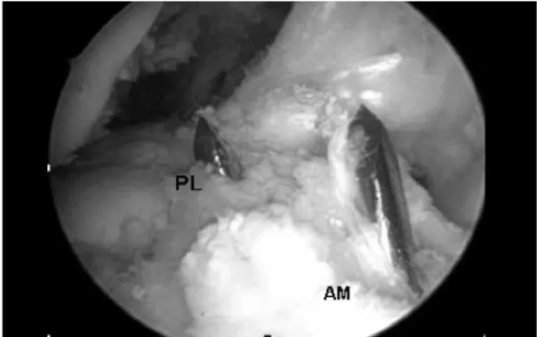
Figure 3 – Arthroscopic view of the anteromedial (AM) and pos-terolateral (PL) bundles, through the anpos-terolateral portal.
Figure 4 – Arthroscopic view of the anteromedial (AM and pos-terolateral (PL) bundles, through the anteromedial portal.
Source: Ápice Ortopedia e Traumatologia
A PROSPECTIVE STUDY WITH TWO-YEAR FOLLOW-UP
A drilled guidewire with a Vicryl 1 wire at two orifices was passed through the anteromedial acces-sory portal, through the posterolateral femoral tun-nel and through the lateral skin of the thigh, while a double end of the Vicryl 1 was kept inside the joint. Next, this end was pulled into the posterolateral tibial tunnel with the aid of a hook, and brought out to the external region of the lower leg. Two wires of dif-ferent colors were used, to make it easier to identify each component.
Following this, the posterolateral graft was passed through the tibial and femoral tunnels, and the Endo-button was overturned, thus providing femoral fixa-tion. The anteromedial graft was then passed through the corresponding tunnels. The Endobutton was over-turned, similarly to the posterolateral graft.
Before fixation, each component of the graft was pretensioned through flexion-extension movements of the knee, 25 times, using manual tension.
We used 25 x 8 metal interference screws to fix the
graft in the anteromedial tunnel, and 30 x 8 screws to fix the graft in the posterolateral tunnel. Absorbable interference screws of similar lengths and diameters could also be used.
The anteromedial graft was fixed with the knee flexed at 45°. The posterolateral graft was fixed with the knee flexed at 15°. In both situations, we under-took manual tensioning of the wires at the tibial end of the graft (Figures 3 and 4).
rehabilitation
We allowed partial body weight-bearing, with the aid of crutches, on the day after the operation, the pro-gression to full weight-bearing was done according to the pain levels. The use of crutches was withdrawn around the seventh day, at which time 90° of flexion The anteromedial femoral tunnel was constructed
at depth in relation to the edge of the posterolateral tunnel. The guidewire could be introduced transtibi-ally, through the posterolateral tunnel, or through the anteromedial accessory portal of the arthroscopy. Our preference was for a transtibial route, which left the femoral tunnels divergent.
Figure 5 – Results from the final assessment using IKDC 2000. 0
10 20 30 40 50 60 70 80
75.86
22.41
1.72
0
A B C D
can usually be achieved. Gentle stretching of the ha-mstrings should be started immediately, to minimize painful adherences.
In a general manner, we followed the same para-meters as described previously(20).
Assessment of the results
The postoperative results were assessed using the protocol of the International Knee Documentation Committee 2000.
RESULTS
For 25 patients, we performed partial medial me-niscectomy in the same surgical procedure as the li-gament reconstruction. In three, we performed partial lateral meniscectomy, and in another nine cases, par-tial resection of both menisci was done. In no case was any reconstructive procedure performed on the joint cartilage.
In group 1 (joint effusion), 54 of the patients’ knees (93.1%) were graded as normal (A) and four (6.89%) as close to normal (B); in group 2 (passive motor deficit), 53 (91.37%) were classified as normal (A) and five (8.62%) as close to normal (B); in group 3 (ligament examination), 52 knees (89.65%) were clas-sified as normal (A), five (8.62%) as close to normal (B) and one (1.72%) as abnormal (C).
In the final assessment, 44 (75.86%) of the pa-tients’ knees were graded as normal (A), 13 (22.41%) as close to normal (B) and one (1.72%) as abnormal (C) (Figure 5).
There were no significant complications and we did not register any cases of infection.
DISCUSSION
The ACL is composed of two anatomical bundles: the anteromedial and posterolateral bundles, according to their tibial insertion(4). Functionally, the anteromedial bundle becomes tense under flexion, which in this position the posterolateral bundle relaxes; conversely, the posterolateral bundle becomes tensioned under extension, while the anteromedial bundle relaxes(21).
Recent studies using navigation for primary re-construction of the ACL using a double bundle have found that the anteromedial and posterolateral com-ponents control both the anterior translation and the rotation during the pivot shift test(22,23).
In the technique described above, the anteromedial portal provided better viewing of the lateral femoral condyle and the femoral insertions of the ACL than did the AL portal, which has traditionally been used for these reconstructions. The arthroscope was insert-ed in the anterominsert-edial portal, while the anterominsert-edial accessory portal was used to make the marks for the ACL insertions in the lateral femoral condyle, and for drilling the posterolateral femoral tunnel.
We preferentially made the anteromedial femoral tunnel through the posterolateral tibial tunnel, because this gave the possibility of producing longer and di-vergent tunnels. In situations in which this has not been possible, we have also made the anteromedial femoral tunnel through the anteromedial accessory portal, which generally results in a shorter tunnel that is parallel to the posterolateral femoral tunnel.
Fu et al(24) reported that accuracy in constructing the anteromedial femoral tunnel through the antero-medial tibial tunnel was only achieved in 10% of the cases. On the other hand, accuracy was achieved through the posterolateral tibial tunnel in 60% of the cases, and through the anteromedial accessory portal in 100% of the times for which this was the access chosen.
35
It is important to highlight that there is no need for special guides, since this is an insertion site technique, thereby ensuring that the reconstruction is not affected by patients’ anatomical variations or by other intra-articular references.
In our assessment using IKDC, four patients (6.89%) reported that they had some swelling. Full joint movement was obtained in the cases of 53 (91.37%) of the knees. Without statistical proof, we observed that with the double-bundle technique, the patients had a tendency to achieve full knee flexion earlier.
In the ligament examinations, the pivot shift test was negative in 44 of the patients’ knees (89.65%); in five cases (8.62%), the result from the pivot shift test was 1+. The patient graded as C (pivot shift 2+) suffered a traumatic failure after slipping on a smooth floor, two months after the operation.
In the final assessment, 44 (75.86%) of our pa-tients’ knees were graded as A, 13 (22.41%) as B and one (1.72%) as C.
In a prospective study without a control group, like in our study, Fu et al(24) assessed 73 patients with a minimum follow-up of two years. They found that 65% had a normal result from the Lachman test and 33% were close to normal. In the pivot shift test, 94% were considered to be normal and 6% were close to normal.
In 2008, Järvelä et al(14) reported that double-bun-dle reconstruction produced better rotational stability. They suggested that this might protect the knee from new lesions that could produce graft failure. In the final assessment using IKDC, 59% of the patients were classified as normal, 36% as close to normal and 4% as abnormal.
Siebold et al(15) also published their results in 2008. In the assessment using IKDC, the group treated with a double bundle presented grade A in 78% of the cases, B in 19% and D in 3%. This last case was a traumatic recurrence of rupture.
In 2010, Aglietti et al(17) assessed 35 patients with chronic ACL injuries who were treated with double-bundle reconstruction. In the final assessment, these authors found that 80% of the patients were classified as normal, 17% as close to normal and 3% as abnormal.
Song et al(25) used a navigation system to measure the intraoperative rotational stability and concluded that reconstruction using a double bundle effectively
reduced the residual pivot shift after ACL reconstruction. Even if the duration of the operation does not in-crease significantly(25), ACL reconstruction surgery using a double bundle is not for surgeons who only occasionally operate on knees. Regarding this topic, Lyman et al(26) concluded that the risk involved in reoperating the ACL is higher in cases treated by sur-geons whose volume of surgery is low.
It is important to emphasize that situations that are more appropriate for ACL reconstruction using a single bundle exist, such as: ACL insertion less than 14 mm, narrow intercondyle (less than 12 mm), open growth plate, grade III arthrosis or higher, multiple ligament lesions and severe bone contusion(19).
Perhaps the most important “side effect” from de-scribing the technique and results from double-bundle reconstruction is the raising of awareness of the need to respect the anatomical ACL insertion sites. In other words, even when a single bundle is used, the femoral insertion should be oblique, done through the antero-medial accessory portal. Femoral tunnel construction using a transtibial route should be avoided. Recon-struction respecting each patient’s anatomy becomes necessary to enable knee function with the recon-structed ACL to be closer to normal.
In this regard, Scanlan et al(27) concluded that graft placement plays a critical role in restoring the normal gait mechanism after ACL reconstruction, and that this may partially explain the incidence of premature arthrosis over the long term.
Despite the biomechanical advantages and the encouraging initial results, there is a need for equip-ment that can quantify rotational slackness and for long-term multicenter prospective studies comparing truly anatomical homogenous techniques for single-bundle or double-single-bundle reconstruction, with pre-cise descriptions, in order to be able prove superior functional results, diminish the likelihood of new meniscal lesions and joint cartilage lesions, and fur-thermore, to diminish the risk of future osteoarthrosis caused by ACL failure.
CONCLUSION
REFERENCES
1. Scavenius M, Bak K, Hansen S, Norring K, Jensen KH, Jorgensen U. Isolated total ruptures of the anterior cruciate ligament--a clinical study with long-term follow-up of 7 years. Scand J Med Sci Sports. 1999;9(2):114-9.
2. Louboutin H, Debarge R, Richou J, Selmi TA, Donell ST, Neyret P, et al. Os-teoarthritis in patients with anterior cruciate ligament rupture: a review of risk factors. Knee. 2009;16(4):239-44.
3. Arnold MP, Kooloos J, van Kampen A. Single-incision technique misses the anatomical femoral anterior cruciate ligament insertion: a cadaver study. Knee Surg Sports Traumatol Arthrosc. 2001;9(4):194-9.
4. Girgis FG, Marshall JL, Al Monajem, AR. The cruciate ligaments of the knee joint. Anatomical, functional and experimental analysis. Clin Orthop Relat Res. 1975;(106):216-31.
5. Freedman KB, D’Amato MJ, Nedeff DD, Kaz A, Bach BR. Arthroscopic anterior cruciate ligament reconstruction: a metaanalysis comparing patellar tendon and hamstring tendon autografts. Am J Sports Med. 2003;31(1):2-11.
6. Yunes M, Richmond JC, Engels EA, Pinczewski, LA. Patellar versus hamstring tendons in anterior cruciate ligament reconstruction: A meta-analysis. Arthros-copy. 2001;17(3):248-57.
7. Andersson D, Samuelsson K, Karlsson J. Treatment of anterior cruciate liga-ment injuries with special reference to surgical technique and rehabilitation: an assessment of randomized controlled trials. Arthroscopy. 2009;25(6):653-85. 8. Yagi M, Wong EK, Kanamori A, Debski RE, Fu FH, Woo SL. Biomechanical
analysis of an anatomic anterior cruciate ligament reconstruction. Am J Sports Med. 2002;30(5):660-6.
9. Petersen W, Tretow H, Weimann A, Herbort M, Fu FH, Raschke M, et al. Bio-mechanical evaluation of two techniques for double-bundle anterior cruciate ligament reconstruction: one tibial tunnel versus two tibial tunnels. Am J Sports Med. 2007;35(2):228-34.
10. Morimoto Y, Ferretti M, Ekdahl M, Smolinski P, Fu FH. Tibiofemoral joint contact area and pressure after single- and double-bundle anterior cruciate ligament reconstruction. Arthroscopy. 2009;25(1):62-9.
11. Yasuda K, Kondo E, Ichiyama H, Tanabe Y, Tohyama H. Clinical evaluation of anatomic double-bundle anterior cruciate ligament reconstruction procedure using hamstring tendon grafts: comparisons among 3 different procedures. Arthroscopy. 2006;22(3):240-51.
12. Yagi M, Kuroda R, Nagamune K, Yoshiya S, Kurosaka M. Double-bundle ACL reconstruction can improve rotational stability. Clin Orthop Relat Res. 2007;(454): 100-7.
13. Muneta T, Koga H, Mochizuki T, Ju YJ, Hara K, Nimura A, et al. A prospective randomized study of 4-strand semitendinosus tendon anterior cruciate liga-ment reconstruction comparing single-bundle and double-bundlentechniques. Arthroscopy. 2007;23(6):618-28.
14. Järvelä T, Moisala AS, Sihvonen R, Järvelä S, Kannus P, Järvinen M. Double--bundle anterior cruciate ligament reconstruction using hamstring autografts
and bioabsorbable interference screw fixation: prospective, randomized, clinical study with 2-year results. Am J Sports Med. 2008;36(2):290-7.
15. Siebold R, Dehler C, Ellert T. Prospective randomized comparison of double--bundle versus singledouble--bundle anterior cruciate ligament reconstruction. Arthros-copy. 2008;24(2):137-45.
16. Ibrahim SAR, Hamido F, Al Misfer AK, Mahgoob A, Ghafar SA, Alhran H. Anterior cruciate ligament reconstruction using autologous hamstring double bundle graft compared with single bundle procedures. J Bone Joint Surg Br. 2009;91(10):1310-5.
17. Aglietti P, Giron F, Losco M, Cuomo P, Ciardullo A, Mondanelli N. Comparison between single-and double-bundle anterior cruciate ligament reconstruction: a prospective, randomized, single-blinded clinical trial. Am J Sports Med. 2010;38(1):25-34.
18. Irrgang JJ, Anderson AF, Boland AL, Harner CD, Kurosaka M, Neyret P, et al. Development and validation of the international knee documentation committee subjective knee form. Am J Sports Med. 2001;29(5):600-13.
19. Van Eck CF, Lesniak BP, Schreiber VM, Fu, FH. Anatomic Single- and Double--Bundle Anterior Cruciate Ligament Reconstruction Flowchart. Arthroscopy 2010;26(2):258-68.
20. Gali JC, Camanho GL. A reabilitação acelerada após reconstrução do ligamen-to cruzado anterior com enxerligamen-to de tendão patelar é segura? Rev Bras Orligamen-top. 1998;33(8):645-50.
21. Amis AA, Dawkins GP. Functional anatomy of the anterior cruciate ligament. Fibre bundle actions related to ligament replacements and injuries. J Bone Joint Surg Br. 1991;73(2):260-7.
22. Ishibashi Y, Tsuda E, Yamamoto Y, Tsukada H, Toh S. Navigation evaluation of the pivot-shift phenomenon during double-bundle anterior cruciate liga-ment reconstruction: is the posterolateral bundle more important? Arthroscopy. 2009;25(5):488-95.
23. Robinson J, Carrat L, Granchi C, Colombet P. Influence of anterior cruciate ligament bundles on knee kinematics: clinical assessment using computer--assisted navigation. Am J Sports Med. 2007;35(12):2006-13.
24. Fu FH, Shen W, Starman JS, Okeke N, Irrgang JJ. Primary anatomic double--bundle anterior cruciate ligament reconstruction: a preliminary 2-year pros-pective study. Am J Sports Med. 2008;36(7):1263-74.
25. Song EK, Oh LS, Gill TJ, Li G, Gadikota HR, Seon JK. Prospective comparative study of anterior cruciate ligament reconstruction using the double-bundle and single-bundle techniques. Am J Sports Med. 2009;37(9)1705-11.
26. Lyman S, Koulouvaris P, Sherman S, Do H, Mandl LA, Marx RG. Epidemiology of anterior cruciate ligament reconstruction. trends, readmissions, and subse-quent knee surgery. J Bone Joint Surg Am. 2009;91(10):2321-8.