Vol.57, n.6: pp. 887-894, November-December 2014 http://dx.doi.org/10.1590/S1516-8913201400874
ISSN 1516-8913 Printed in Brazil
BRAZILIAN ARCHIVES OF BIOLOGY AND TECHNOLOGY
A N I N T E R N A T I O N A L J O U R N A L
Pharmacodynamic and Pharmacokinetic Studies of
β
-Cyclodextrin:Dexamethasone Acetate Complexes in Mice
Patrik Oening Rodrigues
1*Ana Cláudia Becker
2, Viviane Aparecida Dellagnelo
2, Cibele
Nabhan Benetti
1, Eduardo Manoel Pereira
2, Theodoro Marcel Wagner
2and Marcos
Antonio Segatto Silva
31Laboratório de Tecnologia Farmacêutica; Universidade Federal de Mato Grosso do Sul; Campo Grande - MS -
Brasil. 2Departamento de Farmácia; Universidade da Região de Joinville; Joinville - SC - Brasil.3Departamento de Ciências Farmacêuticas; Universidade Federal de Santa Catarina; Florianopolis - SC - Brasil
ABSTRACT
The aim of this study was to evaluate the in vivo activity of the anti-inflammatory and analgesic effects of a suspension of the complex composed of dexamethasone acetate (DMA) with β-cyclodextrin in comparison to a suspension of the pure DMA. Solid complexes prepared by different methods were evaluated in pharmacodynamics and pharmacokinetics studies. The pharmacodynamic effect was investigated although the capacity of the inhibited the inflammation. Models of abdominal constriction, carrageenan-induced paw oedema and formalin induced licking were used. The study of the pharmacodynamic comparison of free DMA and products of β-CD:DMA demonstrated no significant difference in the majority of the tests performed. Plasma concentrations of DMA and DMA:β-CD were assayed by HPLC. A significant (p > 0.05) decrease in the relative bioavailability was obtained with the suspension containing the DMA:β-CD complex as measured by DMA plasma levels. The area under the curve (AUC) of the suspension of DMA was higher than that obtained with the suspension of the complexes. The pharmacokinetic evaluation of dexamethasone carried out on mice in the present study showed that complexed DMA with β-cyclodextrin modifieds some parameters related to the phases of absorption and elimination of this drug.
Key words:
S
olubility, dexamethasone acetate, β-cyclodextrin, bioavailability, anti-inflammatory activity
*Author for correspondence:[email protected]
INTRODUCTION
Dexamethasone acetate (DMA), 9-Fluoro-11β, 17,21-trihydroxy-16α-methylpregna-1,4-diene- 3,20-dione, is a synthetic glucocorticoid drug of the class of steroid hormones widely used in the clinics with low aqueous solubility, which limits its oral absorption. Furthermore, the lack of water solubility reduces the flexibility for drug formulation and administration. DMA is an important drug that exerts various inhibitory effects on the inflammatory processes and as an
Most of the anti-inflammatory effects of DMA are mediated by the modulation of gene expression through either a direct interaction of the glucocorticoids (GCs) receptor to DNA or an interference with the function of transcription factors such as prostaglandin (PG) and leukotrien (Chu et al. 2014). Various techniques have been used to improve the solubility/dissolution rate of poorly water-soluble drugs, among which cyclodextrin (CD) complexation is of particular interest (Wu et al. 2010; Hu et al. 2012). The complexes with β-CD have been prepared by different methods such as kneading, coevaporation, physical mixture and freeze drying (Ruz et al. 2012; George and Vasudevan 2012; Passos et al. 2012; Qiu et al. 2014).
CDs are cyclic oligosaccharides consisting of covalently linked glucopyranose rings. α-, β-, and
γ-CDs are naturally occurring CDs combining six, seven or eight glucopyranose units. The CDs and their derivatives are used in pharmaceutical formulations to enhance the solubility, dissolution rate, stability and bioavailability of poorly water-soluble compounds. The property of enhancing solubility may be explained by the formation of water-soluble inclusion complexes in which the hollow truncated, cone-like CD structure, encapsulates hydrophobic drug molecules in the apolar interior. The hydrophilic region of the CD enables the solubilisation through interaction with water molecules. Encapsulation of a drug molecule affects many of its physicochemical properties and can result in increased aqueous solubility (Shen et al. 2005; Gazpio et al. 2005; Zhang et al. 2009). The purpose of this study was to investigate the bioavailablity of DMA in complex form with β -CD in comparison with the free DMA through the gastrointestinal absorption. It is a drug model with high log P, which demonstrates the potential of using the complexes to increase the solubility.
MATERIAL AND METHODS
The DMA was supplied by Laboratório Farmacêutico do Estado de Santa Catarina - LAFESC (Florianópolis, Brazil) and β-CD was obtained from Roquette (Genay, France).
Preparation of physical mixtures and inclusion complexes
The preparation of solid complexes of DMA and
β-CD were performed by different techniques.
Based on the results of the preliminary phase solubility studies, the molar ratio was kept at 1:1 in all the cases.
Kneading method: Kneaded (KN) product was obtained by adding small amount of water to β-CD placed in a mortar and mixed to obtain a homogeneous paste. Then, DMA powder was slowly added and the mixture was kneaded for 60 min. During the process, few drops of water were introduced to maintain a suitable consistency. The resulting paste was dried in an oven at 45ºC for 24 h. The dried complex was pulverized into a fine powder.
Coevaporation method: Coevaporated (CV) product was obtained by dissolving equimolar amount of β-CD and DMA in suitable volumes of 50% ethanol. The solution was shaken at 25ºC for seven days and the solvent was then removed in an oven at 45°C for 24 h. The obtained solid was pulverized into a fine powder.
Physical mixture method: Physical mixture (PM) product was prepared by homogeneous blending of previously weighed DMA and β-CD in a mortar for 10 min.
Freeze Dryingmethod: Freeze-dried (FD) product was prepared by dissolving the β-CD in ethanol: NaOH 0.1M (1:3; v/v) solution and adding the stoichiometric amount of the drug. The suspension was shaken at 25ºC for 48 h. The resulting solution was frozen by keeping it in a repository at -20ºC and lyophilized in a freeze-dryer (Terroni Fauvel LT 1000/8, Brazil) for 24 h.
The kneading, coevaporation, physical mixture, freeze dried products and free DMA were administrated in the suspension of the 0.5% carboxymethylcellulose (CMC). The vehicle was prepared 24 h before each test and used for seven days after the preparation.
Pharmacodynamic studies
All the studies involving the mice were accomplished in agreement with the Guideline of Experiments with Humans and Animals of the Universidade do Vale do Itajaí – UNIVALI (no 594/2007) and the project was approved for the ethics committee in research with humans and animals.
Analgesic effects of the complexes – the writhing test
described by Besra et al. (1996). Groups of six animals were treated with the suspensions at 2 mg/kg dose of DMA, KN, CV, PM, FD and the control group received 0.5% CMC. After 1 h, acetic acid was administered (i.p.). The number of writhing movements, characterized by the contraction of abdominal muscle together with a stretching of hind limbs, was counted for 20 min.
Carrageenan-induced paw oedema
The procedure used to assess the anti-inflammatory activity was based on the method used by Winter et al. (1962). The animals were treated with the 0.5% CMC suspension (control group) or DMA, KN, CV, PM and FD complexes in the dosage of 2 mg/kg. After 1 h of sample administration, edema was induced by injecting the carrageenan (300 µg/paw diluted in 50 µL in sterile saline) into the plantar side of the left hind paw of the six animals. Three hours later, the mice were sacrificed by cervical dislocation and the right and left paws were weighed. The parameter evaluated was the increase of the weight of the left paw versus the right hind paw, which remained as a control.
Formalin-induced paw licking in mice
The procedure was essentially similar to that described by Correa and Calixto (1993). Formalin solution (30 µL, 2.5% in distillated water) was injected subcutaneously under the surface of the right hind paw in the test groups. The total amount of time spent licking the injected paw was recorded, and was considered as an indicator of pain. The animals were treated with 0.5% suspension of CMC (control group) or with 2mg/kg of DMA, KN, CV, PM and FD complexes. After 1 h, formalin (2.5%) was injected and the responses were observed for 40 min. The first phase of the nociceptive response normally peaked 5 min after formalin injection and the second phase 15-40 min after formalin injection, representing the neurogenic and inflammatory pain responses, respectively (Hunskaar and Hole 1987).
Oral bioavailability study
Swissmice weighing 30–40 g were used. All mice were allowed free access to water, but were fasted for 12 h before drug administration and 4 h after drug administration. The mice were divided into groups of six. The formulations were administered orally via bucco-gastric tube. The DMA, KN, CV,
PM and FD (suspension of 0.5% CMC) was administrated at the dose of 20 mg/kg. After drug administration, the animals were sacrificed and blood samples were collected at the following time intervals: 1, 2, 4, 8 and 24 h. Lastly, the blood samples were heparinized and centrifuged individually. After the extraction process, the blood samples were frozen until HPLC analysis.
HPLC analysis
A modular liquid chromatograph equipped with a Merck Hitachi 7000 IF- Lachrom and gradient pump (L-7100, Merck Hitachi), an autosampler (L-7200, Merck Hitachi) fitted to a 100 µL sampler loop (Rhedyne), a variable wavelength detector (UV- L-7400), a column oven (L-7300, Merck Hitachi) and an interface (D-7000, Merck Hitachi) connected a computer were used. In order to precipitate the proteins, 1.0 mL of methanol was added to each aliquot (0.25 ml) of plasma sample. After vortex-mixing for 1 min, samples were centrifuged at 3000 rpm for 10 min and the supernatant were analyzed.
The DMA assay was conducted using a 5 µm RP-18 (LiChrospher® 100) 125 x 4 mm column and a mobile phase containing 70% acetic acid 1% and 30% methanol at a flow rate of 1 mL/min. Samples were analyzed at 254 nm. Under these conditions, the retention time was 15 min.
Bioavailability parameters
The bioavailability parameters were calculated by plotting the absorption profile of dexamethasone acetate, nd the products of the physical mixture, kneading, coevaporation and freeze-drying. The following pharmacokinetics parameters were evaluated: maximum concentration of the drug in plasma (Cmax), time to reach Cmax (Tmax) and the
area under the curve from 0 to 24 h (AUC 0-24).
The maximum plasma concentrations and the corresponding times were calculated as the mean of the experimental data. AUC values were estimated via trapezoidal summation. Results were considered significant at the 5% critical level (p < 0.05).
RESULTS AND DISCUSSION
thermogravimetry, infrared spectroscopy, optical microscopy and dissolution studies (Ruz et al. 2012; Loh et al. 2014; Mennini et al. 2014) demonstrating that all the methods were efficient in the complexation.
Pharmacodynamic comparison of free DMA
and products of β-CD:DMA
The pharmacodynamic effects of DMA, KN, CV, PM and FD were investigated based on the capacity of suppressing the pain and inflammation induced by the writhing test,formalin test andpaw edema test. The writhing test evaluated the antinociceptive effect of the free DMA and its complexes induzed by acetic acid. The acetic acid causes inflammatory pain by inducing the capillary permeability (Amico-Roxas et al. 1984). Figure 1 shows the effects of DMA, KN, CV, PM and FD in the number inhibition of abdominal constrictions. Although there was a significant difference in comparison to the group that did not receive glucocorticoid, there was no significant difference (p > 0.05) between the animals which received free DMA and the complexes. However, all the groups showed significant difference (p <
0.05) in relation to the control group treated only with vehicle.
Figure 1 - Effect of the free DMA and complexes on the number of writhing after injection of acetic acid 0.7%. All products were administered as suspension in 0.5% (w/v) carboxymethylcellulose. Each bar represents the mean ± Standard Deviation (SD) of six experiments. a p < 0.01 vs DMA. b p < 0.05 vs DMA - Dunnett's Multiple Comparison Test.
Acetic acid induces the pain by increasing the levels of PG-E2 and PG-F2a (Deraedt et al. 1980) at the peritoneal receptors (Bentley et al. 1983) and is a sensitive method in detecting the analgesic
efficacy of the agents (Collier et al. 1968). Results obtained in the present study showed that the formulations exhibited the same anti-nociceptive effect against acetic acid-induced response in mice, inhibiting the peripheral stimulus involved in the induction of nociception signalization (Rang et al. 2012).
Glucocorticoids are widely used as anti-inflammatory and immunosuppressive agents to control several chronic and acute diseases. Their therapeutic activity is due to the enhanced intensity of the physiological effects of endogenous steroids (Bosscher et al. 2000; Payne and Adcock 2001). Glucocorticoids function via the glucocorticoids receptor, which regulates the transcription of several target genes and also mediates glucocorticoids effects indirectly via a negative or positive regulation of transcription factors and other signaling proteins (Payne and Adcock 2001; Wikstrom 2003).
The anti-edematogenic activity to the free DMA and to KN, CV, PM and FD complexes was evaluated by carrageenan-induced paw oedema. The carrageenan-induced paw oedema is an in vivo
model of inflammation (Winter et al. 1963) used to test whether there is activity upon the cyclo-oxygenase products of arachidonic acid metabolism and the production of reactive oxygen species (Smith et al. 1974). Development of oedema induced by carrageenan is commonly correlated with the early exudative stage of inflammation. The results presented in Figure 2 showed that the DMA, and KN, CV and PM complexes reduced significantly (p < 0.05) the paw oedema, in relation to the control group. The freeze dried product did not show the same effect observed in the others complexes, when compared with the control group. There was an increase in the paw weight of animals, which were treated with this formulation.
and suggested that these formulations could also be involved in the reduction of inflammatory precursors, as observed in the effect of the free DMA. It also showed that the anti-inflammatory activity of dexamethasone was still significantly preserved in the complexes.
Figure 2 - Paw edema of the free DMA and complexes for the mass difference of hind paws after injection of carrageenan. All products were administered as suspension in 0.5% (w/v) carboxymethylcellulose. Each bar represents the mean ± S.D of six experiments. a p < 0.01 vs DMA - Dunnett's Multiple Comparison Test.
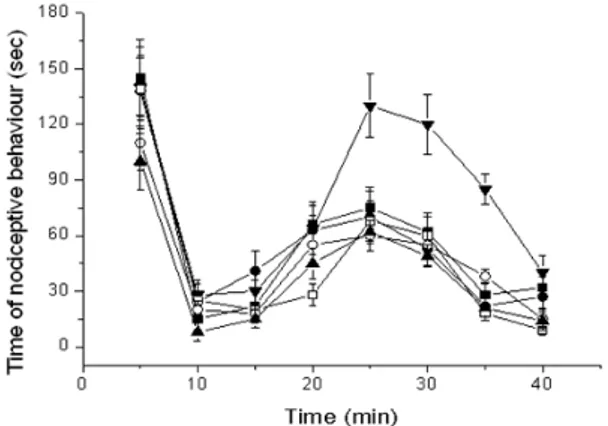
The formalin test assesses the behavioral response of injection of this algogen into the paw of the animals. The response consists of licking and elevation (lifting) of the injected paw, flinching, and also protection of the paw from full pressure when walking or resting. This behavior occurs in a biphasic pattern, with an immediate response after the injection, which comes right after the injection, on the first 5 min, and a second phase beginning from 15 to/40 min after the injection (Porro and Cavazzuti 1993). Drugs that act primarily on the central nervous system inhibit both the phases equally while peripherally acting drugs inhibit the late phase, which is related to the synthesis of inflammatory mediators. The first phase involves the direct activation of nociceptors by the algogenic agent (Tjolsen et al. 1992). Figure 3 shows the influence of the free DMA, KN, CV, PM and FD complexes on the nociceptive behavior of the animals after formalin injection. Both free DMA and its complexes were ineffective in reducing the total time of nociceptive behavior in the first phase (0 at 5 min). In the second phase (15 at 40 min), the drugs administrated also did not show significantive difference in comparison
to the group, which received only formalin (p >
0.05), with exception to the freeze dried complex. Statistically, there was a relevant difference in the FD antinociceptive activity in relation to the free DMA group (p < 0.05). Dexamethasone is a potent inhibitor of the inflammatory mediators, this justifies its reduction of the total amount of time the animals spent showing nociceptive behavior. There was no significant increase indexamethasone associated with cyclodextrin complexes, which suggesteds that the maximum effect of this drug might have been already reached, since the complexes showed no higher efficacy than free DMA.
Figure 3 - Time of nociceptive behavior of free DMA and complexes after injection of formalin. (▼) is carboxymethylcellulose; of (■) free DMA; (●) kneaded; (□) physical mixture; (▲) freeze dried and (○) coevaporated products. All products were administered as suspension of carboxymethylcellulose in 0.5% (w/v). Each point represents the mean ± S.D of six experiments.
In vivo bioavailability study
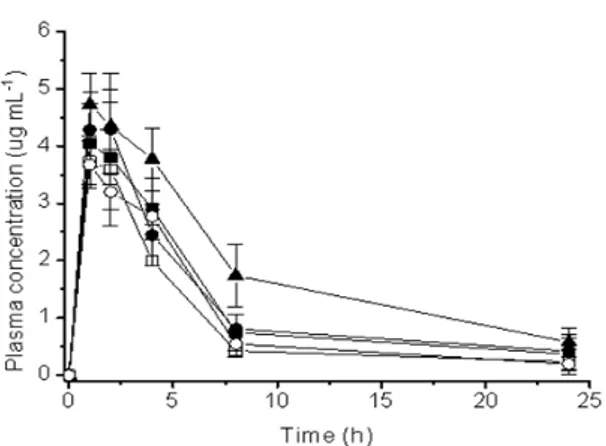
Figure 4 - Plasma levels of DMA after oral administration of (■) free DMA; (●) kneaded; (□) physical mixture; (▲) freeze dried and (○) co-evaporated products. All products were administered as suspension of carboxymethylcellulose in 0.5% (w/v). Each point represents the mean ± S.D of six experiments.
Low DMA concentrations were obtained after oral administration of the DMA: β-CD, compared with the free DMA. Relevant pharmacokinetic parameters corresponding to the plasma concentration profiles are listed in Table 1. Cmax,
Tmax and AUC0–24 h were compared after the
administration of the free DMA and DMA complexes. For all the DMA:β-CD complexes and free DMA, the Tmax were similar, since after the
Tmax everyone had a sharp decline till the peak at
8 h. The pharmacokinetic evaluation of dexamethasone carried out on mice in the present study showed that complexing dexamethasone with β-CD for different methods modified some parameters related to the phases of absorption and elimination. Between 8 at 24 h, the elimination peaks were constant.
The complexation with CD seemeds to have affected the absorption of the drug, which produced lower plasmatic levels of DMA. The complexes kneading, coevaporation, freeze drying and physical mixture demonstrated significant difference (p < 0.05) for Cmax in relation to free
DMA, which showed for the complexes worsening the absorption and bioavailability. Tmax remaineds
constant between the free DMA and the four complexes (p > 0.05). However, the AUC0–24 h had
significant difference (p < 0.05) between the free DMA and products KN, CV, PM and FD. The complexes products KN and PM had better bioavailability with significant difference in comparison to the CV and FD complexes
products. The in vivo evaluation of pharmaceutical drug preparations with poor solubility was performed for a single dose at 20 mg/kg.
Table 1 - Pharmacokinetics parameters (mean ±S.D.) for DMA obtained following oral administration of DMA at 20 mg/kg of body weight to mice (n=6). (DMA) dexamethasone acetate; (KN) kneaded; (PM) physical mixture; (CV) coevaporated and (FD) freeze dried products. All products were administered as suspension in 0.5% (w/v) carboxymethylcellulose.
DMA KN CV PM FD Cmax
(µg mL-1) 4.69± 1.00
2.46± 0.24
0.74± 0.33
2.28± 0.25
0.21± 0.17
T max (h) 1.50±
0.55 1.67± 0.52
1.67± 1.21
1.50± 0.55
1.33± 0.52 AUC
(µg h mL-1) 28.87 ±5.17
13.57 ±2.09
2.11± 0.23
12.67 ±0.93
0.90± 0.05
Invitro results obtained with the same complexes used by Doile et al. (2007) showed that the FD and KN products exhibited higher dissolution rates than the drug alone. The extent of the enhancement of the dissolution rate was dependent on the preparation method, since physical mixture and coevaporated showed lowes dissolution rates (Doile et al. 2007). Comparing the dissolution rate with the bioavailability, the results obtained were contradictory, as the FD product showeds a minor bioavailability in relation to others complexes and free DMA. This decrease might be a result of a reduction in the permeability of the organic biological membranes caused by the complexation process.
Even though there was a reduction in the absorption in relation to free DMA, there was no significant influence in the therapeutic effects of DMA in the different complexed forms. However, this suggesteds that might have a better pharmacological activity, once they could reach the same response as free DMA with lower plasmatic levels. This would be significant because the same effect could be reached with lower levels, which also implied in lower incidence of side effects, as there was less free drug available to reach other tissues.
CONCLUSIONS
modifieds some parameters related to the phases of absorption and elimination of this drug. DMA in the free form displayed significantly higher absorption speed than did the DMA: β-CD complex. Tmax was constant, but Cmax and AUC of
the products complexes were significantly different (p < 0.05) of the free DMA. However, the pharmacodynamic tests evaluated the anti-inflammatory activity, anti-edematogenic and nociceptive of the drug, they showed that the effect of DMA complexed was maintained in relation to the drug not complexed.
ACKNOWLEDGMENTS
We are grateful to Keila A. Fortunato, Mayara M. Doile, Iára C. Schmücker and Sacha K. Schucko for their collaboration with the development of this work.
REFERENCES
Amico-Roxas M, Caruso A, Trombadore S, Scifo R, Scapagnini U. Gangliosides antinociceptive effects in rodents. Arch Int Pharmacodyn Ther. 1984; 272: 103-117.
Bains M, Hall ED. Antioxidant therapies in traumatic brain and spinal cord injury. Biochim Biophys Acta. 2012; 1822: 675-684.
Bentley GA, Newton SH, Starr J. Studies on the anti-nociceptive action of a-agonist drugs and their interaction with opioid mechanisms. Br J Pharmacol. 1983; 79: 125-134.
Besra SE, Sharma RM, Gomes A. Anti-inflammatory effect of petroleum ether extract of leaves of Litchi chinensis Gaertn (Sapindaceae). J Ethnopharmacol. 1996; 54: 1-6.
Bosscher KD, Vanden WB, Haegeman G. Mechanisms of anti-inflammatory action and of immunosuppression by glucocorticoids: negative interference of activated glucocorticoid receptor with transcription factors. J Neuroimmunol. 2000; 109: 16-22.
Chu CC, Hsing CH, Shieh JP, Chien CC, Ho CM, f, Wang JJ. The cellular mechanisms of the antiemetic action of dexamethasone and related glucocorticoids against vomiting. Eur J Pharmacol. 2014; 722: 48-54.
Collier HOJ, Dinneen LC, Johnson CA, Schneider C. The abdominal constriction response and its suppression by analgesic drug in the mouse. Br J Pharmacol. 1968; 32: 295-310.
Correa CR, Calixto JB. Evidence of participation of B1 and B2 kinin receptors in formalin-induced nociceptive response in mouse. Br J Pharmacol. 1993; 110: 193-198.
Deraedt R, Jougney S, Delevalcee F, Falhout M. Release of prostaglandin E and F in an algogenic reaction and its inhibition. Eur J Pharmacol. 1980; 51: 17-24.
Dinis-Oliveira RJ, Duarte JA, Remião F, S´anchez-Navarro A , Bastos ML, Carvalho F. Single high dose dexamethasone treatment decreases the pathological score and increases the survival rate of paraquat-intoxicated rats. Toxicology. 2006; 227: 73-85. Doile MM, Fortunato KA, Schmücker IC, Schucko SK,
Silva MAS, Rodrigues PO. Physicochemical properties and dissolution studies of dexamethasone acetate-β-cyclodextrin inclusion complexes produced by different methods. AAPS PharmSci. 2008; 9: 314-321.
Doile MM, Fortunato KA, Schmücker IC, Schucko SK, Silva MAS, Rodrigues PO. Influência da Complexação com β-ciclodextrina sobre a Liberação do Acetato de Dexametasona a partir de Matrizes Hidrofílicas de Hidroxipropilmetilcelulose (HPMC) e Polioxetileno (PEO). Latin Am J Pharm. 2007; 26: 513-521.
Fiore DC, Hall S, Shoja P. Altitude illness: risk factors, prevention, presentation, and treatment. Am Fam Physician. 2010; 82(9):1103-1110.
Garcia JL, Nakamura L, Leite MP, Rochae MS. Pharmacological analysis of the acute inflammatory process induced in rat’s paw by local injection of carrageenan and by heating. Br J Pharmacol. 1973; 48: 88-96.
Gazpio C, Sánchez M, Zubiri IXG, Vélaz I, Ohárriz CM, Martin C, Zornoza A. HPLC and solubility study of the interaction between pindolol and cyclodextrins. J Pharm Biomed Anal. 2005; 37: 487-492.
Geen KL. Role of endogenous catecholamines in the anti-inflammatory activity of alphaadrenoceptor blockingagents. Br J Pharmacol. 1964; 51: 45-53. Genovese T, Mazzon E,Crisafulli C, Esposito E, Di
Paola R, Muia C, Di Bella P, Bramanti P, Cuzzocrea S. Effects of combination of melatonin and dexamethasone on secondary injury in an experimental mice model of spinal cord trauma. J Pineal Res. 2007; 43:140-153.
George SJ, Vasudevan DT. Studies on the Preparation,Characterization, and Solubility of 2-HP-β-Cyclodextrin-Meclizine HCl inclusion complexes. J Young Pharmacists. 2012; 4: 220-227.
Hunskaar S, Hole K. The formalin test in mice: dissociation between inflammatory and noninflammatory pain. Pain. 1987; 30: 103-114. Li L, Ma P,Wei J,Qian K, Tao L. LC-ESI-MS method
for the determination of dexamethasone acetate in skin of nude mouse. J Chromatogr B. 2013; 933: 44-49.
Loh GOK, Tan YTF, Peh KK. Effect of HPMC concentration on β-cyclodextrin solubilization of norfloxacin. Carbohydr Polym. 2014; 101: 505-510. Mennini N, Bragagni M, Maestrelli F, Mura P.
Physico-chemical characterization in solution and in the solid stateof clonazepam complexes with native and chemically-modified cyclodextrins. J Pharm Biomed Anal. 2014; 89: 142-149.
Payne DN, Adcock IM. Molecular mechanisms of corticosteroid actions. Paediatr Respir 2001; 2: 145-150.
Passos JJ, Sousa FB, Mundim IM, Bonfim RR, Melo R,Viana AF, et al. In vivo evaluation of the highly soluble oral β-cyclodextrin–Sertraline supramolecular complexes. Int. J. Pharm. 2012; 436: 478-485. Porro CA, Cavazzuti M. Spacial and temporal aspects
of spinal cord and brainstem activation in the formalin pain model. Prog Neurobiol. 1993; 41: 565-607.
Qiu N, Cheng X, Wang G, Wang W, Wen J, Zhang Y, et al. Inclusion complex of barbigerone withhydroxypropyl- β-cyclodextrin: Preparation and in vitro evaluation. Carbohydr Polym. 2014; 101:623-630.
Rang HP, Dale MM, Ritter JM. Farmacologia. 7ª Edição. Rio de Janeiro: Elsevier; 2012.
Ruz V, Froeyen M, Busson R, Gonzaleza MM, Baudemprez L, Mooter GD. Characterization and molecular modeling of the inclusion complexes of 2-(2-nitrovinyl) furan (G-0) with cyclodextrines. Int J Pharm. 2012; 439: 275-285.
Smith MJH, Hutchinson AWF, Elliot PNC, Bolam J. Prostaglandin in the anti-inflammatory activity of a human plasma fraction in carrageenan-induced paw oedema in the rat. J Pharm Pharmacol. 1974; 26: 692 - 698.
Shen YL, Yang SH, Wu LM, Ma XY. Study on structure and characterization of inclusion complex of gossypol/beta cyclodextrin. Spectrochimica Acta Part A. 2005; 61: 1025-1028.
Tjolsen A, Berge OG, Hunskaar S, Rosland JH, Hole K. The formalin test: An evaluation of the method. Pain. 1992; 51: 5-17.
Urbańska J, Karewicz A, Nowakowska M. Polymeric delivery systems for dexamethasone. Life Sciences. 2014; 96: 1-6.
Wikstrom AC. Glucocorticoid action and novel mechanisms of steroid resistance: role of glucocorticoid receptor-interacting proteins for glucocorticoid responsiveness. J Endocrinol. 2003; 178: 331-337.
Winter CA, Risley EA, Nuss GW. Carregeenin-induced oedema in hind paw of the rat as assay for anti-inflammatory drugs. Proc Soc Exp Biol Med. 1962; 11: 544-547.
Winter CA, Risley EA, Nuss GW. Anti-inflammatory and antipyretic activities of indomethacin, 1- (p-chlorobenzoyl)- 5- methoxy- 2- methyl- indol- 3- acetic acid. J Pharmacol Exp Ther. 1963; 141: 369-373.
Wu H, Liang H, Yuan Q, Wang T, Yan X. Preparation and stability investigation of the inclusion complex of sulforaphane with hydroxypropyl-β-cyclodextrin. Carbohydr Polym. 2010; 82: 613-617.
Zhang M, Li J, Zhang L, Chao J. Preparation and spectral investigation of inclusion complex of caffeic acid with hydroxypropyl-β-cyclodextrin. Spectrochimica Acta. Part A. 2009; 71:1891-1895.