Gpr116 Receptor Regulates Distinctive
Functions in Pneumocytes and Vascular
Endothelium
Colin Niaudet1*, Jennifer J. Hofmann2☯, Maarja A. Mäe1☯, Bongnam Jung1☯,
Konstantin Gaengel1☯, Michael Vanlandewijck1, Elisabet Ekvärn2¤
, M. Dolores Salvado3, Annika Mehlem2, Sahar Al Sayegh1, Liqun He1, Thibaud Lebouvier1, Marco Castro-Freire1, Kan Katayama2, Kjell Hultenby4, Christine Moessinger2, Philip Tannenberg2,5,
Sara Cunha2, Kristian Pietras2,6, Bàrbara Laviña1, JongWook Hong2, Tove Berg2,
Christer Betsholtz1,2
1Department of Immunology, Genetics and Pathology, Rudbeck Laboratory, Uppsala University, Uppsala, Sweden,2Division of Vascular Biology, Department of Medical Biochemistry and Biophysics, Karolinska Institute, Stockholm, Sweden,3Physiological Chemistry II, Department of Medical Biochemistry and Biophysics, Karolinska Institute, Stockholm, Sweden,4Department of Laboratory Medicine, Division of Clinical Research Center, and Karolinska Institute, Stockholm, Sweden,5Department of Molecular Medicine and Surgery, Division of Vascular Surgery, Karolinska Institute, Stockholm, Sweden,6Lund University, Department of Laboratory Medicine, Medicon Village, Lund, Sweden
☯These authors contributed equally to this work.
¤ Current Address: Octapharma AB, SE-112 75, Stockholm, Sweden
*colin.niaudet@igp.uu.se
Abstract
Despite its known expression in both the vascular endothelium and the lung epithelium, until recently the physiological role of the adhesion receptor Gpr116/ADGRF5 has remained elusive. We generated a new mouse model of constitutive Gpr116 inactivation, with a large genetic deletion encompassing exon 4 to exon 21 of theGpr116gene. This model allowed us to confirm recent results defining Gpr116 as necessary regulator of surfactant homeosta-sis. The loss of Gpr116 provokes an early accumulation of surfactant in the lungs, followed by a massive infiltration of macrophages, and eventually progresses into an emphysema-like pathology. Further analysis of this knockout model revealed cerebral vascular leakage, beginning at around 1.5 months of age. Additionally, endothelial-specific deletion of Gpr116 resulted in a significant increase of the brain vascular leakage. Mice devoid of Gpr116 developed an anatomically normal and largely functional vascular network, surprisingly exhibited an attenuated pathological retinal vascular response in a model of oxygen-induced retinopathy. These data suggest that Gpr116 modulates endothelial properties, a previously unappreciated function despite the pan-vascular expression of this receptor. Our results support the key pulmonary function of Gpr116 and describe a new role in the central nervous system vasculature.
OPEN ACCESS
Citation:Niaudet C, Hofmann JJ, Mäe MA, Jung B, Gaengel K, Vanlandewijck M, et al. (2015) Gpr116 Receptor Regulates Distinctive Functions in Pneumocytes and Vascular Endothelium. PLoS ONE 10(9): e0137949. doi:10.1371/journal.pone.0137949
Editor:Nades Palaniyar, The Hospital for Sick Children and The University of Toronto, CANADA
Received:June 4, 2015
Accepted:August 24, 2015
Published:September 22, 2015
Copyright:© 2015 Niaudet et al. This is an open access article distributed under the terms of the Creative Commons Attribution License, which permits unrestricted use, distribution, and reproduction in any medium, provided the original author and source are credited.
Data Availability Statement:All relevant data are within the paper and its Supporting Information files.
Introduction
The adhesion receptors are the most recently described of the 5 classes of seven transmem-brane receptors. In mammals, each of the 33 members of this class is characterized by the pres-ence of long N-terminal domains harboring various adhesion motifs and a cleavable so-called
GPS domain [1]. To date, specificin vivofunctions have been assigned to only a handful of the
adhesions receptors. In a few cases, targeted loss-of-function mutations have revealed critical roles in key biological processes.
So far, the three Cadherin-EGF LAG Seven-pass G-type Receptor (Celsr, now ADGRC [2])
genes have been the most extensively studied of the adhesion receptors. Various experimental approaches ranging from mutagenesis to pharmacological modulation in organ explants show that Celsr proteins control planar cell polarity in several developmental processes, including
neural tube closure [3], hair follicle formation [4] and lung morphogenesis [5] (for Celsr1/
ADGRC1), ciliogenesis (for Celsr2/ADGRC2) [6], pancreas formation [7] and axon guidance
[8] (for Celsr3/ADGRC3).
In the nervous system, three different adhesion receptors have been shown to take part in essential developmental processes. Gpr126/ADGRG6 is required for Schwann cell maturation
and nerve myelination [9], a role conserved between zebrafish and mouse [10]. The
involve-ment of two heterotrimeric G proteins downstream of Gpr126 have also been demonstrated
[11]. Gpr56/ADGRG1 shapes the perisylvian gyri through control of neocortex progenitors
[12]. Mutations in the human gene,GPR56, cause a malformation of the cerebral cortex known
as bilateral frontoparietal polymicrogyria [13]. Finally, Gpr124/ADGRA2 has proven essential
for angiogenesis in the neural tube and forebrain, and its global or specific endothelial deletion lead to hemorrhagic glomeruloid vascular malformations in the developing central nervous
system (CNS) [14][15][16].
Pioneering work identified Gpr116, recently renamed ADGRF5 [2], as a new adhesion
receptor expressed in the alveolar wall of rat lung [17]. Gpr116 was subsequently recognized as
part of cluster VI, a group of 5 related receptors [1] whose tissue expression patterns have
recently been mapped [18]. During late gestational stages, expression of Gpr116 was shown to
increase in the lungs, suggesting a role in organogenesis [19][20]. Recently, three independent
models of knockout mice forGpr116have been used to investigate its function in the lung [19]
[20][21]. The absence of this gene leads to increased levels of saturated phosphatidylcholine
(Sat PC), an essential component of surfactant, from 1 week of age onward [20]. This
pheno-type was also confirmed in older knockouts and was described as part of a global increase in all
surfactant phospholipids at 4 weeks of age [19], accompanied by a shift in their carbon
satura-tion at 8 weeks [21]. Surfactant proteins were also increased inGpr116knockout mice [19][20]
[21]. The increased surfactant production was paralleled by expansion of alveolar
macro-phages, starting from 3 and peaking at 12 weeks of age [20]. In adult mice, this expansion
developed into a severe pulmonary phenotype, with massive increases in surfactant
phospho-lipids and proteins, as well as hyperplasia and hypertrophy of lipid-laden macrophages [19]
[20][21]. Importantly, the lung defect of the fullGpr116knockout could be reproduced by cell
type-specificGpr116knockout in type II pneumocytes, suggesting that Gpr116 plays a direct
role in the surfactant producing cells [21]. However, this role is not yet completely understood.
We previously identifiedGpr116as part of a cluster of 58 genes specifically expressed in
blood vessel endothelium. Besides known classical endothelial markers, this cluster contained a
handful of new, but poorly characterized GPCRs, one of which wasGpr116[22]. Other data
sup-ported the expression of Gpr116 in endothelial cells in early mouse embryos [23], as well as in
normal adult and tumor tissues [21][20][24], and in circulating endothelial cells [25]. Its
endo-thelial expression, however, remains debated, especially in the lungs[19][21]. Additionally, up
Competing Interests:The authors have declared that no competing interests exist.
until this point, the only reported defect linked to the loss of Gpr116 is lung pathology caused by
surfactant overproduction [21] and a possible adipose tissue deregulation [26]. However, whether
Gpr116 exerts a functional role in the vascular endothelium is still unknown.
The CNS endothelial cells acquire a high level of specialization [27]. Continuous complexes
of tight junctions and reduced transcellular vesicular transport are the key features of the
endo-thelium establishing the blood-brain-barrier (BBB) at embryonic day 15.5 [28]. Opening of the
BBB represents a critical step in drug delivery to the brain. On the other hand, BBB disruption
also plays a pivotal role in many neurological disorders [29]. It is frequently accompanied by
cellular hypertrophy of the astrocytes and an increase in their intermediate-filament glial
fibril-lary acidic protein (GFAP) expression, a hallmark globally referred to as“reactive astrogliosis”
[30][31][32].
The retina became a powerfulin vivomodel to study vascular patterning of the CNS
vascu-lature in both developmental and pathological situations [33][34]. The retinal vasculature
develops postnatally, relying on sprouting angiogenesis. In mice, the primary vascular plexus expands progressively to reach the entire retina by postnatal day (P) 7 with a structured net-work of arteries, veins and capillaries. In a broad range of retinopathies, vascular tufts represent the final stage, and can be modeled in mice by a short challenge of one-week-old pups to hyper-oxia, leading to vaso-obliteration. Furthermore, upon return to normhyper-oxia, the vasculature will
form neovascular tufts extending toward the vitreous. The model of“oxygen-induced
retinopa-thy”(OIR) allows for quick assessment of the involvement of new pathways in the modulation
of proliferative vascular disease [33][34].
In the present study, we report on a new murine model ofGpr116gene inactivation, in
which a large deletion was generated, encompassing the predicted key functional domains of the Gpr116 receptor. Phenotypic analysis of these mice confirmed the severe deregulation of pulmonary surfactant in the absence of Gpr116. This alteration of surfactant quality temporally precedes a massive accumulation of foamy macrophages. In addition to this lung phenotype,
our data also suggest a role for Gpr116 in the vascular endotheliumin vivo, sinceGpr116
knockout mice display increased vascular leakage, and a normalized vascular response in the OIR model.
Materials and Methods
Ethics statement
Animal housing, as well as the experiments performed, were in accordance with Swedish legis-lation and were approved by the local animal ethics committees prior to experimentation. The protocols included in this study were approved by the Stockholm’s North Committee on the Ethics of Animal Experiments (permit numbers N33/10 and N15/12) and by the Uppsala Committee (permit number C224/12). All efforts were made to minimize animal suffering, and all surgery procedures were performed under anesthesia (Ketamin (75mg/kg) and Dexdo-mitor (0.5mg/kg)).
Generation of
Gpr116
knockout mice and genotyping
TheGpr116−/−Velocigene mouse line was generated by Regeneron using a VelociGene
approach[35]. 33 heterozygous mice were generated, of which 7 breeding pairs were used to
generate theGpr116knockout colony. The experiments were performed using mice with a
Knockout and heterozygous pups were identified through genotyping PCR (Fig 1E) using the following primers: forward: 5´-GGGTAACGTGCTCTCTCTGC-3´, reverse wildtype: 5´-TGAACTCCTGGATACTAGCC-3´ or reverse knockout: 5´-TCATTCTCAGTATTGTTT
TGCC-3´). The predicted amplicon sizes are 325 base pairs (bp) for theGpr116wild type
band, and 401 bp for the knockout allele.
For amplification of the Lacz reporter gene, the following primers were used: forward 5'-GG TAAACTGGCTCGGATTAGGG-3' and reverse: 5'-TTGACTGTAGCGGCTGATGTTG-3'. The predicted amplicon size is 210 bp.
Gpr116
AEC and EC knockout generation
Gpr116Δexon17mice, generated by Novartis [19], were crossed to mice expressing either
consti-tutive Sftpc-Cre [36], or the inducible VE-cad:Cre ER [37]. Offsprings were intercrossed to
generate mice homozygotes for the floxedGpr116allele, and carrying one copy of either
Sftpc-Cre or VE-cad:Sftpc-Cre ER, referred as“Gpr116AEC KO”and“Gpr116ECKO”, respectively. In the
latter case, pups were induced by tamoxifen gavage of the dam from postnatal day 1 (P1) to P3.
ROSA
mT/mGx Tie2-Cre mice generation
ROSAmT/mGfemales (Jackson Stock 007576) were crossed to Tie2-Cre males [38] to generate ROSAmT/mGx Tie2-Cre.
PDGF-B
ret/retmice generation
PDGF-Bret/retmice were generated as previously described [39] and used as a positive control
for dextran leakage experiment [40].
Oxygen induced retinopathy (OIR)
OIR was induced according to the protocol established by Smithet al. [33]. Pups at P7 and
their nursing dam were transferred to a chamber (A-30274-P, Biospherix) with an oxygen con-centration maintained at 75% (ProOx Model 110, Biospherix). At P12, pups and their dam were returned to room oxygen levels and pups were sacrificed at P17.
Tracer injections
High-molecular weight dextran injection was performed to visualize lumen formation of the
retina, as described previously [41], in combination with lectin injection to assess functional
vessels and proper blood flow. Briefly, FITC-dextran (2,000 kDa, Sigma Aldrich, FD2000S)
and Alexa Fluor 647conjugated lectin (Griffonia simplicifoliaGS-IB4lectin) were prepared in
PBS at the concentration of 25 mg/ml and 1μg/ml, respectively. P21Gpr116knockout and
lit-termate control were given anesthesia, and intracardiac injection of FITC-dextran and lectin solution was performed. After 3 min, the eyes were enucleated, fixed in 4% paraformaldehyde (PFA), and retinal tissue was processed (see below).
To assess cerebral vascular leakage, 1 kDa Alexa Fluor 555-conjugated cadaverine or 70 kDa tetramethylrhodamine-conjugated dextran (Life Technologies) was injected intravenously into the tail vein in adult mice (1.5 or 18 months). After 2 hours the anesthetized animals were
perfused for 5 min with Hanks’balanced salt solution (HBSS), brains and lung tissues were
Fig 1. Vascular expression and genetic ablation of theGpr116gene in mouse.A.Gpr116mRNA expression in the published organ-specific EC mRNA dataset [45]. B.Gpr116mRNA expression assessed by qRT-PCR in EC from 3-weeks-old and 3-months-old ROSAmT/mGx Tie2-Cre mice. Results are
normalized by brain EC expression. Error bars represent SD. (n = 3 mice per genotype). C.Gpr116mRNA expression in the published brain-specific vascular and EC mRNA dataset [46]. D. Schematic representation of the area targeted by homologous recombination in theGpr116locus. Dotted lines indicate the regions of homology in between the Gpr116 locus and the cassette. The dark grey arrow indicates the position of WT primers: both are located in the untranslated region of exon 21, but the area recognized by the forward primer is lost in the mutant allele. The light grey arrow represents the knockout primer, specific for the cassette. Critical Gpr116 domains (SEA, IgG, GAIN and transmembraine, TM) are indicated above the corresponding encoding exons. E. Example of genotyping PCR products on genomic DNA (toe) fromGpr116WT, heterozygous and knockout littermates. WT primers amplify a 325-bp fragment in the 3´UTR exon 21 ofGpr116gene representing the wild type allele. The 401 bp band is specific for the mutant allele. F. Example of genotyping PCR products using genomic DNA (toe) fromGpr116WT, heterozygous and knockout littermates. LacZ primers amplify a 210 bp fragment in LacZ gene present in the insert replacing exon 4 to 21. G.Gpr116exon 17–18 mRNA expression assessed by qRT-PCR inGpr116WT, heterozygous and knockout organs at P4 (n = 3 mice per genotype). H.Gpr116exon 2–3 mRNA expression assessed by qRT-PCR inGpr116WT, heterozygous and knockout organs at P4 (n = 3 mice per genotype). I. mRNA detection by RNAscope in brain cortical capillary vessels fromGpr116WT (top row), knockout (middle row) and ROSAmTmGX Tie2-Cre mice (lower row) at 3 weeks. On the left column, note that only the probe signal (red) and the nuclear staining (blue) are visible. On
the right column, an endothelial staining (green) is merged to the probe and the nuclear signal: a CD31 antibody staining is on the two upper rows, while Tie-2 Cre mediated GFP is on the lower row. (n = 1 mouse per genotype).
540/590 nm). After HBSS perfusion, tracer extravasation into brain parenchyma was visualized
with a Leica stereomicroscope [40].
Organ homogenates and FACS
Organs were digested in collagenase (0.5 mg/ml) for 20 min at 37°C. Lungs were digested
according to Rawling´s protocol [42] with slight modification. Mice were anesthetized, and
perfused with HBSS through the right ventricle of the heart. The trachea was exposed and a 20-gauge blunt needle was inserted into the trachea. A digestion solution (collagenase/dispase, Roche, 0.8 U/ml, DNAse I, Life Technologies, 35 U/ml, and heparin, Sigma, 0.5 mg/ml in PBS) was used to inflate the lungs, which were then minced and exposed to external digestion in the same solution. After inhibition of the collagenase activity by 20% FCS-containing DMEM (Life Technologies), the homogenates were filtered on a 100 micron mesh, pelleted, and resuspended in DMEM containing 0.2% FBS.
To assess the cellular morphology in the lung homogenates, cells were then seeded on glass coverslip, cultivated 24 hours in RPMI medium containing 10% FBS (Life technologies), and fixed in 4% PFA for 5 min at room temperature (RT), permeabilized using permeabilisation buffer (0.1% BSA and 0.05% Triton X-100 in PBS) for 10 min at RT, and incubated with pri-mary antibodies (CD45, BD Pharmingen, 1 hour, RT, 1:500) followed by secondary antibodies (Jackson, 2 hours, RT, 1:500) or phalloidin-Alexa 647 (PromoFluor, Mediqip, 1 hour, RT, 1:1000) as well as Hoechst (Sigma, 1:1000) and mounted in ProLong mounting medium (Life Technologies).
To analyze autofluorescence in homogenates from 4-weeks-old mice, cell suspensions were incubated with Alexa Fluor 780- and BV421-conjugated antibodies against CD45 and CD11b, respectively (eBiosciences, 15 min, RT, 1:100), or Alexa Fluor 780 or BV421 isotope control, respectively (BD Bioscience). Cells were then washed in 10 ml of DMEM with 0.1% FBS, centrifuged, resuspended and sorted using a BD FACSAria III (BD Biosciences). Selection for autofluorescent cells was based on their emission in the green (excitation 488 nm) and red (excitation 561 nm) spectrum. The gated autofluorescent cells were then analyzed for the expression of CD45. Positive cells were gated based upon a threshold set by Alexa 780 isotope control staining. Finally, CD45 positive cells selected previously were investigated for the expression of CD11b. Selection of positive cells was obtained by investigating maximum emis-sion strength of cells stained with a BV421 isotype control.
To analyze autofluorescence analysis in lung homogenates from aged mice, cells were fixed with 1% PFA and analyzed using a BD FACSCantoII (BD Biosciences).
Brain endothelial cell isolation, culture and staining
Brain endothelial cells (EC) were isolated as described in [22] and [43] with modification.
Brain tissue was digested in collagenase (Sigma, 0.5 mg/ml), for 20 min at 37°C,
serum-inacti-vated by adding FBS-containing medium (Life Technologies) and filtered through a 70μm
1:500) as well as Hoechst (Sigma, 1:1000) and phalloidin-Alexa Fluor 555 (PromoFluor, Medi-qip, 1:1000) and mounted in ProLong mounting medium (Life Technologies).
Extraction of Bronchoalveolar Lavage Fluid (BALF)
Deeply anesthetized animals were perfused for 5 min with HBSS, before cannulating the tra-chea with a 0.58 mm diameter polyethylene tube (Cat# 427410, Becton Dickinson) connected
to a syringe. 200μl aliquots of PBS were used to fill the lungs and collected. The samples were
cleared by centrifugation for 10 min at 1000 g, before further analysis.
Measurement of Protein Contents and Saturated Phosphatidylcholine
(SatPC)
BALF was obtained as described above. The protein concentration was determined using a BCA Protein Assay (Pierce). For SatPC measurement, lipids were extracted by 10 min incuba-tion of BALF with methanol/chloroform (1:2). After centrifugaincuba-tion for 10 min at 1000 g, the
organic phase was collected and evaporated to dryness under a N2flow, prior to analysis with
PC Assay kit (Cell BioLabs).
Western blot analysis of BALF and lungs
For western blots, one snap-frozen lung lobe was lysed in lysis buffer (50 mM Tris-HCl pH 7.5, 150 mM NaCl, 0.5% deoxycholate, 0.5% SDS, 0.1% NP-40, 0.1% Triton X-100, supplemented with PhosSTOP phosphatase inhibitor and Complete protease inhibitor cocktail (both Roche Diagnostics), using a Precellys24 homogenizer and CK14 tubes (Bertin Technologies SAS, Montigny le Bretonneux, France). Tissue debris were removed from the lysates by
centrifuga-tion at 4°C for 30 min at 10,000 rpm 10μg of total proteins were separated by SDS-PAGE on
4–12% gradient gels (Biorad) and transferred to PVDF membranes (Immobilon-P, Millipore). For the detection of proteins, the following primary antibodies were used: goat anti-SP-C, rab-bit anti-SP-A (Santa Cruz Biotechnologies, 1:500), rabrab-bit anti-Actin (Cell Signaling
Technolo-gies, 1:2000), rabbit anti-calnexin ([44], 1:5000).
Quantitative RT-PCR analysis on organs and isolated endothelial cells
Total RNA was isolated from frozen whole organs using the RNeasy minikit, and from sorted EC using the RNeasy microkit (QIAGEN). For the P2 lung, first strand cDNA was synthesized
from 0.5–1μg total RNA using iScript cDNA Synthesis Kit (Bio-Rad). Real-Time quantitative
PCR (qRT-PCR) was performed using KAPA SYBR FAST qPCR Kit Master Mix (2x) Univer-sal (KAPA Biosystems) in Rotor-Gene Q (Qiagen) Real-Time PCR thermal cycler according to
the manufacturers’instructions. Housekeeping genes were chosen using the TATAA biocenter
reference gene panel and GenEx data analysis tool. Expression levels were normalized to the
expression of RPL19 and B2m. The primers used were as follows:Gpr116(AAGAACAGGAC
ATCCGCTCA/AAAACCTTTCCACGGAGTGC),SP-A(TGATCACATGCTGCCTACCA/
ATATGGGCACAGGTCTGGAG),SP-B(GCAGAACTCTGATCAAGCGG/TGGCATCCTC
AGTGGAACAT),SP-C(AGCATCCCTAGTCTTGAGGC/CGGACTCGGAACCAGTAT
CA),SP-D(AACACCTGCACCCTAGTCAT/CCTGGAGGTCCACTTAGTCC),
housekeep-ing genesRPL19(GGTGACCTGGATGAGAAGGA/TTCAGCTTGTGGATGTGCTC) and
B2m (CTGACCGGCCTGTATGCTAT/CCGTTCTTCAGCATTTGGAT).
For P4 whole organs and sorted EC, cDNA was synthesized using the iScript cDNA
Synthe-sis Kit (Bio-Rad) and Real-Time quantitative PCR was carried out with the TaqMan1
Gene
System (Bio-Rad). The following probes were used: Gpr116 (Assay: Mm01269028_m1, FAM, spanning exon 17–18 and Assay: Mm01269033, FAM, _m1 spanning exon boundary 2–3, Life Technologies) and Gapdh (Asssay: qMmuCEP0039581, Cy5, Bio-Rad).
Organ histology
Gpr116wild type and knockout mice were anesthetized, and intracardiac perfusion of HBSS was performed. For lung histology, the trachea was exposed and a 20-gauge blunt needle was inserted, perfused with a 4% PFA solution to inflate the lungs, which were post-fixed overnight in 4% PFA solution.
For cryosections, fixed organs were then bathed overnight in a 30% sucrose solution, embedded in OCT (Neg-50, Richard Allan, Thermo Scientific), and sectioned at 10 microns (lung) or 16 microns (brain, liver, kidney) on a CryoSTAR NX70 cryostat (Thermo). For immunofluorescence, sections were thawed, permeabilized for one hour in blocking buffer (1% BSA and 0.5% Triton X-100 in PBS), incubated overnight with primary antibodies, ADRP (Fitzgerald, 1:1000) and CD31 (R&D Systems, 1:1000), washed in PBS and incubated two hours with Alexa Fluor 647-conjugated secondary antibodies (Jackson, 1:1000), or
Cy3-conju-gatedα-smooth muscle actin (Sigma, 1:1000) and Hoechst (Sigma, 1:1000), then mounted in
ProLong mounting medium.
For paraffin sections, lungs were included in paraffin and sectioned at 8 microns using a HM355S microtome (Thermo). After deparaffinization, endogenous peroxydases were
quenched using 1% H2O2and permeabilized, then incubated with primary antibody against
CD68 (AbSerotech, 1:1000) for three hours, followed by incubation with HRP-coupled anti-rat secondary antibody (GE Healthcare, 1:1000). Sections were washed, and the signal was revealed with a DAB kit (Vector Laboratories), and counterstained with Mayer´s hematoxylin. Finally, slides were dehydrated and mounted in Neo-Mount (Merck).
For brain vibratome sections, fixed brains were sectioned at 50 microns on a Microm HM650V vibratome (Thermo Scientific). Sections were blocked and permeabilized overnight, incubated 2 days with primary antibodies: CD31, GFAP (Dako, 1:100), Glut-1 (Millipore,
1:300), PDGFRβ(Bioscience, 1:200), ZO-1 (Zymed, 1:200), Alexa Fluor 488-conjugated
Clau-din-5 (Life technologies, 1:500), VE-cadherin (eBiosciences, 1:200), one day with
fluorophore-conjugated secondary antibodies or Cy3-fluorophore-conjugatedα-smooth muscle actin (Sigma, 1:1000)
and mounted in ProLong mounting medium (Life Technologies, 1:500).
For transmission electron microscopy, lungs were fixed in 2% glutaraldehyde and 1%
para-formaldehyde in 0.1 M sodium cacodylate buffer containing 0.1 M sucrose and 3 mM CaCl2
(pH 7.4) for 30 minutes at RT, followed by 24 hours at 4°C. The lungs were then rinsed in 0.15
M sodium cacodylate buffer containing 3mM CaCl2(pH 7.4), post-fixed in 2% osmium
tetrox-ide in 0.07 M sodium cacodylate buffer containing 1.5 mM CaCl2(pH 7.4) for 2 hours at 4°C,
dehydrated in ethanol followed by acetone and embedded in LX-112 (Ladd, Burlington, VT). Semithin sections were made and stained with toluidine blue and used for light microscopic analysis. Ultrathin section were prepared on a Leica Ultracut UCT (Leica, Wien, Austria) and contrasted with uranyl acetate, followed by lead citrate and examined in a Tecnai 10 transmis-sion electron microscope at 80 kV (FEI Company). Digital images were made using a Mega-View III digital camera (Soft Imaging System, GmbH, Münster, Germany).
Ear whole-mount staining
with primary antibody (CD31, 1:500), washed in PBS and incubated two hours with
Alexa-con-jugated secondary antibodies (Jackson, 1:1000), Cy3-conAlexa-con-jugatedα-smooth muscle actin (Sigma,
1:1000) and Hoechst (Sigma, 1:1000), then flat mounted in ProLong mounting medium.
Retinal whole-mount staining
Retinas were fixed in 4% PFA in PBS, dissected, permeabilized at 4°C overnight, rinsed in PBS,
washed twice in PBlec (1% Triton-X100, 0.1 mM CaCl2, 0.1 mM MgCl2, 0.1 mM MnCl2in
PBS, pH 6.8), and incubated overnight at 4°C with either FITC-conjugated (Sigma-Aldrich
#L2895) or Alexa Fluor1647-conjugated isolectin B4 (Invitrogen), as well as primary
antibod-ies (CD31, NG2, Erg (Abcam), ASMA (Sigma), all 1:1000). After 3 washes in PBS, retinas were incubated for 2 hours at RT with secondary antibodies, 1:400 diluted in PBS, 0.5% BSA and 0.25% Triton X-100. Retinas were flat-mounted in ProLong Gold mounting medium (Life Technologies) and analyzed using a Leica SP8 confocal microscope. Images were assembled using Adobe Photoshop CS5 (Adobe Systems) and the ones shown are either single Z-section or maximum intensity projections of several Z-stacks. Avascular and total retina areas were
quantified with ImageJ software (http://rsb.info.nih.gov/ij/).
In situ
RNA hybridization
In situRNA hybridization was performed using RNAscope technology (Advanced Cell Diag-nostics) following the manufacturer’s protocol with minor modifications. The probe was designed to recognize nucleotide 1892 to 2795, spanning exon 13 to 17, a sequence entirely suppressed in the knockout allele, where from exon 4 to 21 are excised. Briefly, fresh-frozen
brains tissues were cut into 16μm sagittal sections and mounted on SuperFrost Plus glass
slides. After dehydration, slides were subjected to RNAscope Multiplex Fluorescent Assay.
First, slides were incubated in Pretreat 3 solution for 20 min at RT. RNAscope probes (Gpr116
andPECAM)were then hybridized for 2 h at 40°C, followed by amplification steps according to the manufacturer´s instructions. The fluorescent signal from RNA probes was visualized
and captured using a Leica TCS SP8 confocal microscope (Leica Microsystems). Allin situ
hybridization images presented are 2D maximum intensity projections of ~3μm Z-stacks.
Confocal microscopy
All confocal images were acquired on a Leica SP8 confocal equipped with a tunable WWL, and processed using Photoshop (Adobe). For quantifications, images were analyzed using ImageJ software (NIH).
Gpr116
mRNA expression in published dataset
To identifyGpr116mRNA expression levels in the organ-specific isolated EC, we utilized the
microarray data set from the published study [45]. The array raw data were downloaded from
NCBI GEO database (accession number: GSE47067), and were normalized using the PLIER algorithms (Affymetrix Technical Note. Guide to Probe Logarithmic Intensity Error
Estima-tion.http://affymetrix.com/support/technical/technotesmain.affx). ForGpr116mRNA
expres-sion in the brain-specific isolated EC, we extracted from the processed data in the
supplementary table in the published brain transcriptome [46].
Statistical analysis
Data are expressed as mean ± SD. In datasets containing two distinct groups, statistical
significant. In dataset containing three distinct groups, statistical comparisons among groups
were performed using one-way ANOVA followed by Tukey´s post-hoc test and P<0.05 was
considered statistically significant. On the figures, the error bars represent SD, and P<0.05 is
represented as
, P<0.005 as, P<0.0005 as, P<0.00005 asand“ns”stands for“no
significant difference”.
Results
Analysis of
Gpr116
expression patterns and generation of
Gpr116
knockout mice
We previously identifiedGpr116as part of the endothelial core transcriptomein vivoin mice
[22]. This approach relied on the separation of a vascular cluster from the lung into endothelial
and non-endothelial (i.e. epithelial) transcripts, using vascular transcriptomes from other
tis-sues for comparison [22]. Using this approach, potentially erroneous conclusions could be
obtained for genes expressed in epithelial cells in the lung and endothelial cells in other tissues
[19][21]. While this appears to be the case for Gpr116, its endothelial expression has remained
uncertain. However, the recent establishment of organ-specific endothelial transcriptomes [45]
confirmsGpr116expression in all vascular beds of adult solid organs (Fig 1A). To further
sup-port the endothelial expression ofGpr116, ROSAmT/mGand Tie2-Cre mice were crossed to
obtain ROSAmT/mGx Tie2-Cre offspring. As expected, all cells express a membrane-localized
red fluorescence protein (TdTomato) except for the endothelium, where instead the Tie2-Cre
promoter leads to expression of a membrane-localized green fluorescence protein (GFP). GFP+
cells from 3-weeks-old ROSAmT/mGx Tie2-Cre mice were FACS sorted for each organ, and
quantification ofGpr116mRNA was performed (Fig 1B). In the case of the brain, transcription
profiling of endothelial cells isolated by FACS from Tie2-GFP reporter mice also showed
Gpr116expression in endothelial cells [46](Fig 1C). Interestingly, the levels ofGpr116varied among the different vascular beds, with a higher expression in the brain/heart/lung cluster, as
well as in the kidney (defined in ref [45]). To localizeGpr116vascular expression in the brain,
Gpr116mRNA-expressing cells were assessed using the RNAscopein situhybridization tech-nique. For this analysis, we used tissues from Gpr116 knockout mice which are missing the Gpr116 sequence from exon 4 to 21. Since the probe targets exon 13 to 17, these tissues acted as a control for its specificity. We confirmed a general vascular endothelial pattern of
expres-sion forGpr116(shown in the cortical sub-region inFig 1I). Overall,Gpr116exhibited a
spe-cific, but non-exclusive endothelial distribution.
To delineate the functions of Gpr116in vivo, a new mouse model of genetic inactivation of
Gpr116was generated. ES cells with 129S6SvEv/C57BL6F1 background were generated through a VelociGene approach, in which a 32 kbp region spanning exons 4 to 21 was replaced
by a lacZ lox-Ub1 promoter- EM7 Neomycin-lox cassette (Fig 1D). The recombination
effi-ciency was assessed in the ES cells through a“loss-of-native-allele”assay [35], and a clone with
a score of 0.87 was used to generate theGpr116knockout mouse line. Heterozygous and
homo-zygous knockout mice identified by PCR (schematized inFig 1D, typical results inFig 1E),
were born in strict Mendelian ratios. The inserted transmembrane domain-lacZ (TM-lacZ,
[47]) gene was detected in knockout biopsies at the genomic level (Fig 1F). Unfortunately,
no LacZ mRNA was detected by qRT-PCR nor by a functional enzymatic activity of the ß-galactosidase, which prevented the intended use of this mouse strain as a reporter of Gpr116 expression (data not shown). The measurement of mRNA from total organs of wild type, het-erozygous and knockout postnatal day (P4) littermates confirmed the efficiency of the
inactiva-tion strategy.Gpr116mRNA was undetectable in knockout animals using primers spanning
the wild type, as expected. Importantly, levels of Gpr116 mRNA coding for exons 2 and 3 were severely reduced in knockout animals, which might explain the absence of expression of the
LacZ reporter (Fig 1H).
Gpr116
deletion results in a massive pulmonary surfactant accumulation
Despite having expected numbers at birth,Gpr116knockout mice were affected by a higher
rate of spontaneous death than wild type littermates, starting around 14 months of age (data not shown). Autopsies of aged mice (between 12 and 18 months) revealed a striking change in appearance of several organs. The most prominent phenotype was observed in the lungs of the
knockout animals, with a strong increase of lung size and weight, as well as pale coloring (Fig
2A and 2B). Heart and spleen were also frequently enlarged in old knockout animals (Fig 2C to
2F). Interestingly, the onset of those phenotypes was noticeable rather early as evidenced in
one-month-old animals (S1 Fig). Bronchoalveolar lavage fluid (BALF) of knockout mice was
drastically altered in appearance, being whitish and cloudy compared to the rather transparent
wild type BALF, and resembling the change in overall appearance of the lungs (Fig 2G).
Analy-sis of the knockout BALF demonstrated an increase in saturated phosphatidylcholine (Fig 2H),
total protein content (Fig 2I) and surfactant proteins SP-A, and SP-C (Fig 2J), compared to
lit-termates controls.
The changes in surfactant levels were accompanied by the abundant presence of phagocytic cells in the airspace. Morphological analysis by electron microscopy, as well as hematoxylin-eosin staining, revealed dilated cells that were filled with phagocytic material and were often
occupying a major proportion of the airspace (Fig 2K and 2L). ADRP staining indicated that
the accumulated material inside the cells was at least partially composed of lipids (Fig 2M), and
the cells resembled histologically“foamy”macrophages that are typically present. in
atheroscle-rotic lesions. Furthermore, the foamy macrophages exhibited a strong autofluorescence, in the visible range, which was quenched by glycerin treatment (visible range is 435 nm—724 nm:
spectrum shown inFig 2Nand the appearance of the cells shown inFig 2O). Observing the
macrophages accumulating in aortic roots ofApoEnull mice, identical autofluorescent
proper-ties were demonstrated by these cells (data not shown). Therefore, we hypothesized that such autofluorescent properties could be used as a quantitative tool. Analysis of unstained cells from
Gpr116knockout lung homogenates sorted by FACS, indeed displayed an increase in large,
autofluorescent cells (Fig 2P).
The severity of the pulmonary phenotype in aged animals prompted us to examine early lung dysfunction. To preclude the possibility of an early surfactant deficiency that could lead to a complex phenotype later in life, we assessed surfactant mRNA level in P2 mouse lung and
found no changes inGpr116knockouts compared to wild type littermates (S2A Fig). To
distin-guish among the various features of the pulmonary phenotypes inGpr116knockouts, we
assessed their emergence in postnatal lungs. At 2 weeks, knockout lungs appeared normal, the alveolar morphology was normal and foamy macrophages could not be detected. However, the BALF from 2-weeks-old knockout mice was already whitish in color and showed a clear
increase in protein content (S2B Fig). This alteration of BALF persisted at 3 and 4 weeks of age
and, at this point, was accompanied by changes in the size of alveolar macrophages which
grad-ually increased with age (S2C and S2D Fig). At 4 weeks, cells in the alveolar space of the
knock-out lungs started to display the typical autofluorescent pattern described in the older lungs, and
the majority of these cells were CD45-positive immune cells (S2E Fig). The autofluorescent,
CD45-positive cells were prominent in single cell suspensions of 4 weeks-old-mice lungs (S2F
Fig). Accordingly, when those suspensions were FACS sorted, Gpr116 knockout mice
Gpr116 wild type mice (S2G Fig). The gated autofluorescent cells selected from the knockout population were analyzed for the expression of CD45 and the results indicate that immune
cells contribute to the majority of the autofluorescence (92.4% ±1.9) (S2H Fig). Furthermore,
approximately half of the cells were identified as CD11b-positive, a pan-macrophage marker (S2I Fig).
The involvement of macrophages as well as the various cell types expressing Gpr116 prompted us to employ a genetic tool, consisting of a Gpr116 allele where exon 17 has been
flanked by LoxP sequences [19] to delete Gpr116 gene in a cell-specific manner. Inactivation of
the floxed allele by Cre recombinase expressed from the type II pneumocyte-specific SP-C
pro-moter, termed“Gpr116 AEC KO”mice, resulted in altered surfactant at 4 weeks, with an
increase in protein content (S2J Fig). The alveolar macrophages in Gpr116 AEC KO lungs
appeared enlarged in size and contained a cytoplasm full of phagocytosed elements. However, these features seemed delayed when compared to the full knockout animals, both at the level of
the protein content in BALF (2.11μg/μL ± 0.2, for the full knockout, vs 1.04μg/μL ± 0.1,
P<0.005), and the average size of macrophages (20.2μm2± 0.6, n = 399 for the full knockout,
vs 9.3μm2± 0.3, n = 248 for the SP-C Cre knockout, P<0.0001). Overall, the SP-C
Cre-medi-ated Gpr116 knockout phenocopied the main features of the full knockout, in regards to the lung phenotype, despite a later progression that may be a result of incomplete gene inactiva-tion. Nonetheless, these results demonstrate that the lack of Gpr116 in the type II pneumocytes caused the surfactant defect. On the contrary, genetic ablation of Gpr116 in the endothelium
(“Gpr116 ECKO”) did not result in any obvious alteration in alveolar morphology (S2K Fig).
Taken together, these results indicate that the loss of Gpr116 leads to an early and rapid surfac-tant deregulation, which progresses into a massive macrophage infiltration and severe pulmo-nary emphysema in aged knockout mice.
Gpr116
deletion does not disrupt vascular patterning and perfusion
The early and broad expression pattern ofGpr116in the endothelium has yet to be linked to a
specific vascular role or function.Gpr116knockout mice do not display any overt defect in
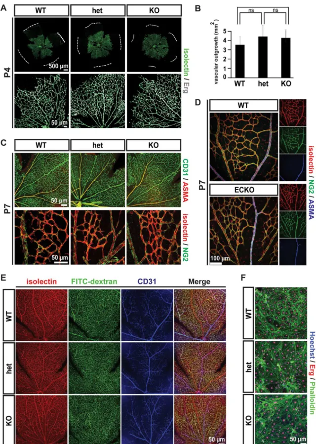
early blood vessel patterns generated by sprouting angiogenesis, as evidenced by examination of the retinal vasculature. The vascular networks at P4 were indistinguishable in morphology (Fig 3A), and occupied equivalent areas in wild type, heterozygous and knockout retinas (Fig
3B). Additionally, the subsequent recruitment of pericytes and arterio-venous differentiation
in retinas appeared normal at P7 (Fig 3C). Endothelial-specific ablation of Gpr116 (Gpr116
ECKO) animals showed normal retinal vascular patterning and mural cell recruitment assessed
by immunofluorescence staining in NG2 and ASMA (Fig 3D). Additionally, Gpr116 knockout
Fig 2. Massive accumulation phenotype in lungs of agedGpr116-/-mice.A. Bright field image of the inflated lung fromGpr116WT, heterozygous and knockout littermates. B. Weights of whole lungs over total body weight fromGpr116WT, heterozygous and knockout littermates (n5 mice per genotype). C. Bright field images of heart fromGpr116WT, heterozygous and knockout littermates. D. Weights of the heart (left) over total body weight fromGpr116WT, heterozygous and knockout littermates (n5 mice per genotype). E. Bright field images of the spleen fromGpr116WT, heterozygous and knockout littermates. F. Weights of the spleen (left) over total body weight fromGpr116WT, heterozygous and knockout littermates (n5 mice per genotype). G. BALF collected fromGpr116WT, heterozygous and knockout littermates (The picture shown is representative of 3 mice for each genotype). H. Quantification of saturated phosphatydilcholine in BALF by ELISA (n = 3 mice per genotype). I. Quantification of protein content in BALF by BCA assay (n = 3 mice per genotype). J. Surfactant proteins detection in BALF by western blot. Molecular weights are indicated on the right. (n = 2 mice per genotype). K. Bright field images of the lung, after hematoxylin and eosin staining. The black arrowheads indicate alveolar macrophages (the image is representative of 4 mice for each genotype). L. Electron microscopy view ofGpr116wildtype and knockout lungs (n = 2 mice for each genotype). M. Confocal images of lung sections stained with ADRP (white) and nuclear stain (Hoechst, blue). Note that a red autofluorescent signal appears in knockout lungs. (the image shown is representative of 2 mice for each genotype). N. Confocal images of lung sections stained with nuclear marker Hoechst (blue) to show autofluorescent cells accumulated in the alveolar space, either in the green or red channel (the image is representative of 3 mice for each genotype). O. Autofluorescence emission spectrum of macrophages in the old knockout lung, upon 405 nm excitation (the image is representative of 2 mice). P. Detection of autofluorescent cells fromGpr116knockout lung by FACS (n = 2 mice per genotype)
did not exhibit overt abnormalities in the CNS vasculature (S3A and S3B Fig), or other vascular
beds (intestinal villi,S3C Fig, kidney glomerulus,S3D Fig, and outer ear,S3E Fig). To
deter-mine if the vessels lacking Gpr116 were functional, the distribution of a 2,000 kDa dextran tracer was assessed. After 3 minutes of circulation, FITC-dextran was evenly distributed throughout the entire retinal vascular bed, from arteries to capillaries and veins, and from the
central optic nerve to the periphery (Fig 3E). These results indicate a proper perfusion of the
retinal vascular network and, given the large size of the tracer, precludes any significant lumen
defect inGpr116knockout mice. A proper perfusion and lumen formation were also verified in
various organs including brain cortical vessels (S3F Fig), liver (S3G Fig), intestine (S3H Fig),
and glomeruli (S3I Fig). Endothelial cells isolated from the brain of knockout animals
estab-lished a cell monolayer with a density and an actin cytoskeleton indistinguishable from the
pat-tern of the wild type control (Fig 3F). Together, these data suggest that Gpr116 is dispensable
for retinal vasculature development and functionin vivo, and the establishment of brain
endo-thelial cell monolayerin vitro.
Gpr116
deletion leads to blood-brain-barrier disruption
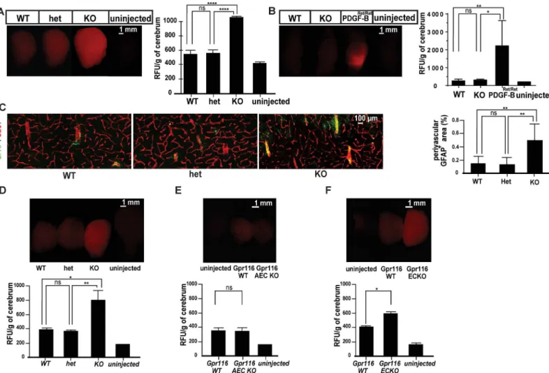
To provide a primary assessment of the barrier properties of the Gpr116 deficient vascula-ture, we injected a 1 kDa fluorescent tracer, Alexa Fluor 555-cadaverine, and examined its tis-sue distribution. Alexa Fluor 555-cadaverine extravasates into most tistis-sues, but normally is excluded from the CNS, the vasculature of which is endowed with specific barrier properties,
termed the blood-brain barrier (BBB). As shown inFig 4A, Gpr116 deficient vessels in
12-months-old animals failed to fully retain the 1 kDa Alexa Fluor 555-cadaverine tracer after tail vein injection, leading to tracer accumulation in the brain parenchyma. However, the BBB impairment was restricted to small molecular weight tracers. Lysine-fixable, 70 kDa tetramethylrhodamine-conjugated dextran did not accumulate into the knockout brains,
in comparison to PDGF-BRet/Retmice where the loss of pericytes leads to leakage of high
molecular weight molecules (Fig 4B)[40]. Consistent with sustained BBB impairment,
immu-nostaining for the intermediate filament protein GFAP in order to visualize astrocytes, showed increased GFAP-positive areas around blood vessels in 18-months-old knockouts,
suggesting astrogliosis (Fig 4C). Tracer leakage across the BBB could also be detected in
young (1.5 month) full knockout animals (Fig 4D). Interestingly, no BBB leakage was
observed in 2-months-oldGpr116AEC KO mice (Fig 4E) which showed a strong lung
phe-notype (S2K Fig). These data argue against the BBB leakage inGpr116knockout mice as a
secondary consequence of the pulmonary pathology, and raise the possibility that endothelial cell-driven Gpr116 is responsible for the BBB leakage. Genetic ablation of Gpr116 under the
control of VE-Cadherin (VE-cad:Cre ER [37]), an endothelial specific inducible promoter,
resulted in a significant increase of the 1 kDa Alexa Fluor 555-cadaverine leakage in the
2-months-old mouse brains (Fig 4F). Of note, pericyte coverage (S3B Fig) and patterning of
the endothelial junctions (S4 Fig) were comparable to littermate control in the knockout
brain cortex. These results establish the first evidence for endothelial cell-autonomous
func-tion(s) of Gpr116in vivo.
were used to visualize endothelium, and NG2 (green) and ASMA (red) to detect mural cells (the images shown are representative of 3 mice for each genotype). D. Vascular patterning in P7 retinas fromGpr116ECKO and littermates controls. Isolectin (red) is used to visualize endothelium, and NG2 (green) and smooth muscle actinα(ASMA, blue) to detect mural cells (the images show are representative of 2 mice per genotype). E. Isolectin (red) and FITC-dextran (green) distribution in P21 retinas fromGpr116WT, heterozygous and knockout littermates. CD31 (green) is used to stain the endothelium, and nuclei are stained with Hoechst (blue) (the images shown are representative of 3 mice per genotype). F. Monolayers formed by isolated endothelial cells from Gpr116WT, heterozygous and knockout brain. Endothelial cells (CD31) and nuclei (Hoechst) are indicated in green and blue, respectively (the pictures shown are representative of 3 mice for each genotype)
Gpr116
deletion modulates pathological angiogenesis in retina
Pathological angiogenesis, especially in ischemic situations, is characterized by formation of dilated and leaky vessels, driven by increased growth factor production. In mice, the oxygen-induced retinopathy (OIR), mimicking human Retinopathy of Prematurity, is a commonly
adopted model of induction and study of pathological angiogenesis [33]. Altered responses in
the OIR model have been observed in several mutants with normal developmental angiogene-sis, suggesting that pathological and developmental angiogenesis may, in part, engage different
mechanisms [48][49][50], leading us to ask whether Gpr116 might play a role in pathological
neovascularization.
Fig 4. Blood brain barrier breakdown inGpr116-/-mice.A. Whole brain images taken after 1kDa cadaverine perfusion (left) and associated quantification of extravasated cadaverine (right) in agedGpr116WT, heterozygous and knockout mice (n5 mice for each genotype). B. Whole brain images taken 70 kDa tetramethylrhodamine dextran perfusion (left) and quantification of extravasated tracer (right) inGpr116WT and heterozygous andGpr116ECKO mice (n = 3 for wild type and ECKO, n = 2 forPDGF-Bret/ret, n = 1 for uninjected control). C. Confocal images of cerebral cortex from agedGpr116WT, heterozygous and knockout mice. Astrocytes (GFAP) appear in green, endothelial cells (CD31) in red (the images are representative of 4 mice per genotype) and associated quantification of perivascular associated astrocytes in agedGpr116WT, heterozygous and knockout mice (n = 4 mice for each genotype, 2 sections at least quantified per genotype). D. Whole brain fluorescence images taken after Alexa 555-cadaverine circulation (upper) and quantification of extravasated cadaverine (lower) in 1.5-month-oldGpr116knockout (n = 3 mice per genotype). E. Whole brain fluorescent images taken after cadaverine circulation (upper) and associated quantification of extravasated cadaverine (lower) in 2-months-oldGpr116AEC KO (n = 6 mice per genotype). F. Whole brain fluorescent images taken after cadaverine circulation (upper) and quantification of extravasated cadaverine (lower) in 2-months-oldGpr116ECKO (n = 7 mice per genotype)
P7 pups were, therefore, submitted to a classical OIR protocol: pups were exposed to 75%
hyperoxia from P7 until P12 [33][51], to normal air between P12-17, followed by retinal
analy-sis at P17. As expected, the wild type retinas showed large areas of vaso-obliteration (Fig 5A)
and formed pathological neovascular tufts (Fig 5E). In knockout mice, however, despite a
ste-reotypical vaso-obliteration around the optic nerve at P12 [33](Fig 5A, quantified inFig 5C),
physiological revascularization occurred much faster than in wild type retinas, causing an
almost complete recovery of the avascularization at P17 (Fig 5B, quantified inFig 5D).
Interest-ingly, this rapid vascular regrowth was accompanied by an almost complete vascular
normali-zation, with almost undetectable intra-vitreal pathological tufts (Fig 5E). Finally, heterozygous
mice, in which vessels express only half of the normal amount of Gpr116, showed an interme-diary phenotype with the ratio of the avascular area over the total area reflecting a value mid-way between knockout and wild type retinas, suggestive of a dose-dependent effect of Gpr116 during pathological angiogenesis.
Discussion
Most adhesion receptors lack defined ligands and known downstream signaling pathways. This limits the ability to validate the functional consequences of the mutagenesis of the recep-torsin vivoto mRNA and protein expression. This limitation makes it particularly important to confirm phenotypic results by several convergent and independent knockout approaches.
In the case of Gpr116, two different gene deletion strategies have recently been applied in order to generate knockout mice. Two full knockouts have been reported in which exon 2 was targeted. Exon 2 encodes the signal peptide, and its deletion would therefore be expected to inhibit translocation of a nascent protein to the endoplasmic reticulum and further trafficking
to the cell membrane [21][20]. In another approach, the mutation deletes exon 17, which
encodes one of the transmembrane domains [19]. The knockout model reported here is an
extensive, constitutive knockout encompassing exon 4 to part of exon 21 of theGpr116gene,
which deletes the potential extracellular adhesion domains, the GPS, as well as all transmem-brane domains and a large portion of the C-terminal intracellular domain.
Using this extensive knockout model, we also found that Gpr116 deficiency led to a specific pulmonary phenotype in adult mice, characterized by a profound alteration of surfactant com-position, with increased protein and saturated PC content. The other remarkable feature of knockout lungs was a massive accumulation of hypertrophic, and sometimes necrotic, macro-phages. These observations complement the previously published findings regarding the
Gpr116gene deletion in mice aged two months or more, where lipid accumulation is associated with enlarged macrophages and alveoli. Moreover, we describe previously unappreciated func-tions of Gpr116 in vascular homeostasis and pathophysiology.
The lung dysfunction occurred in an age-dependent fashion in young knockout animals. Previous models identified some transcriptionally altered genes in late embryonic lung (RyR2,
[19], and inflammatory cytokines, [52]). The first regulated surfactant components were noted
at one (SatPC, [20]) and two weeks (SP-A,[21]) of age. The more extensive evidence showed
that, at 4 weeks, the surfactant protein and lipid composition was deregulated. However, the precise sequence of the defects varies among the models, leading to two different interpreta-tions of the pathway regulated by Gpr116. In the exon 17 mutant, satPC synthesis is increased,
in line with an overactivation of thede novopathway. In the exon 2 mutants, all surfactant
pro-teins are increased in BALF, followed at 6 weeks by a profound modification in the phospho-lipid composition of the surfactant, compatible with an alteration of the surfactant recycling
pathway. It is important to note that, at 4 weeks, the alveolar structure is already modified [19]
quite severe (S2C Fig). Our results demonstrate thatGpr116knockouts already have a clear surfactant alteration at 2 weeks, which was progressively followed by an accumulation of enlarged macrophages at 3 weeks. Here, we propose a time window in which each component of the lung phenotype can be studied. The initial surfactant deregulation occurs during the first
two postnatal weeks, and thus determining whether thede novoor the recycling pathway is
affected could be explored at those time points. It is noteworthy that the onset of surfactant alteration also clearly preceded the macrophage accumulation and it is consistent with the idea that macrophage enlargement is a secondary event. This concept is reinforced by the fact that Gpr116 deletion in type II pneumocytes, using a specific SP-C Cre promoter, recapitulates the
lung abnormalities (show herein at one month, as well as in one year old-mice in ref [21]),
albeit with a somewhat diminished severity in the inducible mouse model.
The range of cell types in which Gpr116 plays a critical role still remains to be fully deter-mined. It is all the more difficult to predict in the absence of defined modules downstream of the receptor. In this regard, the importance of Gpr116 in the vascular endothelium, one of the major sites of Gpr116 expression as demonstrated both herein, as well as in previous work, remains enigmatic.
We confirmed previous reports showing the extensive expression of Gpr116 in vascular endothelium, including in the lungs, where its presence in the endothelium in addition to the
epithelium has been challenged [19].
Consistent with this extensive vascular expression, the loss of Gpr116 caused a vascular leakage in the brain of 1.5-month-old animals. The absence of any leakage in 2-months-old
Gpr116AEC KO mice argues against a systemic vascular effect driven by the pulmonary sur-factant accumulation. The leakage persisted in older animals, where it correlated with increased astrogliosis. The cause of the leakage, which was restricted to small molecular weight molecules and was of moderate amplitude compared to another leakage model where the BBB opens as a
consequence of pericyte deficiency [40], has not been explored in the present study. It is not
likely that the BBB disruption inGpr116knockout mice was caused by increased transcytosis,
which affects transport of large molecular weight tracers. However, major alterations in the junction distribution of the CNS endothelial cells were not detected and thus, could not completely rule out the former explanation.
The second vascular phenotype that we found inGpr116knockouts was a strikingly
“nor-malized”appearance of the retinal vasculature after OIR challenge. This normalization
occurred after a phase of vaso-obliteration comparable to that of wild type controls, indicating that Gpr116 might not impinge on oxygen sensing and the NO synthase pathways. The revas-cularization of the knockout retina happened much faster than the wild type retina, but did not develop the pathological tufts that are a hallmark of this challenge model and the feature of the equivalent syndrome in premature human infants. The complete revascularization of avascular areas argued against a strong anti-angiogenic activity of Gpr116, as seen in the case of severe
blockade of VEGF [53]. The absence of tuft formation is, in turn, one of the more remarkable
findings in comparison with previously described OIR phenotypes (such as in [54]), and seems
as extensive as the A2A receptor and N-CAM mutants [49][50]. It is also the only phenotype
reported to date inGpr116knockouts that presents a graded response, with intermediate effect
Fig 5. Normalized pathological angiogenesis inGpr116-/-retinas.A. Confocal images of post-OIR retinas fromGpr116WT, heterozygous and knockout littermates at P12 (the images shown are representative of 5 mice per genotype). B. Confocal images of post-OIR retinas fromGpr116WT, heterozygous and knockout littermates at P17 (the images shown are representative of 5 mice per genotype). C. Quantification of the avascular area on the post-OIR retinas fromGpr116WT, heterozygous and knockout littermates at P12 (n = 5 mice at least per genotype). D. Quantification of the avascular area on the post-OIR retinas fromGpr116WT, heterozygous and knockout littermates at P17 (n7 mice at least per genotype). E. Confocal images of post-OIR tufts (blue arrows) inGpr116WT, heterozygous and knockout littermates at P17 (the images shown are representative of 5 mice per genotype)
in the heterozygous littermates. The background of the Gpr116Δexon17mice (BALB/c, which is highly resistant to OIR) precluded a confirmation that the recovery is an intrinsic endothelial property. Therefore, the role of an accessory cell type, such as astrocytes, Müller cells or retinal
pigmented epithelium cannot be ruled out at this point [55]. Furthermore, even if the results
were to be verified usingGpr116knockout backcrossed on a C57BL6 background, a potential
neighboring effect of modifying genes would be very hard to eliminate. Only the age at which pups are analyzed (P17) argues against the OIR recovery as a secondary consequence of the lung phenotype.
In conclusion, our results strengthen the description of the critical role played by Gpr116 in surfactant regulation: the lack of Gpr116 in type II pneumocytes results in a fast alteration of surfactant composition, with onset occurring at around 1 week after birth. The accumulation of enriched surfactant leads to secondary events where the accumulation of foamy, lipid over-loaded macrophages is prominent.
Our work also provides a foundation for exploring the function of Gpr116 outside of the pneumocytes. The severity of the lung phenotype implies that a number of features of full knockout mice might be derived from the general pathological situation in the lung. Still, use of young full knockout, as well as cell type-specific knockout, provides means to untangle the dis-crete functions of the receptor in different cell types. As a proof-of-concept, we show here that Gpr116 controls a number of functions of the vascular endothelium, one of its main sites of expression. As the vascular functions assessed here are CNS-specific, we cannot yet draw global
conclusions about endothelial Gpr116 functions. Clearly, other vascular challenges toGpr116
knockout are warranted, as are more specific studies of the mechanisms of vascular permeabil-ity and angiogenic sprouting. As for the lung phenotype, ligand identification and distribution as well as discerning the regulated molecular pathways will be critical in order to understand the role of Gpr116 in vascular endothelium.
Supporting Information
S1 Fig. Heart and spleen enlargement in young Gpr116-/-mice.A. Bright field image of the heart from 1-month-old Gpr116 WT, heterozygous and knockout littermates. B. Weights of the heart over total body weight from 1-month-old Gpr116 WT, heterozygous and knockout
littermates (n4 mice per genotype). C. Bright field image of the spleen from 1-month-old
Gpr116 WT, heterozygous and knockout littermates. D. Weights of the spleen over total body weight from 1-month-old Gpr116 WT, heterozygous and knockout littermates (n4 mice per genotype).
(TIF)
S2 Fig. Sequential development of pulmonary impairment in Gpr116-/-mice.A. Quantifica-tion of surfactant mRNA by RT-PCR in lungs from Gpr116 WT, heterozygous and knockout
littermates at P2 (n4 mice per genotype). B. BALF appearance and protein content quantified
each genotype). F. Confocal images of pre-FACS cells purified from 1-month-old Gpr116 WT, heterozygous and knockout mouse lungs. On the left panel, the cell cytoskeleton is delineated by phalloidin-Alexa 647 (white). On the right panel, immune cells appear in white (CD45). On both panel, cellular autofluorescence is shown in green, and nuclei in blue (Hoechst) (the image shown is representative of 3 mice for each genotype). G. Pseudo-colored scatter plots from FACS purified single cell suspensions from 4-weeks-old-mouse lung. Cell suspensions were incubated with Alexa 780- and BV421-conjugated antibodies against CD45 and CD11b, respectively. Selection for autofluorescent cells is based on their emission in the green (excita-tion 488 nm) and red (excita(excita-tion 561 nm) spectrum. Note that Gpr116 knockout mice display a larger population of autofluorescent cells compared to Gpr116 heterozygotes or Gpr116 WT mice (the plot is representative of 3 mice for each genotype). H. The gated autofluorescent cells selected in (G) are analyzed for CD45 expression. CD45- positive cells are gated based upon a threshold set by Alexa 780-isotope control staining. I. CD45+ cells selected in (H) were investi-gated for CD11b expression. A selection of CD11b-positive cells was obtained by investigating maximum emission strength of cells stained with a BV421 isotype control. Note that the cells display a high autofluorescence in blue (excitation 405 nm). J. BALF appearance and protein content in 1-month-old Gpr116 AEC KO (n = 4 mice per genotype). K. Bright field views of lung from 1-month- or 2-months-old Gpr116 Gpr AEC KO (top rows) and 6-months old Gpr116 ECKO (lower row) stained with hematoxylin and eosin. View from 3-months-old lung from full knockout animal is displayed for comparison (the pictures shown are representative of 2 mice for each genotype).
(TIF)
S3 Fig. Development of central and peripheric vascular beds in Gpr116-/-mice.A. Vascula-ture of the 1-month-old brain cortex from Gpr116 WT, heterozygous and knockout litter-mates. Glut-1 (green) is used to visualize endothelium, and ASMA (red) to distinguish the major vessels (the images shown are representative of 2 mice per genotype). B. Vasculature of the 1-month-old brain cortex from Gpr116 WT, heterozygous and knockout littermates.
CD-31 (green) is used to visualize endothelium, and PDGFRβ(red) to reveal the pericytes coverage
S4 Fig. Vascular junction pattern in Gpr116-/-mouse brain cortical vessels.A. Adherens junction staining in 1-month-old brain cortex from Gpr116 WT, heterozygous and knockout littermates. VE-cadherin (red) is used to visualize adherens junctions, and podocalyxin (green) to delineate the vessels. A higher magnification of VE-cadherin is shown on the right side of the figure (the images shown are representative of 2 mice per genotype). B. Tight junction pat-terning in 1-month-old brain cortex from Gpr116 WT, heterozygous and knockout littermates. Claudin-5 (red) is used to visualize tight junctions, and CD31 (green) to delineate the vessels. A higher magnification of the claudin-5 is shown on the right side of the figure (the images shown are representative of 2 mice per genotype). C. Tight junction staining in 1-month-old brain cortex from Gpr116 WT, heterozygous and knockout littermates. Another tight junction marker, Zo-1 (red) is used and CD31 (green) to delineate the vessels. A higher magnification of the ZO-1 is shown on the right side of the figure (the images shown are representative of 2 mice per genotype).
(PDF)
S5 Fig. Graphical abstract.
(PDF)
Acknowledgments
The authors thank Cecilia Olsson, Pia Peterson, Jana Chmielniakova and Helen Leksell for
technical assistance, the Biovis platform (http://www.biovis.medfarm.uu.se/) for bright field
imaging and FACS sorting, and Josefin Skogsberg for providing withApoEnull aortic roots.
The authors would like to aknowledge Taija Mäkinen and her group, especially Ines Martinez-Corral and Maria Ulmvar, for sharing mice lines, providing reagents and thorough technical advice.
Author Contributions
Conceived and designed the experiments: CN CB TB KP. Performed the experiments: CN JJH KG MAM EE BJ MDS MV AM SAS LH TL MCF KK KH CM PT SC BL JWH. Analyzed the data: CN JJH MAM BJ. Contributed reagents/materials/analysis tools: TL TB. Wrote the paper: CN CB.
References
1. Bjarnadóttir TK, Fredriksson R, Höglund PJ, Gloriam DE, Lagerström MC, Schiöth HB. The human and mouse repertoire of the adhesion family of G-protein-coupled receptors. Genomics. 2004; 84: 23–33. doi:10.1016/j.ygeno.2003.12.004PMID:15203201
2. Hamann J, Aust G, Araç D, Engel FB, Formstone C, Fredriksson R, et al. International Union of Basic and Clinical Pharmacology. XCIV. Adhesion G protein-coupled receptors. Pharmacol Rev. 2015; 67: 338–367. doi:10.1124/pr.114.009647PMID:25713288
3. Curtin JA, Quint E, Tsipouri V, Arkell RM, Cattanach B, Copp AJ, et al. Mutation of Celsr1 disrupts pla-nar polarity of inner ear hair cells and causes severe neural tube defects in the mouse. Curr Biol CB. 2003; 13: 1129–1133.
4. Devenport D, Fuchs E. Planar polarization in embryonic epidermis orchestrates global asymmetric mor-phogenesis of hair follicles. Nat Cell Biol. 2008; 10: 1257–1268. doi:10.1038/ncb1784PMID:
18849982
5. Yates LL, Schnatwinkel C, Murdoch JN, Bogani D, Formstone CJ, Townsend S, et al. The PCP genes Celsr1 and Vangl2 are required for normal lung branching morphogenesis. Hum Mol Genet. 2010; 19: 2251–2267. doi:10.1093/hmg/ddq104PMID:20223754
6. Tissir F, Qu Y, Montcouquiol M, Zhou L, Komatsu K, Shi D, et al. Lack of cadherins Celsr2 and Celsr3 impairs ependymal ciliogenesis, leading to fatal hydrocephalus. Nat Neurosci. 2010; 13: 700–707. doi:
7. Cortijo C, Gouzi M, Tissir F, Grapin-Botton A. Planar cell polarity controls pancreatic beta cell differenti-ation and glucose homeostasis. Cell Rep. 2012; 2: 1593–1606. doi:10.1016/j.celrep.2012.10.016
PMID:23177622
8. Tissir F, Bar I, Jossin Y, De Backer O, Goffinet AM. Protocadherin Celsr3 is crucial in axonal tract devel-opment. Nat Neurosci. 2005; 8: 451–457. doi:10.1038/nn1428PMID:15778712
9. Monk KR, Naylor SG, Glenn TD, Mercurio S, Perlin JR, Dominguez C, et al. A G protein-coupled recep-tor is essential for Schwann cells to initiate myelination. Science. 2009; 325: 1402–1405. doi:10.1126/ science.1173474PMID:19745155
10. Monk KR, Oshima K, Jörs S, Heller S, Talbot WS. Gpr126 is essential for peripheral nerve development and myelination in mammals. Dev Camb Engl. 2011; 138: 2673–2680. doi:10.1242/dev.062224 11. Mogha A, Benesh AE, Patra C, Engel FB, Schöneberg T, Liebscher I, et al. Gpr126 functions in
Schwann cells to control differentiation and myelination via G-protein activation. J Neurosci Off J Soc Neurosci. 2013; 33: 17976–17985. doi:10.1523/JNEUROSCI.1809-13.2013
12. Piao X, Hill RS, Bodell A, Chang BS, Basel-Vanagaite L, Straussberg R, et al. G protein-coupled recep-tor-dependent development of human frontal cortex. Science. 2004; 303: 2033–2036. doi:10.1126/ science.1092780PMID:15044805
13. Bae B-I, Tietjen I, Atabay KD, Evrony GD, Johnson MB, Asare E, et al. Evolutionarily dynamic alterna-tive splicing of GPR56 regulates regional cerebral cortical patterning. Science. 2014; 343: 764–768. doi:10.1126/science.1244392PMID:24531968
14. Anderson KD, Pan L, Yang X, Hughes VC, Walls JR, Dominguez MG, et al. Angiogenic sprouting into neural tissue requires Gpr124, an orphan G protein-coupled receptor. Proc Natl Acad Sci U S A. 2011; 108: 2807–2812. doi:10.1073/pnas.1019761108PMID:21282641
15. Cullen M, Elzarrad MK, Seaman S, Zudaire E, Stevens J, Yang MY, et al. GPR124, an orphan G pro-tein-coupled receptor, is required for CNS-specific vascularization and establishment of the blood-brain barrier. Proc Natl Acad Sci U S A. 2011; 108: 5759–5764. doi:10.1073/pnas.1017192108PMID:
21421844
16. Kuhnert F, Mancuso MR, Shamloo A, Wang H-T, Choksi V, Florek M, et al. Essential regulation of CNS angiogenesis by the orphan G protein-coupled receptor GPR124. Science. 2010; 330: 985–989. doi:
10.1126/science.1196554PMID:21071672
17. Abe J, Suzuki H, Notoya M, Yamamoto T, Hirose S. Ig-hepta, a novel member of the G protein-coupled hepta-helical receptor (GPCR) family that has immunoglobulin-like repeats in a long N-terminal extra-cellular domain and defines a new subfamily of GPCRs. J Biol Chem. 1999; 274: 19957–19964. PMID:
10391944
18. Prömel S, Waller-Evans H, Dixon J, Zahn D, Colledge WH, Doran J, et al. Characterization and func-tional study of a cluster of four highly conserved orphan adhesion-GPCR in mouse. Dev Dyn Off Publ Am Assoc Anat. 2012; 241: 1591–1602. doi:10.1002/dvdy.23841
19. Bridges JP, Ludwig M-G, Mueller M, Kinzel B, Sato A, Xu Y, et al. Orphan G protein-coupled receptor GPR116 regulates pulmonary surfactant pool size. Am J Respir Cell Mol Biol. 2013; 49: 348–357. doi:
10.1165/rcmb.2012-0439OCPMID:23590306
20. Fukuzawa T, Ishida J, Kato A, Ichinose T, Ariestanti DM, Takahashi T, et al. Lung surfactant levels are regulated by Ig-Hepta/GPR116 by monitoring surfactant protein D. PloS One. 2013; 8: e69451. doi:10. 1371/journal.pone.0069451PMID:23922714
21. Yang MY, Hilton MB, Seaman S, Haines DC, Nagashima K, Burks CM, et al. Essential regulation of lung surfactant homeostasis by the orphan G protein-coupled receptor GPR116. Cell Rep. 2013; 3: 1457–1464. doi:10.1016/j.celrep.2013.04.019PMID:23684610
22. Wallgard E, Larsson E, He L, Hellström M, Armulik A, Nisancioglu MH, et al. Identification of a core set of 58 gene transcripts with broad and specific expression in the microvasculature. Arterioscler Thromb Vasc Biol. 2008; 28: 1469–1476. doi:10.1161/ATVBAHA.108.165738PMID:18483404
23. Takase H, Matsumoto K, Yamadera R, Kubota Y, Otsu A, Suzuki R, et al. Genome-wide identification of endothelial cell-enriched genes in the mouse embryo. Blood. 2012; 120: 914–923. doi:10.1182/ blood-2011-12-398156PMID:22535667
24. Masiero M, Simões FC, Han HD, Snell C, Peterkin T, Bridges E, et al. A core human primary tumor angiogenesis signature identifies the endothelial orphan receptor ELTD1 as a key regulator of angio-genesis. Cancer Cell. 2013; 24: 229–241. doi:10.1016/j.ccr.2013.06.004PMID:23871637
![Fig 1. Vascular expression and genetic ablation of the Gpr116 gene in mouse. A. Gpr116 mRNA expression in the published organ-specific EC mRNA dataset [45]](https://thumb-eu.123doks.com/thumbv2/123dok_br/18337774.351622/5.918.59.844.114.770/vascular-expression-genetic-ablation-expression-published-specific-dataset.webp)