to the Hepatic Innate Immune Response via TLR3
Caroline L. Wilson1., Jelena Mann1., Meagan Walsh1
, Maria J. Perrugoria1, Fiona Oakley1,
Matthew C. Wright1, Chiara Brignole2, Daniela Di Paolo2, Patrizia Perri2, Mirco Ponzoni2, Michael Karin3, Derek A. Mann1*
1Institute of Cellular Medicine, Faculty of Medical Sciences, Newcastle University, Newcastle upon Tyne, United Kingdom,2Experimental Therapy Unit, Laboratory of Oncology, Istituto Giannina Gaslini, Genoa, Italy,3Laboratory of Gene Regulation and Signal Transduction, Department of Pharmacology and Pathology, School of Medicine, University of California San Diego, La Jolla, California, United States of America
Abstract
Toll-like Receptor 3 (TLR3) is a pathogen pattern recognition receptor that plays a key role in innate immunity. TLR3 signalling has numerous functions in liver, both in health and disease. Here we report that TLR3 is expressed by quiescent hepatic stellate cells (HSC) where it functions to induce transcription and secretion of functional interferons as well as a number of other cytokines and chemokines. Upon transdifferentiation into myofibroblasts, HSCs rapidly loose the ability to produce interferon gamma (IFNc). Mechanistically, this gene silencing may be due to Polycomb complex mediated repression via methylation of histone H3 lysine 27. In contrast to wild type, quiescent HSC isolated fromtlr3knockout mice do not produce IFNc in response to Poly(I:C) treatment. Therefore, quiescent HSC may contribute to induction of the hepatic innate immune system in response to injury or infection.
Citation:Wilson CL, Mann J, Walsh M, Perrugoria MJ, Oakley F, et al. (2014) Quiescent Hepatic Stellate Cells Functionally Contribute to the Hepatic Innate Immune Response via TLR3. PLoS ONE 9(1): e83391. doi:10.1371/journal.pone.0083391
Editor:Lena Alexopoulou, Centre d’Immunologie de Marseille-Luminy, CNRS-Inserm, France
ReceivedJune 6, 2013;AcceptedNovember 4, 2013;PublishedJanuary 8, 2014
Copyright:ß2014 Wilson et al. This is an open-access article distributed under the terms of the Creative Commons Attribution License, which permits unrestricted use, distribution, and reproduction in any medium, provided the original author and source are credited.
Funding:The present study was supported by the National Institutes of Health (NIH) grants (U01AA018663, P50AA11199, R24AA12885), Newcastle Biomedical Research Centre, the Wellcome Trust (WT086755MA to D.A.M. and M.K.), Medical Research Council grants (MK/K001949/1 to D.A.M., J.M., F.O. and G0700890 to D.A.M., M.C.W. and F.O.) and the European Commission FP7 program grant ‘INFLA-CARE’ (EC Contract No. 223151; http://inflacare.imbb.forth.gr/ to D.A.M., M.P., C.B., D.D.P. and P.P.). The funders had no role in study design, data collection and analysis, decision to publish, or preparation of the manuscript.
Competing Interests:The authors have declared that no competing interests exist.
* E-mail: [email protected]
.These authors contributed equally to this work.
Introduction
Hepatic stellate cells (HSC) are specialised pericytes of the liver sinusoids found in the Space of Disse`, a basement membrane-like structure located between columns of hepatocytes and sinusoidal endothelial cells [1]. The most characterised function of HSCs is the ability to transdifferentiate from a quiescent phenotype into highly proliferative, contractile and wound-healing myofibroblast [2]. This so-called activated HSC (aHSC) produces vast quantities of fibril-forming collagens and promotes net deposition of fibrotic extracellular matrix, a process important for repair following infection or trauma to the liver. However, in the chronically injured liver, this normal physiological process may become dysregulated and lead to the development of fibrosis [1].
The role of quiescent HSCs (qHSC) has received much less attention. A known key function is storage of Vitamin A which is found in numerous intracellular droplets [3,4]. Additionally, several investigators have described that qHSC possess multiple thorn-like cytoplasmic extensions which can protrude into the sinusoidal space or make direct contact with hepatocytes. These membrane projections have been shown to function as a leading edge for the qHSC and play a role in sensing of extracellular factors that influence HSC phenotype. Given the anatomical location of qHSCs and their morphology, they have the potential to operate as sinusoidal sentinels, detecting mechanical or
biochemical alterations in hepatocytes, endothelial cells, within the Space of Disse` or even within the sinusoidal spaces [4].
The innate immune system serves as a ‘‘first defence’’ responding to acute tissue trauma or infection by mounting protective anti-microbial and wound-healing response. These responses are mediated by a variety of immune cells including recruited neutrophils, mast cells, eosinophils, natural killer (NK) cells and tissue macrophages. The molecular triggers for innate immunity are pattern recognition receptors (PRRs) [5] including the IL-1 Receptors [6] (reviewed in [7]), members of the Toll-like Receptor (TLR) family [8] and Nucleotide Oligomerisation Domain (NOD)-like receptor families [9]. A total of 13 distinct mammalian TLRs (TLR1-13) have been identified to date and each responds to specific ligands of microbial origin or from the intracellular contents of damaged or dying host cells. Upon engagement by their ligands, the TLRs trigger a cascade of intracellular signalling pathways that culminate in induction of genes encoding interferons, cytokines and chemokines required for the recruitment and activation of innate immune cells [10].
Sekiet al, have described how TLR4 on activated mouse HSC is required for sensitising the cells to TGFb1, specifically by promoting down-regulation of the TGFb1 pseudo-receptor Bambi [13]. The observation that mice lacking TLR4 or its downstream adaptor Myd88 were resistant to fibrosis induced by a variety of different injury mechanisms highlighted the importance of innate immune receptor in hepatic wound-healing response.
In the present study, we have addressed the question of which TLRs are expressed and functional on qHSC. We focused on TLR3, a sensor of dsRNA, which is expressed at relatively high levels on both qHSC and aHSC but has distinct functions in these two phenotypic states. Our data suggest that engagement of TLR3 on qHSC results in induction of interferons (a,b and c) and cytokine (IL-6) gene expression. However, aHSC lose the capacity to induce interferons in response to TLR3, but retain TLR3-induction of IL-6. Our data suggest that qHSC may play a significant contributing role to TLR3-mediated interferon-depen-dent innate immune responses.
Materials and Methods
Ethics statement
All animal experiments were undertaken in accordance with appropriate licences for animal experiments which were issued/ approved by local ethical committee and UK Home Office.
Animals and models of liver disease
Chronic CCl4liver injury model. Fibrogenesis was induced by 4-week CCl4 treatment of 250 g adult Sprague-Dawley rats. Rats were injected intraperitoneally (IP) twice weekly with CCl4/ olive oil mix in a 1:3vol/vol ratio at 2ml/g body weight. Twenty-four hours after the final CCl4 administration, animals were sacrificed and blood and tissues harvested.
Acute CCl4liver injury. Single dose of CCl4was given by IP injection prepared as CCl4/olive oil in a 1:1vol/vol ratio at 2ml/g body weight to both rats and mice. Animals were sacrificed at varying times after injury (as stated in figures and figure legends) and tissues harvested for histological and molecular analysis.
SDS-PAGE and Immunoblotting
SDS-PAGE and immunoblotting was performed as previously described [15]. Primary antibodies recognising TLR3 (AnaSpec), IRAK1, TRAF6 (Santa Cruz) were used at 1:1000 dilution and GAPDH (Abcam) at 1:2000. Membranes were probed with the appropriate secondary antibody (anti-mouse Sigma; anti-rabbit Cell Signalling Technologies) and proteins visualised using chemiluminescence (Pierce).
Immunocytochemistry
Formalin fixed, paraffin embedded tissue was dewaxed and rehydrated in decreasing concentrations of ethanol. Sections underwent antigen retrieval in citric saline solution and were subsequently permeabilized with 0.1% saponin in 0.5% bovine serum albumin (BSA). Toll-like receptor 3 primary antibody (AnaSpec) was used in a concentration of 1:1000 and incubated at room temperature for 1 hour. Coverslips were mounted in DAPI-containing fluorescent mounting media.
Quantitative Reverse Transcriptase-Polymerase Chain Reaction (qRT-PCR)
Total RNA was purified from isolated cells or whole liver using the RNeasy Mini Kit (Qiagen, UK). cDNA was generated using random hexamer primers and MMLV reverse transcriptase
enzyme (Promega, UK). Quantitative PCR was performed on an ABI 7500 with a 3 step amplification program: 20 sec at 94uC, 40 cycles of 20 sec at 55uC, 30 sec at 72uC and 5 sec at 94uC. All reactions were normalised to GAPDH and relative level of transcriptional difference calculated using the following equation: 1/(2A)6100. (Primer sequences are listed in Table 1).
Reagents
TLR ligands. Rat and mouse HSCs were incubated with
TLR ligands (InvivoGen) as detailed in figure legends. The ligands and their concentration in cell culture were (unless otherwise stated) - TLR2 (Lipoteichoic (LTA), 100 ng/ml), TLR3 (Poly (I:C), 1mg/ml), TLR4 (lipopolysaccharide (LPS), 100 ng/ml), TLR5 (flagellin, 1mg/ml), TLR7/8 (Imiquimod, 1mg/ml) and TLR9 (stimulatory CpG ODN, 10mg/ml). IL-1a was used at a concentration of 2 ng/ml (Peptroech 211-11A), IFNcat 100 ng/ ml (Peprotech 315-05). Transcriptional inhibitors, actinomycin D and 5,6-Dichlorobenzimidazole Riboside (DRB) were purchased from Sigma Aldrich (A9415 and D1916 respectively). The C13-GT was used at a concentration of 20mg/gram of body weight. Clodronate-liposomes were a kind gift from Professor Mirco Ponzoni and injected intraperitoneally at a concentration of 25mg/gram of body weight.
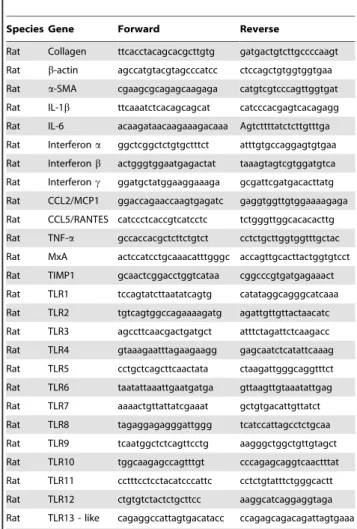
Table 1.Table of primers.
Species Gene Forward Reverse
Rat Collagen ttcacctacagcacgcttgtg gatgactgtcttgccccaagt
Rat b-actin agccatgtacgtagcccatcc ctccagctgtggtggtgaa
Rat a-SMA cgaagcgcagagcaagaga catgtcgtcccagttggtgat
Rat IL-1b ttcaaatctcacagcagcat catcccacgagtcacagagg
Rat IL-6 acaagataacaagaaagacaaa Agtcttttatctcttgtttga
Rat Interferona ggctcggctctgtgctttct atttgtgccaggagtgtgaa
Rat Interferonb actgggtggaatgagactat taaagtagtcgtggatgtca
Rat Interferonc ggatgctatggaaggaaaga gcgattcgatgacacttatg
Rat CCL2/MCP1 ggaccagaaccaagtgagatc gaggtggttgtggaaaagaga
Rat CCL5/RANTES catccctcaccgtcatcctc tctgggttggcacacacttg
Rat TNF-a gccaccacgctcttctgtct cctctgcttggtggtttgctac
Rat MxA actccatcctgcaaacatttgggc accagttgcacttactggtgtcct
Rat TIMP1 gcaactcggacctggtcataa cggcccgtgatgagaaact
Rat TLR1 tccagtatcttaatatcagtg catataggcagggcatcaaa
Rat TLR2 tgtcagtggccagaaaagatg agattgttgttactaacatc
Rat TLR3 agccttcaacgactgatgct atttctagattctcaagacc
Rat TLR4 gtaaagaatttagaagaagg gagcaatctcatattcaaag
Rat TLR5 cctgctcagcttcaactata ctaagattgggcaggtttct
Rat TLR6 taatattaaattgaatgatga gttaagttgtaaatattgag
Rat TLR7 aaaactgttattatcgaaat gctgtgacattgttatct
Rat TLR8 tagaggagagggattggg tcatccattagcctctgcaa
Rat TLR9 tcaatggctctcagttcctg aagggctggctgttgtagct
Rat TLR10 tggcaagagccagtttgt cccagagcaggtcaactttat
Rat TLR11 cctttcctcctacatcccattc cctctgtatttctgggcactt
Rat TLR12 ctgtgtctactctgcttcc aaggcatcaggaggtaga
Rat TLR13 - like cagaggccattagtgacatacc ccagagcagacagattagtgaaa
tlr2/23 knockout mice
The knockout mice were obtained from Dr Lars Eckmann, University of Southern California, San Diego, USA. Authors wish to thank Dr Eckmann for his help and support with breeding and supplying the mice. The knockout mice were originally generated as described in [16].
Hepatic Stellate Cell isolation
Rat and mouse HSCs were isolated from 250 g male Sprague-Dawley rats and 25–30 g adult male mice respectively, by sequential perfusion with collagenase B (Roche) and pronase (Roche) and quiescent HSCs separated by discontinuous density centrifugation in 11.5% Optiprep (Sigma Aldrich D1556). Mouse and rat primary HSCs were maintained at 37uC (5% CO2) in Dulbecco’s Modified Eagle’s Media supplemented with 16% foetal bovine serum (FBS), 100 U/ml penicillin, 100mg/ml streptomy-cin, 2 mM L-glutamine (Life Technologies). Culturing of freshly isolated quiescent HSC on tissue culture plastic leads to their activation over a period of 7 to 10 days and spontaneous acquisition of a myofibroblast phenotype, which is thought to be highly representative ofin vivoHSC activation.
Murine Macrophage isolation
Bone marrow cells were isolated from the femurs of C57/Blk6 mice as previously described [17]. Briefly, cells were differentiated into macrophages by culturing for 7 days with media supplement-ed with 5% horse serum (Sigma Aldrich H1270) and 10% of L929 cell line conditioned media (that contains M-CSF).
Enzyme linked Immunosorbent Assay
Culture supernatant concentration of IL-6 was measured using a rat IL-6 Quantikine ELISA according to manufacturer’s instructions (R&D Systems Minneapolis, MN).
Fluorescence activated cell sorting (FACS)
Cells were incubated at 4uC for 1 hr with 24G2 antibody that prevents non-specific Fc receptor binding, followed by 1 hr incubation at 4uC with anti MHC class II-FITC conjugated antibody (eBioscience 11-0920-82). Following incubation, cells were washed and resuspended in 2% FBS in phosphate-buffered saline. Up to 10,000 events were analysed on FACScan/FACS Canto II (BD, Oxford, UK) using Flowjo software (FlowJo, Inc).
Chromatin Immunoprecipitation Assay
Antibodies used for immunoprecipitation were purchased from: histone H3 di methyl K27 (H3K27me2, Abcam) and Histone H3 trimethyl K27 (H3K27me3, Diagenode). 10mg of each antibody or appropriate irrelevant antibody control were used in each ChIP reaction as described previously [18]. Primers used for detection of relevant rat genomic sequences were: IFNc 243 kb sense 59 -aaggtcaagccataacattc-39and antisense 59- cagggatgaacaaggaccag-39; IFNc20.5 kb sense 59- cttttgtaaccgaacgccttc-39and antisense 59- cttttacttcacaccatttg-39; IFNc0.4 kb sense 59- tcggtgaggtgttcgtt-gac-39 and antisense 59- aagaatgaaaaccatgaagg-39 and IFNc 1.1 kb sense 59- gagttgagtttatttgtgg-39 and antisense 59 -ctgtggagttttgttgaatg-39. Each PCR reaction was performed in triplicate and the analysis was repeated three times from independent ChIP experiments. A signal intensity value for each sample was calculated from the average of the experiments. Average values of eluates were normalized to average values of control antibody sample and expressed as fold enrichment above background (i.e. control antibody) [18].
Statistical Analysis
Data are expressed as means 6 standard error of the mean (SEM). All P values were calculated using a two-tailed paired or unpaired Student t test. Statistically significant data is represented in figures where *, **, and *** denote P values of,0.05,,0.01 and,0.001, respectively.
Results
TLR3 is expressed and functional in quiescent and activated HSC
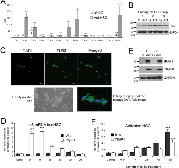
To determine relative expression of TLRs between qHSC and aHSC we measured transcript expression for TLR1-13 in freshly isolated or culture-activated rat HSC. TLR2, 3, 7, 8 and 13 transcripts were all induced with culture activation and were expressed at relatively high levels compared with other TLRs (Figure 1A). The highest expressed TLR transcripts in qHSC were TLR3 (27.57615.16 RLTD) and TLR13 (14.5468.09 RLTD) (p,0.05 compared with other TLR transcripts). Western blot analysis confirmed that TLR3 protein is expressed in both qHSC and aHSC, however no significant change in expression occurred with activation (Figure 1B). Furthermore, we confirmed presence of TLR3 in qHSC (culture day 1) by immunocytochemistry (Figure 1C). As no previous studies have reported a role for TLR3 in qHSC we determined if they are responsive to the TLR3 ligand Poly(I:C). As illustrated in Figure 1D, qHSCs treated with Poly(I:C) underwent a transient increase in IL-6 mRNA transcript that peaked between 2 (9.7 RLTD, p,0.001) and 4 (10.54 RLTD, p,0.001) hours following treatment (Figure 1D). By contrast qHSC were unresponsive to IL-1a (Figure 1D) which may be explained by absence of key downstream signalling factors TRAF6 and IRAK1 in quiescent HSC, which are subsequently induced during HSC activation (Figure 1E). As expected, aHSC were fully responsive to IL-1atreatment which stimulated the expression of IL-6 and TIMP-1 indicating a profibrogenic phenotype (Figure 1F). Maximal induction of IL-6 and TIMP-1 occurred after 8 hours of IL-1a treatment (7.6961.14 RLTD, 4.360.62 RLTD, respectively p,0.001)
that it is unlikely that TLR3 functions as a modulator of the fibrogenic activities of HSCs.
Activation of HSC decreases TLR3-mediated interferon response and subsequent cytokine production
TLR3 plays a fundamental anti-viral role by producing interferons (IFN) in response to viral dsRNA [19]. Given that
HSC express functional TLR3, we were intrigued to determine if the activation of the receptor can trigger IFN production in these cells. For this purpose we isolated HSC from 3 groups of rats; control group which was given olive oil vehicle (qHSC); rats administered CCl4 acutely for 48 hours (transitionary HSC) and rats receiving a chronic CCl4injury for 4 weeks (myofibroblastic aHSC). HSC were subsequently treated for up to 24 hours with Poly(I:C) prior to measurement of gene expression by qRT-PCR. Figure 1. Quiescent and activated HSCs express Toll like receptors.(A) mRNA levels of TLR1-13 were quantified by qRT-PCR in three separate preparations of primary rat qHSCs (day 0) and day 10 transdifferentiated myofibroblasts. Data are expressed as relative level of transcriptional difference (RLTD) to TLR1 mRNA expression (n = 3). (B) Thirty micrograms of whole cell protein extract from three separate preparations of quiescent rat HSCs (culture day 1) or activated myofibroblasts (culture day 10) were separated by SDS-PAGE and immunoblotted for TLR3 and GAPDH. (C) Toll-like receptor 3 was visualised in the cytoplasm of rat qHSCs (ex vivo) culture day 1 (bar represents 75mm). (D) Quiescent rat HSCs were treated with Poly(I:C) (1mg/ml) or IL-1a(2 ng/ml) for up to 24 hours; IL-6 mRNA was measured and normalised tobactin (n = 3). (E) Thirty micrograms of whole cell protein extract from two separate preparations of quiescent HSCs or activated myofibroblasts (culture day 10) were separated by SDS-PAGE and immunoblotted for IRAK1, TRAF6 and GAPDH. (F) Activated rat myofibroblasts were treated with IL-1a(2 ng/ml) for up to 24 hours; IL-6 and TIMP1 mRNA were measured and normalised tobactin (n = 3). (*p,0.05, ***p,0.001).
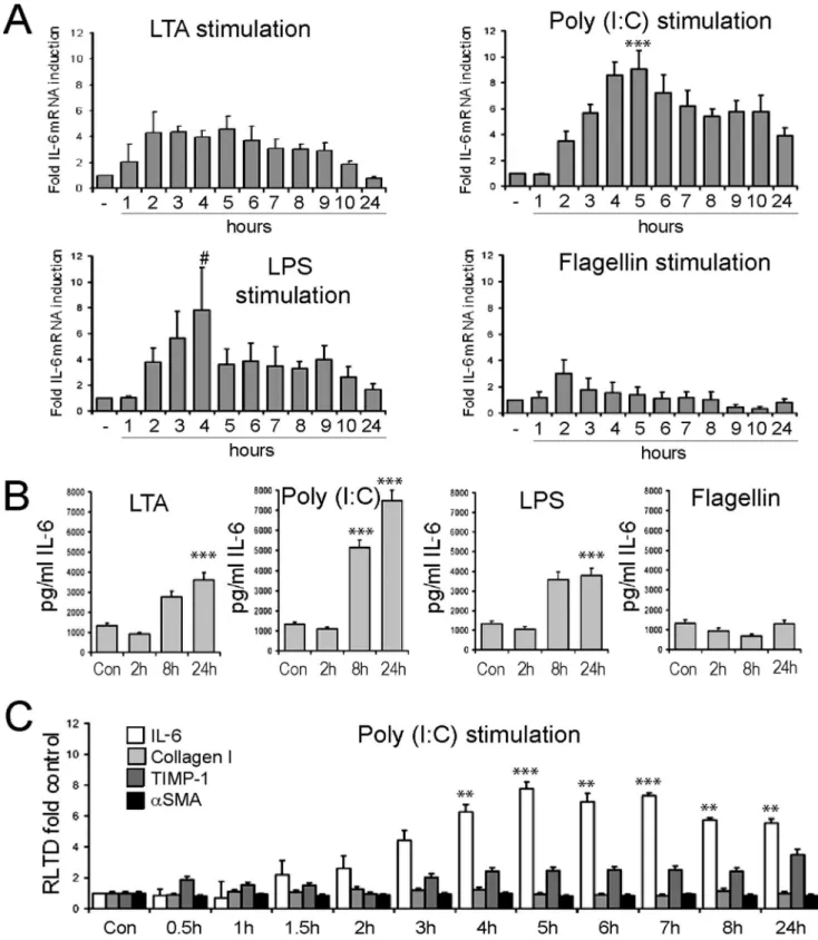
Figure 2. Hepatic stellate cells are responsive to stimulation by TLR ligands.(A) mRNA level of IL-6 was quantified by qRT-PCR in four separate preparations of activated rat HSCs (culture day 10) treated with TLR2 ligand (LTA, 100 ng/ml), TLR3 ligand (Poly(I:C), 1mg/ml), TLR4 ligand (LPS, 100 ng/ml) or TLR5 ligand (Flagellin, 1mg/ml) for up to 24 hours (n = 4). Expression level was normalised tobactin. (B) Secreted IL-6 protein was measured by ELISA in conditioned media collected from activated HSCs treated with TLR ligands as in (A) following 2 h, 8 h or 24 h of stimulation (n = 4). (C) mRNA levels of IL-6, TIMP1,aSMA and collagen I were quantified by qRT-PCR in four separate preparations of activated rat HSCs (culture day 10) treated with TLR3 ligand (Poly(I:C), 1mg/ml) for up to 24 hours. Expression level was normalised to b actin. (#p,0.1, *p,0.05, **p,0.01***p,0.001).
HSC from untreated rats responded to Poly(I:C) with induction of transcripts for IFNa, b and c (Figure 3A–C). However, HSC isolated from acute and chronic CCl4-injured animals failed to induce IFN gene expression to similar levels seen in quiescent HSCs (Figure 3A–C). Additionally, we see failure of induction of key cytokines CXCL10, TNF-a, MxA, CXCL1/KC and IL-1bin transitionary and activated HSCs compared with quiescent HSCs (Figure 3D). Similar to Wang and colleagues, we also found indoleamine 2,3-dioxygenase was induced after stimulation of qHSCs with Poly(I:C) [20], and this was subsequently reduced in transitionary and activated HSCs (Figure 3D). However, these findings were not universal and we found no change in induction of cytokines such as CTGF, IL-10, MCP1 and CCL5 (Figure S1). In all studies, HSC were pre-plated in order to remove contaminating Kupffer cells (KC), however it remained possible any remaining minor contamination with KC was responsible for the observed TLR3 induction of IFN gene expression. To address this issue, HSC were isolated from rats treated with clodronate-liposomes for 48 hours to clear KC from the liver. Isolated qHSC
from clodronate-treated rats were then exposed to Poly(I:C) for 2, 8 and 24 hours prior to measurement of secreted IFNc in the culture media. Media from control qHSC contained low levels of IFNc, by contrast media from qHSC treated for 8 or 24 hours with Poly(I:C) contained greater than 100 pg/ml levels of IFNc (p,0.01 for both 8 and 24 hours)(Figure 3E). As a control for these experiments we also determined IFNcproduction by qHSC isolated from rats administered carrier liposomes only (Figure 3F), these cells responded to Poly(I:C) by producing only slightly higher levels of IFNc than qHSC from rats exposed to clodronate-liposomes. Hence any KC contaminant in the qHSC cultures makes only a minor contribution to the overall level of TLR3-induced IFNc in the culture model. To confirm that qHSC produce bioactive IFNccapable of modulating immune responses, macrophages were exposed to media conditioned by qHSC exposed to Poly(I:C) for 8 hours and induction of MHC class II was measured by FACS. Macrophage class II expression was increased in media from Poly(I:C) treated qHSC compared with untreated media, though levels remain less than in macrophages Figure 3. Activation of HSC decreases TLR3 mediated interferon and cytokine response.(A,B,C) HSCs were isolated from control, acute CCl4treated rats (single injection), or chronic CCl4treated rats (4 weeks twice weekly injections); cells were seeded onto plates and treated with
Poly(I:C) (1mg/ml) for up to 24 hours. Interferona,bandcwere measured and normalised tobactin. Data are expressed as RLTD. (D) Interferon-inducible cytokines CXCL10, TNF-a, MxA, CXCL1/KC, IL-1band indoleamine 2,3-dioxygenase were measured and normalised tobactin (n = 4). Data are presented as fold change, Poly(I:C) stimulated to unstimulated. Secreted IFNcwas measured by ELISA in the media of cultured HSCs isolated from rats treated with either chlodronate-liposomes (E) or empty liposome control (F). (G) Expression of MHC class II on macrophages cultured in qHSC conditioned media. Flow cytometric analysis of control macrophages (untreated), or macrophages incubated with control media, untreated qHSC conditioned media, Poly(I:C) treated qHSC conditioned media or 100 ng/ml recombinant IFNcas positive control. (*p,0.05, **p,0.01, ***p,0.001). doi:10.1371/journal.pone.0083391.g003
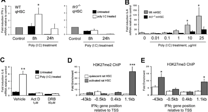
Figure 4. Interferon gamma and IL-6 expression are TLR3 mediated and loss of ability to induce IFNcin aHSCs is associated with
transcriptional repression by Polycomb complex.(A) WT andtlr32/2activated mHSCs were treated with Poly(I:C) (1mg/ml) for 8 and 24 hours;
subsequently IFNcmRNA expression was measured by qPCR. Data are normalised to GAPDH and expressed as fold induction relative to control. (B) IL-6 mRNA expression was measured in WT andtlr32/2activated mHSCs in response to increasing concentrations of Poly(I:C). (C) Wild type qHSCs
were treated with transcriptional inhibitors Act D (1mM) and DRB (80mM) for 24 hours and IL-6 mRNA expression analysed by qRT-PCR. Data are normalised to GAPDH and expressed as fold induction relative to untreated control. (D and E) One hundred micrograms of crosslinked chromatin from quiescent and activated rat HSC was incubated with 10mg of anti-trimethyl and anti-dimethyl H3K27 antibody and ChIP assay performed. Data are expressed as fold enrichment relative to IgG control. (*p,0.05, **p,0.01, ***p,0.001).
cultured with media supplemented with maximal dose of recombinant IFNc(Figure 3G).
As dsRNAs and Poly(I:C) are able to induce innate immune responses via alternative pathways from those triggered by TLR3 (e.g. RIG-1 and MDA5) we wished to confirm that the IFN and IL-6 responses we have described can be specifically attributed to TLR3 signalling. We therefore compared responses to Poly(I:C) between freshly isolated, quiescent wild type and tlr32/2 HSC. Treatment of wild type qHSC with Poly(I:C) was again associated with a time-dependent induction of IFNcwhich was not detected with tlr32/2 qHSC (Figure 4A) (2.8360.8 RLTD WT control p = 0.048). For measurement of the IL-6 response, we performed a detailed Poly(I:C) dose-response with aHSC, ranging from 10 ng/ ml to 25mg/ml (Figure 4B). For wild type aHSC, IL-6 induction was observed at 1mg/ml (2-fold), 10mg/ml (6-fold) and 25mg/ml (10-fold). By contrast a modest 3-fold induction of IL-6 transcript was observed with the highest 25mg/ml dose for tlr32/2 cells which is likely to be non-specific at this very high concentration of Poly(I:C). Abrogation of TLR3 signalling resulted in a significant, but incomplete reduction of IL-6 mRNA expression. We cannot exclude the possibility that MDA5 or RIG-1 may play a role in this setting, however, it is worth noting a complete absence of any additional IL-6 production in tlr32/2 HSCs at 1mg/ml of Poly(I:C) which was the concentration used throughout all other experiments. Treatment of wild type qHSC with transcriptional inhibitors actinomycin D and 5,6-Dichlorobenzimidazole Ribo-side (DRB) confirmed that TLR3 mediated activation of IL-6 expression is regulated at the transcriptional level (Figure 4C). To investigate an explanation for the inability of aHSC to mount an IFNc response following engagement of TLR3 we employed chromatin immunoprecipitation (ChIP) to determine if chromatin structure is modified at the IFNcgene. Di- and tri-methylation of lysine 27 on Histone 3 (H3K27) is associated with transcription-ally-repressed chromatin mediated by Polycomb proteins [21]. Activated HSC acquire the repressive dimethyl-H3K27 signature within the downstream coding region of the IFNc gene (Figure 4D). Additionally, relative to qHSC, aHSC show enrichment of trimethyl-H3K27 in both the upstream promoter and downstream coding regions (Figure 4E). These data suggest that remodelling of the HSC epigenome during their transdiffer-entiation to a myofibroblastic phenotype includes Polycomb-mediated silencing of the IFNcgene.
Discussion
The innate immune response of the liver is essential for the clearance of pathogenic microbes and initiation of the hepatic wound-healing response to liver trauma or toxic damage. TLR3 is an important component of the innate immune system providing a sensor for dsRNA originating from RNA viruses or leakage from damaged host cells [22]. In liver, TLR3 is expressed on parenchymal and non-parenchymal cells as well as infiltrating immune cells. Activation of TLR3 signalling in the liver leads to inflammation and injury through induction of NK cell activation and accumulation [23].
Augmentation of mouse liver-associated natural killer activity by biologic response modifiers occurs largely via rapid recruitment of large granular lymphocytes from the bone marrow [24]. TLR3 activated NK cells produce IFNcwhich has been shown to induce apoptosis of activated HSC and to inhibit their proliferation. This in turn limits further progression of liver fibrosis [25,26]. TLR3 is further involved in numerous processes within the liver, ranging from regeneration, viral hepatitis infection as well as autoimmune disease (reviewed in [27]).
TLR3 along with TLR4 has been implicated in the control of HCV replication via stimulation of IFNc[28,29]. Furthermore, TLR3 signalling has been implicated as an important controller of CD8+T cell infiltration via the induction of IFNcand chemokines such as CXCL9 [28]. This latter pathway has been proposed to promote liver damage and possibly facilitate the development of autoimmune hepatitis [30]. These observations suggest important but complex functions for TLR3 in liver homeostasis and immunity, as such it will be critical to define the cellular events that regulate TLR3 responses if TLR3 signalling is to be targeted therapeutically.
Non-parencymal liver cells that have previously been shown to express TLR3 in the liver include resident KC and liver sinusoidal endothelial cells (LSECs) [29,31]. Here, we provide evidence that rodent HSC express TLR3 in both their quiescent and activated phenotypes. Of note our data are in contrast to a recently published study which reported an absence of TLR3 in quiescent HSC. The reasons for this discrepancy are unclear but may relate to differences in isolation procedures, or that while we mainly focus our studies on rat HSC, the previous study utilised mouse HSC [32]. Importantly we have also demonstrated an unexpected innate immune function for qHSC, since they can express type I and type II interferons in response to Poly(I:C) treatment and in a TLR3-dependent manner. Interestingly this property of qHSC is lost during transdifferentiation to their activated phenotype, despite the cells retaining TLR3 expression and ability to elevate their production of IL-6 in response to Poly(I:C) treatment. By focusing on the IFNc gene we were able to show that aHSC acquire transcriptionally repressive chromatin modifications that may in part explain loss of IFNcproduction (Figure 4D and E). Presumably this loss of IFNcresponse protects aHSC from the previously documented anti-fibrogenic and pro-apoptotic actions of IFNc, which if produced in an autocrine manner would act to suppress their fibrogenic function [33].
Recent reports suggest that the HSC phenotype is more plastic than previously thought, particularly in the context of liver injuries that may periodically resolve and recur. It is proposed that aHSC that avoid apoptosis during resolution of fibrogenesis can revert to a more quiescent phenotype (iHSC), although in a state where they are primed to activate more efficiently than naive qHSC [34,35]. Given that iHSC may be within microenvironments where liver damage is not fully resolved, it would be interesting to determine if their phenotype reversion recovers their ability to express IFNs in response to dsRNA. Chronic production of IFNs by these cells in significant numbers and in the context of recurring liver damage may promote immune dysfunction of relevance to acute and chronic pathologies.
In summary, we have discovered an unexpected role for TLR3 in qHSC as a stimulator of type I and type II interferon expression in response to Poly(I:C) treatment. We propose that prior to activation, qHSC may contribute to the induction of the hepatic innate immune response to injury or infection. Further investiga-tion of the funcinvestiga-tion of TLR3 on qHSC may therefore lead to strategies for modulating the recruitment and activation of innate immune cells during acute viral infections and drug-induced liver injuries.
Supporting Information
MCP1, and RANTES (CCL5) in transitionary or activated HSCs compared with control.
(TIF)
Author Contributions
Conceived and designed the experiments: JM, CLM, DAM, CLW. Performed the experiments: JM, MW, CLM, MJP, FO, CLW. Analyzed the data: JM, MW, CLM, DAM, CLW. Contributed reagents/materials/ analysis tools: MCW, CB, DDP, PP, MJP, MK. Wrote the manuscript: JM, MW, DAM, CLW.
References
1. Friedman SL (2008) Hepatic stellate cells: protean, multifunctional, and enigmatic cells of the liver. Physiol Rev 88(1):125–72.
2. Friedman SL (2008) Mechanisms of hepatic fibrogenesis. Gastroenterology 134(6):1655–69.
3. Hendriks H, Verhoofstad W, Brouwer A, De Leeuw A, Knook D (1985) Perisinusoidal fat-storing cells are the main vitamin A storage sites in rat liver. Exp Cell Res 160(1):138–49.
4. Blomhoff R, Wake K (1991) Perisinusoidal stellate cells of the liver: important roles in retinol metabolism and fibrosis. FASEB J 5(3):271–7.
5. Gordon S (2002) Pattern recognition receptors: doubling up for the innate immune response. Cell 111(7):927.
6. Medzhitov R, Preston-Hurlburt P, Kopp E, Stadlen A, Chen C, et al. (1998) MyD88 is an adaptor protein in the hToll/IL-1 receptor family signaling pathways. Mol Cell. 2(2):253–8.
7. Kawai T, Akira S (2010) The role of pattern-recognition receptors in innate immunity: update on Toll-like receptors. Nat Immunol 11(5):373–84. 8. Ozinsky A, Underhill DM, Fontenot JD, Hajjar AM, Smith KD, et al. (2000)
The repertoire for pattern recognition of pathogens by the innate immune system is defined by cooperation between toll-like receptors. Proc Natl Acad Sci U S A 97(25):13766–71.
9. Inohara N, Nunez G (2003) NODs: intracellular proteins involved in inflammation and apoptosis. Nat Rev Immunol. 3(5):371–82.
10. Aderem A, Ulevitch RJ (2000) Toll-like receptors in the induction of the innate immune response. Nature 406(6797):782–7.
11. Paik YH, Schwabe RF, Bataller R, Russo MP, Jobin C, et al. (2003) Toll-like receptor 4 mediates inflammatory signaling by bacterial lipopolysaccharide in human hepatic stellate cells. Hepatology 37(5):1043–55.
12. Paik YH, Lee KS, Lee HJ, Yang KM, Lee SJ, et al. (2006) Hepatic stellate cells primed with cytokines upregulate inflammation in response to peptidoglycan or lipoteichoic acid. Lab Invest 86(7):676–86.
13. Seki E, De Minicis S, O¨ sterreicher CH, Kluwe J, Osawa Y, et al. (2007) TLR4 enhances TGF-bsignaling and hepatic fibrosis. Nat Med 13(11):1324–32. 14. Watanabe A, Hashmi A, Gomes DA, Town T, Badou A, et al. (2007) Apoptotic
hepatocyte DNA inhibits hepatic stellate cell chemotaxis via toll-like receptor 9. Hepatology 46(5):1509–18.
15. Perrugorria M, Wilson C, Zeybel M, Walsh M, Amin S, et al. (2012) Histone Methylatransferase ASH1 ochestrates fibrogenic gene transcription during myofibroblast transdifferentiation. Hepatology 56(3):1129–1139
16. Alexopoulou L, Holt AC, Medzhitov R, Flavell RA (2001) Recognition of double-stranded RNA and activation of NF-kappaB by Toll-like receptor 3. Nature 413(6857):732–8.
17. Wilson CL, Hine DW, Pradipta A, Pearson JP, van Eden W, et al. (2012) Presentation of the candidate rheumatoid arthritis autoantigen aggrecan by antigen-specific B cells induces enhanced CD4(+) T helper type 1 subset differentiation. Immunology 135(4):344–54
18. Mann J, Chu DCK, Maxwell A, Oakley F, Zhu NL, et al. (2010) MeCP2 controls an epigenetic pathway that promotes myofibroblast transdifferentiation and fibrosis. Gastroenterology 138(2):705–14. e4.
19. Kawai T, Akira S (2006) Innate immune recognition of viral infection. Nat Immunol 7(2):131–7.
20. Wang N, Liang Y, Devaraj S, Wang J, Lemon SM, et al. (2009) Toll-like receptor 3 mediates establishment of an antiviral state against hepatitis C virus in hepatoma cells. J Virol 83(19):9824–34.
21. Cao R, Zhang Y (2004) SUZ12 is required for both the histone methyltrans-ferase activity and the silencing function of the EED-EZH2 complex. Mol Cell 15(1):57–67
22. Guillot L, Le Goffic R, Bloch S, Escriou N, Akira S, et al. (2005) Involvement of toll-like receptor 3 in the immune response of lung epithelial cells to double-stranded RNA and influenza A virus. J Biol Chem 280(7):5571–80. 23. Wiltrout RH, Mathieson BJ, Talmadge JE, Reynolds CW, Zhang S, et al. (1984)
Augmentation of organ-associated natural killer activity by biological response modifiers. Isolation and characterization of large granular lymphocytes from the liver. J Exp Med 160(5):1431–49.
24. Wiltrout RH, Pilaro AM, Gruys ME, Talmadge JE, Longo D, et al. (1989) Augmentation of mouse liver-associated natural killer activity by biologic response modifiers occurs largely via rapid recruitment of large granular lymphocytes from the bone marrow. J Immunol 143(1):372–8.
25. Radaeva S, Sun R, Jaruga B, Nguyen VT, Tian Z, et al. (2006) Natural Killer Cells Ameliorate Liver Fibrosis by Killing Activated Stellate Cells in NKG2D-Dependent and Tumor Necrosis Factor–Related Apoptosis-Inducing Ligand– Dependent Manners. Gastroenterology 130(2):435–52.
26. Jeong WI, Park O, Radaeva S, Gao B (2006) STAT1 inhibits liver fibrosis in mice by inhibiting stellate cell proliferation and stimulating NK cell cytotoxicity. Hepatology 44(6):1441–51.
27. Yin S, Gao B (2010) Toll-like receptor 3 in liver diseases. Gastroenterol Res Pract 2010.
28. Li K, Chen Z, Kato N, Gale M, Lemon SM (2005) Distinct poly (IC) and virus-activated signaling pathways leading to interferon-bproduction in hepatocytes. J Biol Chem 280(17):16739–47.
29. Broering R, Wu J, Meng Z, Hilgard P, Lu M, et al. (2008) Toll-like receptor-stimulated non-parenchymal liver cells can regulate hepatitis C virus replication. J Hepatol 48(6):914.
30. Lang KS, Georgiev P, Recher M, Navarini AA, Bergthaler A, et al. (2006) Immunoprivileged status of the liver is controlled by Toll-like receptor 3 signaling. J Clin Invest 116(9):2456.
31. Wu J, Meng Z, Jiang M, Zhang E, Trippler M, et al. (2010) Toll-like receptor-induced innate immune responses in non-parenchymal liver cells are cell type-specific. Immunology 129(3):363–74.
32. Byun J-S, Suh Y-G, Yi H-S, Lee Y-S, Jeong W-I (2013) Activation of toll-like receptor 3 attenuates alcoholic liver injury by stimulating Kupffer cells and stellate cells to produce interleukin-10 in mice. J Hepatol 58(2): 342–349 33. Saile B, Eisenbach C, Dudas J, El-Armouche H, Ramadori G (2004)
Interferon-c acts proapoptotic on hepatic stellate cells (HSC) and abrogates the antiapoptotic effect of interferon-a by an HSP70-dependant pathway. Eur J Cell Biol 83(9): 469–76.
34. Kisseleva T, Cong M, Paik Y, Scholten D, Jiang C, et al. (2012) Myofibroblasts revert to an inactive phenotype during regression of liver fibrosis. Proc Natl Acad Sci U S A 109(24): 9448–9453.