Cassette Encoding an Extended-Spectrum
b
-Lactamase
Daniel Aubert, Thierry Naas*, Patrice Nordmann
Service de Bacte´riologie-Virologie, INSERM U914 ‘‘Emerging Resistance to Antibiotics,’’ LabEx LERMIT, Hoˆpital de Biceˆtre, Assistance Publique/Hoˆpitaux de Paris, Faculte´ de Me´decine Universite´ Paris-Sud, Paris, France
Abstract
Theveb1gene cassette encodes the extended spectrumb-lactamase, VEB-1 that is increasingly isolated from worldwide Gram-negative rods.Veb1is commonly inserted into the variable region of different class 1 integrons in which it is always associated with a downstream-located aadB gene cassette encoding an aminoglycoside adenylyltransferase. In
Pseudomonas aeruginosa, the majority of veb1-containing integrons also carry an insertion sequence, IS1999 that is inserted upstream of theveb1gene cassette and disrupts the integron specific recombination site,attI1. Investigation of the recombination properties of the sites surrounding veb1 revealed that insertion of IS1999 reduces significantly the recombination frequency ofattI1and thatveb1 attCis not efficient for recombination in contrast toaadB attC. Subsequent sequence optimisation ofveb1 attCby mutagenesis, into a more consensualattCsite resemblingaadB attC, successfully improved recombination efficiency. Overall, this work gives some insights into the organisation of veb1-containing integrons. We propose that IS1999 and the nature of veb1 attCstabilize the veb1gene cassette environment likely by impairing recombination events upstream or downstream ofveb1, respectively.
Citation: Aubert D, Naas T, Nordmann P (2012) Integrase-Mediated Recombination of the veb1 Gene Cassette Encoding an Extended-Spectrum b -Lactamase. PLoS ONE 7(12): e51602. doi:10.1371/journal.pone.0051602
Editor:Sergey Korolev, Saint Louis University, United States of America
ReceivedMarch 14, 2012;AcceptedNovember 7, 2012;PublishedDecember 10, 2012
Copyright:ß2012 Aubert et al. This is an open-access article distributed under the terms of the Creative Commons Attribution License, which permits unrestricted use, distribution, and reproduction in any medium, provided the original author and source are credited.
Funding:The research leading to these results has received funding from INSERM, from the Ministe`re de l’Education Nationale et de la Recherche (UPRES-EA3539), and from the European Community’s Seventh Framework Programme FP7/2010–2013 under grant agreement no. 241742 (TEMPOtest-QC). The funders had no role in study design, data collection and analysis, decision to publish, or preparation of the manuscript.
Competing Interests:Patrice Nordmann is a PLOS ONE Editorial Board member. This does not alter the authors’ adherence to all the PLOS ONE policies on sharing data and materials.
* E-mail: [email protected]
Introduction
Class 1 integrons are increasingly reported as a reservoir for antibiotic resistance genes in Gram-negative rods [1,2]. These structures possess two conserved regions located on each side of a variable region consisting of integrated gene cassettes [1–3] (Figure 1A). The 59conserved segment (59-CS) classically includes a gene encoding a site-specific recombinase of the DNA integrase family,intI1, the cassette integration site,attI1, and the promoter Pc, which is oriented toward the integration point of the gene cassettes and is responsible for gene cassette expression [2–6]. Class 1 integrons may not always contain the entire 39conserved segment (39-CS), which typically includes along with an open reading frame of unknown function (orf5), the truncated disinfec-tant (qacED1) and the sulfonamide (sul1) resistance determinants [1–3]. Gene cassettes from the variable region are composed of a gene, usually an antibiotic resistance gene, and a downstream recombination site known asattCsite or 59-base element (59-be) [1–3]. Gene cassettes are independent units that are most often promoter-less. Consequently, gene expression levels depend on the cassette position in the integron. The further the cassette is relative to Pc, the lower its expression will be [4].
Gene cassettes are non-replicative mobile elements that exist under a free circular or integrated linear form [3,7]. Site-specific recombination leading to gene cassette excision or integration is catalyzed by the integraseIntI1, which recognizes two structurally distinct sites, attI1 and attC [7,8]. The attI1 site is particularly
conserved and includes four integrase binding domains (Figure 1B). A pair of inversely oriented binding sites is located at the core site and two other integrase binding sites in direct repeat (DR1, DR2) are located further upstream [2,9,10]. A fullattI1site containing four integrase binding domains is required for optimal recombination with anattCsite [11,12]. TheattCsites that are associated with the gene cassettes are more complex and weakly related to each other [2]. They differ greatly in sequence and length but contain two pairs of inversely oriented integrase-binding domains (1L-2L and 2R-1R) [13,14] (Figure 1C and 1D). Recombination mediated by IntI1 involves recognition of the bottom strand of theattCsite [15]. Upon folding into a hairpin structure, single-strandedattCsites present an almost canonical core site consisting of 2L-2R and 1L-1R duplexes separated by a bulged area [2,16–20]. HoweverattCsite recognition and proper interaction with the integrase are not dependent on canonical DNA but on the position of two extrahelical bases that interact with the integrase and originate from symmetrical folding of the bottom strand ofattC[19].
Integrase-mediated recombinations between two attC sites, between two attI1 sites, and between an attI1 site and an attC
site have been documented, the latter being the most efficient [8,21]. During recombination the crossover point is located between the G and TT of the 7-bp core site motif, GTTRRRY, found at the 39end of the recombination sites [2,14].
Among the antibiotic resistance genes that are integron-located, the extended spectrumb-lactamase (ESBL)blaVEB-1gene has been
worldwide [22–26]. At least seven different types of
veb1-containing integrons were identified based on cassette content [22,23,26]. It is more than likely that the differentveb1-containing integrons evolved from a common ancestor, however they have maintained interesting characteristics. In all cases, veb1 is associated with a downstream-located aadBcassette encoding an aminoglycoside acetyltransferase (Figure 1A) [26]. An insertion sequence (IS1999) is inserted in the majority of theveb1-containing integrons characterized in P. aeruginosa [26,27]. Upon insertion, IS1999disrupts the integron-specific recombination site,attI1, but provides an outward-directed promoter Pout, which increases
blaVEB-1expression inP. aeruginosa(Figure 2A) [28]. Moreover,veb1
has always been reported as the first cassette within the variable region of IS1999-containing integrons carrying different cassette arrays [26,27].
The stability of theveb1environment was puzzling given the fact that the variable region of an integron is normally in constant evolution since it is subject to gene cassette rearrangement, loss and acquisition [3]. Cointegration assays were performed to determine the relative recombination efficiency of the different recombination sites present in theveb1vicinity (i.e.attI1, disrupted
attI1, veb1 attCandaadB attC). This work revealed thatveb1 attCand the disrupted attI1 site of IS1999-containing integrons are not
efficient for recombination and consequently might preserve the associations veb1-aadB and IS1999-veb1 from being disrupted, respectively.
Results
Theveb1 attCsite is not efficient for recombination
Integrase-mediated recombination involving integrons located on multicopy plasmids can generate different recombination products including: (i) free circular DNA molecules comprising one or more gene cassettes, resulting from recombination between the gene cassette attC and either attI1 or another attC site from another gene cassette [7]; (ii) cointegrates, resulting from recombination between two copies of the same plasmid. Different cointegrates can be formed depending on the sites that are available for recombination; (iii) gene duplications, which can arise by either insertion of a second gene copy encoded on a previously excised gene cassette or by formation and resolution of coin-tegrates (Figure S2). While the abundance of circular intermedi-ates is very low, plasmid cointegrintermedi-ates and gene duplications are predominantly formed during recombination when intI1 is overexpressed [29]. Moreover, under these conditions gene
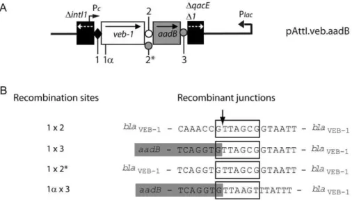
Figure 1. Schematic representations of theveb1-containing integron fromP. aeruginosa14 and of the recombination sites[26].(A) veb1-containing integron fromP. aeruginosa14. The 59and the 39-CSs are underlined. ORFs are shown as boxes with an arrow indicating the orientation of the coding sequence. The promoter Pcis indicated by a broken arrow. The black diamond representsattI1and circles representattC sites. (B–D) Recombination sites. Sequences related to the 7-bp core site are boxed; their relative orientations are indicated with arrows. The crossover points are marked by vertical arrows. The region derived from the downstream-located cassette at the recombination point is in lower case. The extra-helical bases (EHBs) as defined by Bouvieret al. [18] are marked with an asterisk. (B)attI1: the nucleotides belonging toattI1are indicated in white on a black background. The experimentally determined strong and weak IntI1-binding sites and the pair of direct repeats, DR1 and DR2, as well as the location of the IS1999insertion inattI1are indicated. (C)veb1 attCand (D)aadB attC: 7-bp putative core sites (1L, 2L, 2R and 1R) related to the core site consensus as defined by Stokes et al. [14] are shown. TheaadBgene cassette is boxed in grey.
cassettes that are unnecessary for bacterial growth are often excised and lost from the variable region [21].
The relative recombination efficiency of the sites present within different veb1-containing constructs was tested (Table 1 and Figure S1). Upon induction of integrase expression, circular DNA molecules (including plasmids, cointegrates and excised genes cassettes) were purified, digested with BspEI and separated according to their size on an agarose gel. The bottom part of the gel containing only small DNA molecules (,2-kb) was analysed andveb1-containing products were detected by hybridization using ablaVEB-1specific probe (Figure 3A). Our experimental conditions
specifically allow for detection of recombination events that involved anattI1and anattCsite but do not distinguish whether the product is an excised cassette, a gene cassette duplication or a cointegrate (Figure S2).
Plasmid pAttI.veb contains a fullattI1site, theveb1cassette and a truncated aadB cassette, while pAttI.veb.aadB contains, in addition toattI1 andveb1, a full-lengthaadBcassette. WhileattI1
and veb1 attC sites are theoretically the only sites available for recombination in pAttI.veb, pAttI.veb.aadB offers an additional recombination site, aadB attC. Recombination between attI1and
veb1 attC leading to either veb1 cassette excision, duplication or plasmid cointegration should produce, after BspE1 digestion, a
veb1-containing DNA fragment of 1.1-kb. (Figure S2 and S3). However, no such product was observed with pAttI.veb and pAttI.veb.aadB, suggesting that veb1 attC is not efficient for recombination (Figure 3A lanes 1 and 2). Instead, a 1.6-kb product was clearly detected using circular DNA extracts fromE. coli (pAttI.veb.aadB) (Figure 3A lane 2). Additional experiments confirmed that the 1.6-kb product was integrase-mediated since it was only detected when IntI1 was expressed (Figure 3A lanes 2 and 9).
A more sensitive approach using PCR amplification was used. Circular DNA molecules were purified, digested and separated on an agarose gel as before but agarose gel slices were cut at the expected migration of the 1.1-kb and 1.6-kb DNA fragments. Gel extracted DNAs were then subjected to PCR using outward-directedblaVEB-1 specific primers. These primers are located on
each side of the BspE1 restriction site and specifically allow for
amplification of veb1-containing DNA fragments that were
linearized (excised gene cassettes) or released upon BspE1
restriction from cointegrates and plasmids with veb1 duplication (Figure S2 and S3).
PCR amplification using DNA extracted from the 1.1-kb gel slices fromE. coli(pAttI.veb) andE. coli(pAttI.veb.aadB) yielded a 0.7-kb PCR product (Figure 3B lanes 1L and 2L). Sequencing indicated that it corresponded toveb1and further analysis of the recombinant junction confirmed that recombination occurred precisely between attI1 and veb1 attC (Figure 1B, 1C and 4).
Figure 2. Schematic representation of theveb1-containing integron fromP. aeruginosa15 and of the disruptedattI1recombination site[26].(A)veb1-containing integron fromP. aeruginosa15. ORFs are shown as boxes with an arrow indicating the orientation of the coding sequence. Promoters Pcand Poutare indicated by broken arrows. Disruption ofattI1by IS1999is represented by a split black diamond; circles representattCsites. The inverted repeats of IS1999are shown as empty triangles. (B) Sequence of the disruptedattI1site (DattI1). The nucleotides belonging to IS1999andattI1are indicated in grey and in white on a black background, respectively. Sequences related to the 7-bp core site are boxed; their relative orientations are indicated with arrows. The crossover point is marked by vertical arrow. The region derived from the downstream-located cassette (veb-1) at the recombination point is in lower case. The 7-bp from IS1999replacing the fourth integrase binding site fromattI1are shown in a dashed box.
doi:10.1371/journal.pone.0051602.g002
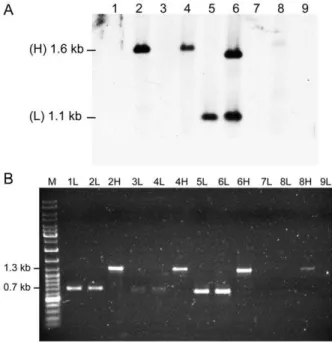
Figure 3.BlaVEB-1-specific hybridization and PCR amplifications ofveb1-containing fragments.(A)BlaVEB-1-specific hybridization of small BspEI-digested DNA fragments from E. coli DH10B (p112.Kan) harboring pAttI.veb (1), pAttI.veb.aadB (2), pAttI.IS.veb (3), pAttI.IS.ve-b.aadB (4), pAttI.veb* (5), pAttI.veb*.aadB (6), pVeb (7), pVepAttI.IS.ve-b.aadB (8) and fromE. coli DH10B (pTRC99A.Kan) harboring pAttI.veb.aadB (9) Locations of the low (L) 1.1-kb and high (H) 1.6-kb signals are indicated. (B) PCR amplifications ofveb1-containing fragments using the outward directed primers VEBINV3-VEBINV2 and ca. 1.1-kb (L) and 1.6-kb (H) DNAs that were gel-extracted based on (A), as template.
Similarly, PCR amplifications using the 1.6-kb product fromE. coli
(pAttI.veb.aadB) yielded a 1.3-kb fragment (Figure 3B lane 2H). Sequencing revealed that it contained both veb1 and aadB gene cassettes and that recombination occurred precisely betweenattI1
andaadB attC(Figure 1B, 1D and 4). The predominant formation of the 1.6-kb product also indicated that aadBremained mostly adjacent toveb1after recombination.
These results underline the weak activity of veb1 attC for recombination and the stability of the associationveb1-aadB.Both
veb1 attC and aadB attC can recombine with attI1. However in contrast toveb1 attC, aadB attCis highly efficient for recombination
as it is favored over veb1 attC during integrase-mediated
recombination.
Plasmids pVeb and pVeb.aadB containing a truncatedattI1site with only the 7-bp core site motif (GTTAGCG) at the junction
with the veb1 gene cassette were tested for recombination
(Figure 1B and S1). Surprisingly, recombination products were detected in circular DNA extracts fromE. coli(pVeb.aadB) albeit at low levels (Figure 3A lane 8 and 3B lane 8H). Sequencing revealed that recombination occurred between aadB attC and a secondary site (1a) instead of the expected GTTAGCG motif of the veb1 gene cassette (Figure 4). This secondary recombination site (GTTAAGT) is homologous to a consensus core site GTTRRRY and is located 32-bp downstream of the translation
initiation codon of blaVEB-1. Thus, recombination resulted in a
truncatedveb1gene cassette associated toaadB(Figure 4). Despite several attempts, we were not able to detect by PCR any product containing the truncatedveb1 cassette alone (Figure 3B lanes 7L and 8L). As expected, the 7-bp core site is not sufficient to support precise recombination withattC.
Sequence optimisation ofveb1 attCimproves recombination
The aadB attCsite fits closely to the consensus sequence of an
attCsite [2,14]. It is 60-bp long and made of two nearly perfect inverted repeats, which are bounded by sequences matching precisely the consensus RYYYAAC/GTTRRRY (Figure 1D).
Moreover, the bottom strand of aadB attC contains the two
extrahelical bases (T-N6-G) found in the most easily excisableattC
sites [30] (Figure 5). Therefore it is not surprising thataadB attC
worked efficiently in our recombination assays. Folding of the bottom strand of veb1 attC revealed a characteristic secondary structure with the three structural elements common toattCsites [2] (Figure 5). However,veb1 attCpresents two striking differences as compared toaadB attC: (i) theveb1 attCsite (133-bp) has a longer variable terminal structure (VTS) and (ii) the two putative Table 1.Strains and plasmids.
Strain or plasmid Relevant characteristicsa Source and/or reference
Strains
P. aeruginosa
Strain#14 Clinical isolate containing class 1 integron,veb1 [26]
Strain#15 Clinical isolate containing class 1 integron, IS1999,veb1,aadB [26]
E. coli
DH10B F–mcrAD(mrr-hsdRMS-mcrBC)W80lacZDM15DlacX74recA1endA1araD139
D(ara leu) 7697galUgalKrpsLnupGl–
Life Technologies
DH10B-Rif DH10B, RifR Laboratory stock
Plasmids
R388 33kb IncW plasmid containing class 1 integron In3 (dfrB2, orfA), Tra+, TmpR [38]
p112 pTRC99A::intI1, expression vector for integrase, Ptrc, AmpR [33]
p112.Kan p112 derivative, expression vector for integrase, Ptrc, intI1, KanR This study
pTRC99A.Kan p112.Kan derivative withoutintI1, KanR This study
pBBR1MCS.3 Broad host range cloning vector, TetR [34]
pVeb class 1 integron containingveb1in pBBR1MCS.3, CazR, TetR This study
pVeb.aadB class 1 integron containingveb1, aadBin pBBR1MCS.3, CazR, TetR This study
pAttI.veb class 1 integron containing,attI1, veb1in pBBR1MCS.3, CazR, TetR This study
pAttI.veb.aadB class 1 integron containingattI1, veb1, aadBin pBBR1MCS.3, CazR, TetR This study
pAttI.IS.veb class 1 integron containing disruptedattI1, IS1999, veb1in pBBR1MCS.3, CazR, TetR
This study
pAttI.IS.veb.aadB class 1 integron containing disruptedattI1, IS1999, veb1, aadBin pBBR1MCS.3,
CazR, TetR This study
pAttI.veb* pAttI.veb derivative, sequence modification ofveb1 attCto matchaadB attC (veb1 attC*)
This study
pAttI.veb*.aadB pAttI.veb.aadB derivative, sequence modification ofveb1 attCto matchaadB attC (veb1 attC*)
This study
pAttI.vebD pAttI.veb derivative,veb1with shorterveb1 attC (veb1 attCD) This study
pAttI.veb’ pAttI.veb derivative, sequence modification of the integrase binding sites 2L and 2R fromveb1 attC (veb1 attC’)
This study
aAntibiotic resistance: AmpR, ampicillin; CazR, ceftazidime; KanR, kanamycin; RifR, rifampin; TetR, tetracycline; TmpR, trimethoprim.
integrase binding sites 2L and 2R diverge from the attC site consensus sequence (Figure 1C).
The DNA segment fromveb1 attCthat is located between the 1L and 1R binding sites was replaced with the one fromaadB attC, thus reducing the size and restoring the 2L and 2R consensus binding sites in veb1 attC (veb1 attC*) (Figure 5D). In this configuration, ca. 1.1-kb products were strongly detected with pAttI.veb* or pAttI.veb*.aadB (Figure 3A lane 5 and 6), which indicate that recombination betweenattI1andveb1 attC*occurred. Moreover, in cells harboring pAttI.veb*.aadB,veb1-and
veb1-aadB-containing recombination products were detected at similar levels (Figure 3A lane 6) suggesting thatveb attC*andaadB attChave a similar recombination efficiency. Sequencing confirmed that precise recombination had taken place and involved either the
veb1 attC*site or theaadB attCsite (Figure 4).
These results indicate that the sequence located between the 1L and 1R sites fromveb attCis not optimal for recombination. The
veb1 attCsite was further modified to determine whether the long intermediate region located between the 2L and 2R integrase binding sites or the 2L and 2R sequences that diverged from an
attCconsensus sequence was responsible for the low recombination efficiency ofveb attC. Two differentveb1 attCsites namedveb1 attCD
andveb1 attC’ were generated. The DNA segment located between the 2L and 2R binding sites fromveb1 attCwas reduced to 20-bp as found inaadB attCgiving rise toveb1 attCD(Figure 5E). Inveb1 attC’, the 2L and 2R sequences were modified to match the 2L and 2R sequences found inaadB attC(Figure 5F).
Plasmids pAttI.vebDand pAttI.veb’ were tested for recombina-tion (Figure 6). In contrast toveb1 attC*, veb1 attCDandveb1 attC’ did not allow for the detection ofveb1-containing recombination products by hybridization (Figure 6A lanes 1–2 and 4). However,
veb1-containing recombination products were detected after PCR amplification indicating that veb1 attCD and veb1 attC’ were functional (Figure 6B lanes 1–2). Overall, veb1 attCD, veb1 attC’ and wild type veb1 attC had similar activities indicating that modifications made to reduce the VTS or change the 2L and 2R sequences did not improve the recombination efficiency.
IS1999insertion decreasesattI1recombination efficiency
InP. aeruginosa, theattI1site of manyveb1-containing integrons is disrupted by IS1999in such a way that only the last 34-bp of the site (containing three integrase binding sites) remain adjacent to
veb1 (Figure 2B). Using circular DNA isolated from E. coli
(pAttI.IS.veb.aadB), veb1-containing recombination products that resulted from recombination between the disruptedattI1andaadB attCwere detected by hybridization (Figure 3A lane 4). Recom-bination products involving the disruptedattI1andveb1 attCwere only detected after PCR amplification (Figure 3B lanes 3L and 4L).
These results suggest that the attI1 site disrupted by IS1999
insertion is still functional for recombination. Nevertheless, based on signal intensity, the recombination efficiency of the disrupted
attI1 site seemed significantly lowered in comparison to a full-lengthattI1site (Figure 3A lanes 2 and 4, 3B lanes 1L and 3L).
Recombination frequencies
Activities of the sites present within different veb1-containing constructs (Table 1 and Figure S1) were assayed in vivo by measuring the frequency of recovery of cointegrates formed between the test plasmids and plasmid R388. The self-conjugative plasmid R388 (TmpR, Tra+) includes an integron (In3) that contains thedfrB2cassette conferring resistance to trimethoprim (Tmp) followed by an open reading frame, orfA, of unknown function.
Veb1-containing constructs (conferring resistance to ceftazidime, Caz) were introduced into a rifampin sensitiveE. colicontaining the integrase expressing plasmid and plasmid R388. Upon induction of integrase expression, cointegrates resulting from recombination between the plasmid-located veb-1 integrons and the recipient integron In3 located on plasmid R388 were predominantly formed. Plasmid R388 and cointegrates were transferred by conjugation into E. coli DH10B-Rif (rifampin resistant) and cointegration frequencies were measured as ratios of cointegrates (CazR-RifR) to total R388 transconjugants (TmpR-RifR) (Table 2).
Figure 4. Recombinant junction analysis.(A) Representation of the pAttI.veb.aadB recombinant plasmid. The location of the different crossover points involved in recombination are indicated by a number: (1) 7-bp core site ofattI1-veb1; (1a) secondary recombination site found within the blaVEB-1coding sequence; (2), (2*) and (3) 7-bp site found at theveb1 attC-aadB, veb1 attC*-aadBandaadB attC-qacED1junctions, respectively. (B) Sequencing results of the recombinant junctions from experimentally isolatedveb1-containing recombination products. The numbers shown on the left indicate the recombination sites that were involved to create the recombinant junctions. Sequences related to the 7-bp core site are boxed. The crossover point is marked by vertical arrow. The nucleotides belonging to theaadBgene cassette are boxed in grey.
Plasmid cointegration was IntI1 mediated since CazR-RifR cointegrates were only recovered from strains over-expressing the integrase. In plasmid pVeb,veb1 attCis the only site available for
recombination with either attI1 or an attC site from In3.
Cointegrates (pVeb::R388) were selected at a very low frequency (4.4161025) close to the limit of detection of our assay, thus
reflecting the inefficiency of veb1 attC for recombination. As compared to pVeb, the presence ofaadB attCin pVeb.aadB, led to a 50-fold increase in the cointegration frequency, confirming that
aadB attC is more efficient for recombination than veb1 attC. Comparison between (pVeb::R388) and (pAttI.IS.veb::R388) revealed that the disruptedattI1site is 3-times more efficient for
Figure 5. Bottom strand secondary structures.(A) consensusattCstructure as of Cambray et al. [2]; (B)aadB attC; (C)veb1 attC; (D)veb1 attC*; (E)veb1 attCD; and (F)veb1 attC’. The putative IntI1 binding domains are marked with boxes and the inverted repeats 1L-1R and 2L-2R are indicated.
The protruding G that determines the recombination strand present in 2L and the protruding T that increases the recombination efficiency, are also boxed. CT, conserved triplet; UCS, unpaired central spacer; EHB, extrahelical base; VTS, variable terminal structure. Folded representations were based on secondary structures obtained thanks to the mfold Web Server. The folded structure of theveb1 attC*site was similar to the folded structure of aadB attCand possessed a free energy of230.3 Kcal/mol.
recombination than the 7-bp core site from pVeb. However, insertion of IS1999intoattI1(pAttI.IS.veb) led to a 6-fold decrease in the cointegration frequency, as compared to plasmid pAttI.veb. Sequence optimisation ofveb1 attCintoveb1 attC* led to an almost 20-fold increase in the cointegration frequency of pAttI.veb* as compared to pAttI.veb. Similar cointegration frequencies were obtained with plasmids pVeb.aadB, pAttI.veb.aadB, pAttI.IS.ve-b.aadB and pAttI.veb*.aadB. All these constructs contain the highly efficient recombination siteaadB attCthat was likely mainly involved in cointegration.
For each plasmid tested, several cointegrates were analyzed by PCR to determine which sites (i.e.attI1, veb1 attC, veb1 attC* or
aadB attCfrom theveb1-containing plasmids) were involved in the
recombination. As expected, all the pVeb::R388 and pVe-b.aadB::R388 cointegrates involved veb1 attC and aadB attC, respectively. Cointegrates pAttI.veb::R388 and pAttI.IS.veb::R388 were formed by recombination usingattI1 or veb1 attC. All the pAttI.veb.aadB::R388 and pAttI.IS.veb.aadB::R388 cointegrates involvedaadB attC and retained the aadBcassette in association withveb1. All the pAttI.veb::R388 cointegrates involvedveb1 attC* and the pAttI.veb.aadB::R388 cointegrates had systematically lost theaadBcassette. SinceaadBcassette excision could occur before or after cointegration, we could not determine whether pAttI.ve-b.aadB::R388 arose by cointegration involvingveb1 attC* oraadB attC.
CazR-RifR transconjugants were further screened for tetracy-cline susceptibility to discriminate cointegrates from preciseveb1
insertion into In3. In contrast to cointegration, which results from one recombination event; cassette insertion results from two recombination events (cassette excision-integration or plasmid cointegration-resolution) and occurs at a lower frequency. Despite most colonies (.95%) were resistant to tetracycline, few CazR-RifR-TetS transconjugants were identified. CazR-CazR-RifR-TetS transconjugants were recovered only from donor cells containing pAttI.veb.aadB, pAttI.veb* and pAttI.veb*.aadB plasmids with similar frequencies (Table 2). Each of these constructs has the particularity to carry at least two efficient sites that were used during cassette insertion. More than 95% of the CazR-RifR-TetS transconjugants recovered from cells containing pAttI.veb.aadB had integratedveb1along withaadB, demonstrating thatveb1is co-mobilized with aadB. Using plasmid pAttI.veb*.aadB, both veb1
and veb1-aadB insertions were found, however veb1 inserts were mostly recovered indicating that veb1 recombines efficiently and independently ofaadBwhen it containsveb1 attC*.
Discussion
Recombination activities of the sites surrounding theveb1gene cassette were investigated by using two independent
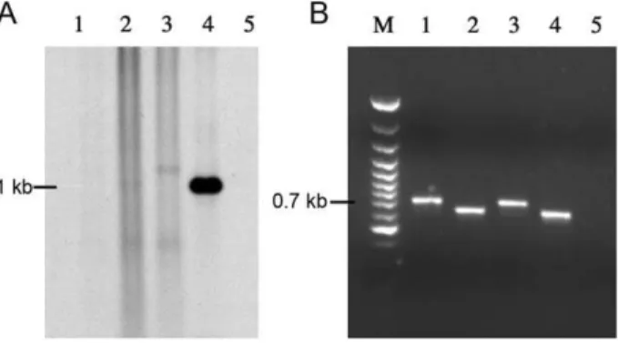
recombina-Figure 6.veb1-containing recombination products depending
on veb1 attC variants.of veb1-containing fragments. (A) BlaVEB-1 -specific hybridization of small BspEI-digested DNA fragments fromE. coli DH10B (p112.Kan) harboring pAttI.veb’ (1), pAttI1.vebD
(2), pAttI.veb (3), pAttI.veb* (4), and from E. coli DH10B (pTRC99A.Kan) harboring pAttI.veb (5). (B) PCR amplifications of veb1-containing fragments using the outward directed primers VEBINV3-VEBINV2 primers and the ca. 1.1 kb DNAs that were gel extracted from (A) as template.
doi:10.1371/journal.pone.0051602.g006
Table 2.Cointegration and cassette integration frequencies.
Plasmida Plasmid cointegrationfrequencyb
Cassette integration frequency
Number of CazR-RifR- TetS
coloniese veb1cassette insertiongene
veb1–aadBgene cassette insertion
pAttI.veb.aadB+pTRC99A.Kan 0 - - -
-pBBR1MCS.3+p112.Kan 0 - - -
-pVeb+p112.Kan 4.4161025(+/21.9661025)c NDd 0 -
-pVeb.aadB+p112.Kan 2.1561023(
+/21.8061024) ND 0 -
-pAttI.Veb+p112.Kan 7.3561024(+/23.2661024) ND 0 -
-pAttI.veb.aadB+p112.Kan 3.3161023(+/21.8961024) 1.4461024 21 1 20
pAttI.IS.veb+p112.Kan 1.2361024(+/23.3661026) ND 0 -
-pAttI.IS.veb.aadB+p112.Kan 3.7961023(+/2.1061024) ND 0 -
-pAttI.veb*+p112.Kan 1.3761022(+/22.5261023) 1.7061024 6 6
-pAttI.veb*.aadB+p112.Kan 6.2561023(+/27.5361024) 2.0761024
(1.5561024
)f
16 12 4
aDonor strains also contain R388; pTRC99A.Kan did not expressed IntI1 (negative control); p112.Kan (a pTRC99A.Kan derivative) expressed IntI1. The pVeb, pVeb.aadB,
pAttI.veb, pAttI.veb.aadB, pAttI.IS.veb, pAttI.IS.veb.aadB, pAttI.veb* and pAttI.veb*.aadB were used for recombination (Figure S1). pBBR1MCS.3, cloning vector (negative control).
bThe limit of detection of our assay is,1.5 61025.
cStandard deviations calculated from three independent experiments. dND, Not determinable.
eup to 484 CazR, RifR transconjugants were tested for tetracycline sensitivity. fIntegration frequency of theveb1gene cassette alone.
tion assays. It is noteworthy that experiments were performed inE. coliDH10B, which contains an inactivated form of RecA (RecA1) excluding recombination between homologous sequences. Thus, despite plasmids share some sequence identity, cointegration is unlikely to have occurred by homologous recombination but is truly the result of site-specific recombination mediated by the integrase.
Dissemination and acquisition of a single gene cassette within the variable region of integrons is the result of integrase-mediated recombination between two different sites, one of which is the gene cassetteattC[7]. We demonstrate here thatveb1 attCfound within theveb1gene cassette, is not efficient for recombination implying that veb1 alone is not highly mobilizable. Our cointegration experiments also revealed thataadB attCis favored overveb1 attCas it is almost exclusively involved in recombination when both gene cassettes are present on a plasmid. Despite no selection, aadB
remained associated to wild typeveb1in all the cointegrates tested, while gene cassettes that are unnecessary for bacterial growth, are often unstable and lost from the variable region when IntI1 is overexpressed [21]. Gene cassettes are often considered as independent units but their excision depends greatly on the recombination sites that are flanking them. Therefore, it was anticipated thataadBwould be rarely excised sinceaadBexcision require the involvement ofveb1 attC. Stability of the association
veb1-aadBwithin integrons is likely preserved by the nature ofveb1 attC, which impairs not onlyaadBexcision but also new insertions of gene cassettes in between them. Our cassette integration assay in plasmid R388 showed that the associationveb1-aadB is to the benefit ofveb1, which is mobilized along withaadBthanks to the high recombination efficiency ofaadB attC.
TheaadB attC site is 50 times more efficient for recombination than veb1 attC. Sequence optimisation ofveb1 attC into a more consensual attC site resembling aadB attC (veb1 attC*) improved drastically the recombination efficiency and allowed independent excision ofaadBfromveb1-containing integrons. This experiment revealed also that the features responsible for the weak activity of
veb1 attCreside within the DNA sequence located between the 1L and 1R integrase binding sites. Two major differences between
veb1 attCandaadB attCwere found in this region and concerned: (i) the 2L and 2R integrase binding sites, which sequences inveb1 attC
diverged from the consensus and (ii) the length of the variable terminal structure (VTS). However, modification of one or the other in veb1 attC was not sufficient to improve recombination
activity. While VTS seem to have a minor role in the attC
recombination efficiency, other structural features including the unpaired central spacer (UCS) bulge shape and two extrahelical bases (EHBs) appear to be important for recognition and recombination rate [18]. In contrast to aadB attC, veb1 attC
presents slightly larger UCS. Also, while the EHBs inaadB attCare 6 nucleotides apart (T-N6-G) as found in the attC sites flanking
cassettes that are efficiently excised by IntI1 [30],veb1 attCcontains only one EHB (G). It has been recently demonstrated that the identity and spacing of the EHBs in theattCsites has a pronounced effect on the efficiency of cassette excision [30]. The relevance of these structural differences in the activity ofveb1 attCremains to be established. It is also possible that the recombination efficiency of
veb1 attCis not affected by only one of these features but by several of them.
Several studies have shown that a fullattI1site containing the four integrase binding sites was required for high efficiency recombination withattCand that progressive 59deletions reaching
closer to the 7-bp core site of attI1 leads to decreased
recombination rates [11,12,31]. Insertion of IS1999 into attI1
displaces only the weak integrase binding site located in DR2.
Accordingly, the disrupted attI1 is still sufficient to support recombination albeit at a lower frequency than a fullattI1 site. Analysis of the bases from IS1999replacing part ofattI1did not reveal any obvious 7-bp core site sequence, which could have compensated for the loss of the fourth integrase binding site. Also, by reducing the recombination rate ofattI1,IS1999likely increases the stability ofveb1 and consequently aadBat the first positions within the variable region by impairing integration of new gene cassettes upstream of veb1 and excision of veb1-aadB. However, since the disrupted attI1 still supports recombination, new gene cassettes could be inserted at the first position and benefit from the additional promoter Pout located in IS1999 for their expression
[28].
Several studies have demonstrated that integrase is also able to catalyse recombination between one specific site (attI1orattC) and non-specific secondary sites conforming to the consensus GNT at a very low frequency [12,21,31]. Moreover, limitation ofattI1 to the 7-bp core site is insufficient for determining recombination specificity alone and leads to the formation of cointegrates owing to recombination with secondary sites [12]. Using plasmid pVeb.aadB, we demonstrated upon PCR amplification thataadB attC is able to recombine with the secondary site (GTTAAGT) located within the blaVEB-1 gene. This event resulting in a
truncatedveb1 gene cassette is extremely rare since it has been detected only once in our repeats. The region that is truncated in
blaVEB-1 encodes the first eleven amino acids of the pre-enzyme
VEB-1 and includes the translation initiation codon and most of the signal peptide. Even upon insertion in an environment providing transcription and translation signals, it is unlikely that the protein encoded by the truncated veb1 cassette would be functional in vivo since it would not be properly targeted to the periplasm.
Overall, this work gives some insights into the organisation of
veb1-containing integrons that are widespread among Gram-negative bacteria. It is more than likely that the different veb1 -containing integrons evolved from a common ancestor presenting an early associationveb1-aadB. It is also possible thataadBis at the origin of the veb1 gene cassette recruitment and of the co-mobilization ofveb1-aadBinto class 1 integrons. Even thoughveb1 -containing integrons are still subject to gene cassette rearrange-ments, we propose that IS1999and the nature ofveb1 attCstabilize theveb1 gene cassette environment likely by impairing recombi-nation events upstream or downstream ofveb1, respectively.
Materials and Methods
Bacterial strains, plasmids and culture conditions
Bacterial strains and plasmids used in this study are listed in Table 1. The clinical strains P. aeruginosa 14 and 15 carrying different veb1-containing class 1 integrons were from the Siriraj Hospital, Bangkok, Thailand [26]. The recombination deficient strainE. coliDH10B (Life Technologies, Eragny, France) was used as bacterial host in electroporation experiments. The conjugative plasmid R388 (TmpR, Tra+) includes an integron (In3) that contains thedfrB2cassette conferring resistance to trimethoprim (Tmp) followed by an open reading frame, orfA, of unknown function [32]. Plasmid p112 (a pTRC99A derivative) was a gift from D. Mazel [33]. This plasmid contains theintI1gene under the control of an IPTG-inducible synthetic Ptrcpromoter.E. coli
DH10B harboring various plasmids and E. coli DH10B-Rif
Soy (TS) broth or onto TS agar plates (Sanofi Diagnostics Pasteur, Marnes-La-Coquette, France) with antibiotics when needed.
Antimicrobial agents
The antimicrobial agents and their sources were as follows: ceftazidime, GlaxoSmithKline (Marly-Le-Roi, France); rifampin, Aventis (Paris, France); trimethoprim and tetracycline, Sigma (Saint-Quentin Fallavier, France); kanamycin, Euromedex (Mun-dolsheim, France).
Nucleic acid extractions
Circular DNA molecules were extracted using Plasmid Maxi Kits (Qiagen, Courtaboeuf, France) according to the instructions of the manufacturer. Extractions of whole-cell DNAs were done as described elsewhere [35].
PCR experiments
Taqand Pfu DNA polymerases were from Roche Diagnostics
(Meylan, France) and Promega Corporation (Madison, Wis.), respectively. PCR experiments [36] were performed using the series of primers listed in Table S1. The PCR products were purified using Qiaquick columns (Qiagen). To determine the insertion sites of the veb1 cassette or derivatives into the In3
integron, plasmids from independent E. coli DH10B-Rif
(R388::veb1) transconjugants were extracted and amplifications using combination of primers TMPB, VEBINV2-ORFAB, VEBINV2-QACEB, and TMPA-VEBINV3 were per-formed. The aadB cassette was detected by PCR amplification using the AADBF and AADBB primers.
For each construct tested, five cointegrates resulting from cointegration between R388 and veb1-containing plasmids were analyzed. Amplifications using combination of primers T3-59CS, T7 promoter-39CS and VEBINV2-39CS were performed in order to determine which recombination sites were involved in the formation of the cointegrates.
Cloning experiments and electroporation
T4 DNA ligase, and restriction endonucleases were used according to the recommendations of the manufacturer (Amer-sham Biosciences, Orsay, France). The plasmid p112.Kan was constructed by inserting a HindIII-digested omega fragment (VKm) from plasmid pHP45V-Km [37], made of a kanamycin resistance gene (aph(39)-IIa) flanked by transcriptional and trans-lational termination sequences, into the HindIII site from the multiple cloning site of p112 plasmid (pTRC99A::intI1) [33]. The pTRC99A.Kan plasmid was constructed by removing the 1.2-kb EcoRI-BamHI fragment containing theintI1gene from p112.Kan, filling in its ends with PfuDNA polymerase and followed by self ligation. The inserts of the recombinant plasmids pVeb and pVeb.aadB, corresponded to fragments of 1.6-kb (containing the
veb1cassette) and 1.7-kb (containing theveb1 andaadBcassettes) that were amplified with the pairs of primers VEBCASF/AADBB
and VEBCASF/39CS, respectively and genomic DNA of P.
aeruginosa14 as template (Figure S1). The inserts of the recombi-nant plasmids pAttI.veb and pAttI.veb.aadB corresponded to fragments of 2-kb (containing an entire attI1 site and the veb1
cassette) and 2.1-kb (containing an entireattI1site and theveb1and
aadB cassettes) that were amplified with the pairs of primers
INTIN/AADBB and INTIN/39CS, respectively and genomic
DNA ofP. aeruginosa14 as template (Figure S1). The inserts of the recombinant plasmids pAttI.IS.veb and pAttI.IS.veb.aadB corre-sponded to fragments of 3.4-kb (containing a disruptedattI1site, IS1999, and theveb1cassette) and 3.5-kb (containing a disrupted
attI1 site, IS1999, and the veb1 and aadB cassettes) that were amplified with the pairs of primers INTIN/AADBB and INTIN/ 39CS, respectively and genomic DNA of P. aeruginosa 15 as template (Figure S1). PCR products were purified prior to cloning into the SmaI-restricted pBBR1MCS.3 vector.
Three different modifications of theveb1 attCsite (veb1 attC*,veb1 attCD, andveb1 attC’) were generated. Creation of theveb1 attC* site was performed by using the attCVEB1 and attCVEB2 primers that anneal to the beginning of theveb1 attCsite up to the inverse core site and to the beginning of theaadBgene cassette (core site), respectively. These primers have floating 59 ends, each corre-sponding to a half of theaadB attCsite, and have in common the portion containing the BsaHI restriction site (Figure S1). The recombinant plasmid pAttI.veb* containing veb1 attC* site was constructed as follows: a 1.5-kb fragment amplified with the pair of primers T7 promoter-attCVEB1 and pAttI.veb as template was digested with SpeI-BsaHI. A 0.7-kb fragment was amplified with the primers attCVEB2-T3 and pAttI.veb as template and was digested with BsaHI-PstI. Then, the digested inserts were purified and mixed in a three-way ligation with the PstI-SpeI-restricted pBBR1MCS.3 vector generating pAttI.veb* (Figure S1). The pAttI.veb.aadB and pAttI.veb* recombinant plasmids were digested with SacI endonucleases. The SacI insert of pAttI.veb* was cloned into the SacI-restricted pAttI.veb.aadB plasmid generating pAttI.veb*.aadB (Figure S1).
Similarly, pAttI.vebD(containingveb1 attCD) was constructed as follows: a 1.5-kb fragment amplified with the pair of primers T7 promoter-Shortattc1 and pAttI.veb as template was digested with SacI. A 0.6-kb fragment was amplified with the primers Shortattc2-T3 and pAttI.veb as template and was digested with KpnI. Then, the digested inserts were purified and mixed in a three-way ligation with the SacI-KpnI-restricted pBBR1MCS.3 vector generating pAttI.-vebD(Figure S1). The recombinant plasmid pAttI.veb’ (containing
veb1 attC’) was constructed as follows: a 1.6-kb fragment amplified with the pair of primers T7 promoter-attc2L and pAttI.veb as template was digested with NdeI. The resulting 0.9-kb and 0.7-kb fragments were purified and subsequently digested with AvaI and SacI, respectively. A 0.3-kb fragment was amplified with the primers attc2R-T3 and pAttI.veb as template and was digested with AvaI-XhoI. Then, the digested inserts were purified and mixed before ligation with the SacI-XhoI-restricted pAttI.veb plasmid generating pAttI.veb’ (Figure S1).
Ligation products were electroporated first intoE. coliDH10B as previously described [35]. Selection was performed onto TS-agar plates containing tetracycline (15mg/ml) and ceftazidime (15mg/ml) except for pVeb and pVeb.aadB plasmids that were
selected onto TS-agar plates containing tetracycline (15mg/ml) only. Clones harboring recombinant plasmids pVeb, pVeb.aadB, pAttI.veb, pAttI.veb.aadB, pAttI.IS.veb, pAttI.IS.veb.aadB, pAt-tI.veb*, pAttI.veb*.aadB, pAttI.vebD, and pAttI.veb’ were retained for further experiments (Figure S1).
Sequencing
Sequencing was performed on both strands using laboratory-designed primers on an ABI PRISM 3100 automated sequencer (Applied Biosystems, Les Ullis, France).
Induction of integrase expression
phase in TS-broth containing tetracycline 15mg/ml and
kana-mycin 30mg/ml. The cultures were then diluted 1000-fold into 200 ml TS-broth containing tetracycline 15mg/ml, kanamycin 30mg/ml and grown to exponential phase (OD 600 nm: 0.5). Integrase expression was then induced for 2 h by adding IPTG at a final concentration of 0.6 M.
Detection ofveb1-containing recombination products by hybridization
This assay allows for the detection ofveb-1containing molecules that are the result of a recombination event between attI1 (or disrupted attI1) and an attC site and gives an insight into their recombination efficiency. Total circular DNA content was
extracted from E. coli DH10B (p112.Kan) strains harboring
recombinant plasmids pVeb, pVeb.aadB, pAttI.veb, b.aadB, pAttI.IS.veb, pAttI.IS.veb.aadB, pAttI.veb*,
pAttI.ve-b*.aadB, pAttI.vebD, and pAttI.veb’ and E. coli DH10B
(pTRC99A.Kan) harboring pAttI.veb.aadB after IPTG-induction. Fivemg of circular DNA extracts were digested in duplicate with 10 units of BspEI, which cleaves at a unique site located in theveb1
cassettes (Figure S1). Digested DNA samples were then loaded on two agarose gels (25 cm, 0.7%) and electrophoresed at 45 V for 16 h using Tris-borate-EDTA running buffer [36]. The duplicates that were loaded on the second gel were spaced two wells apart.
The first agarose gel was used for hybridization experiments as follows: The bottom section of the gel that contained small BspEI excision products (,2-kb), was cut and transferred onto a N+
Hybond nylon membrane (Amersham Biosciences). Southern blot hybridizations [36] were performed under high-stringency condi-tions using the ECL nonradioactive labeling and detection kit (Amersham Biosciences). The probe consisted of a PCR-generated fragment internal to blaVEB-1 that was amplified using primers
VEB1A/VEB1B and whole-cell DNA of P. aeruginosa 14 as
template.
The second agarose gel was used to extract BspEI digestedveb1 -containing fragments. Since DNA amounts were very low and could not be detected visually after ethidium bromide staining, DNA location was spotted by superposing the autoradiography film obtained from the first gel after hybridization. Gel slices were cut with a separate disposable scalpel to avoid sample cross-contamination. DNA was extracted using Qiaquick Gel extraction kit (Qiagen) and subjected to PCR amplification using the VEBINV3-VEBINV2 outward-directed primers. These primers are located on each side of the BspE1 restriction site, and thus allowed amplification of the recombinant junction that was created upon recombination (Figure S2 and S3).
Cointegration assay using plasmid R388 and calculation of cointegration frequencies
Precise integration ofveb1gene cassettes into the class 1 integron In3 carried by the conjugative plasmid R388 or cointegration betweenveb1-containing plasmids and R388 was investigated using a mating-out assay as follows. The recombinant plasmids pVeb, pVeb.aadB, pAttI.veb, pAttI.veb.aadB, pAttI.IS.veb, pAttI.IS.ve-b.aadB, pAttI.veb*, pAttI.veb*.aadB and pBBR1MCS.3 cloning vector were freshly electroporated intoE. coliDH10B (p112.Kan; R388); the plasmid pAttI.veb.aadB was freshly electroporated into
E. coli DH10B (pTRC99A.Kan; R388). Twenty-four hours after electroporation, three single colonies (for each plasmid tested) were independently cultured overnight at 37uC in 10 ml TS broth containing tetracycline, 15mg/ml and kanamycin, 30mg/ml. The overnight cultures were diluted 10-fold in fresh TS broth without antibiotic and were cultured under low agitation at 37uC for 1 h
30, prior to a 2 h IPTG-induction (induction ofintI1expression). Mating was performed by incubating 800ml of recipient E. coli
DH10B-Rif and 200ml of the tested strain under low agitation at 37uC for 3 h. Subsequently, the mating mixture was vigorously vortexed, placed on ice, and plated. One hundred microliters aliquots of serial ten fold dilutions were plated onto trimethoprim-(25mg/ml) and rifampin- (200mg/ml) containing plates and
ceftazidime- (15mg/ml) and rifampin- (200mg/ml) containing
plates. For E. coli DH10B (R388; p112.Kan; pBBR1MCS.3)
aliquots were plated onto trimethoprim- (25mg/ml) and rifampin-(200mg/ml) containing plates and tetracycline- (15mg/ml) and
rifampin- (200mg/ml) containing plates. The cointegration frequency was calculated by dividing the number of ceftazidime and rifampin resistant (CazR-RifR) transconjugants by the number of trimethoprim and rifampin resistant (TmpR-RifR) transconjugants. For each plasmid tested, up to 484 CazR-RifR colonies were further screened for tetracycline susceptibility in order to discriminate cointegrates from precise veb1 cassettes integration into In3.
For E. coli DH10B (R388; p112.Kan; pBBR1MCS.3) the cointegration frequency was calculated by dividing the number of tetracycline and rifampin resistant (TetR-RifR) transconjugants by the number of TmpR-RifR transconjugants.
Supporting Information
Figure S1 Schematic representation of the plasmid constructs used in this study. All constructs were cloned into the multiple cloning site of the pBBR1MCS.3 shuttle-vector represented with a solid line. The coding regions are shown as boxes with an arrow indicating the orientation of their transcription. Dashed lines indicate truncated genes. The black diamond, white, grey, white with black dots, and black with white dots circles representattI1,veb1 attC, aadB attC, veb1 attCD, andveb1 attC’, respectively. Theveb1 attC*site is highly similar to theaadB attC site and is also represented by a grey circle. The IS1999
inverted repeats are shown by empty triangles. The broken arrows indicate the Pc, Poutand Placpromoters. Restriction sites used for cloning are indicated on the pAttI.veb.aadB representation. Small arrows (1 and 2) located on each side of the BspEI restriction site represent the positions of the VEBINV3 and VEBINV2 primers, respectively.
(TIF)
Figure S2 Recombination products obtained with pAt-tI.veb. A) Schematic representation of plasmid pAttI.veb. Construct was made in the pBBR1MCS.3 shuttle-vector repre-sented with a solid line. The coding regions are shown as boxes with an arrow indicating the orientation of their transcription. Dashed lines indicate truncated genes. The black diamond and white circle representattI1andveb1 attC, respectively. The broken arrow indicates the Pcpromoter. Small arrows (1 and 2) located on each side of the BspEI restriction site represent the positions of the VEBINV3 and VEBINV2 primers, respectively. B–D) Recombi-nation products. Each possible recombiRecombi-nation product is shown. The sites involved in the recombination and the size of the relevant BspEI digestion products are indicated. B)Veb1 cassette excision product. C) Cointegrates. Double lines represent scale breaks. D) Gene duplications. Veb1 duplication can arise from integration (attIxveb1 attCorveb1 attCxveb1 attC) of an excisedveb1
gene cassette, or by resolution (veb1 attCxattI1orattI1xattI1) of cointegrates. In any case, the 1.1-kb fragment is only recovered after BspEI digestion when recombination betweenattI1andveb1 attChas occurred.
Figure S3 Recombination products obtained with pAt-tI.veb.aadB.A) Schematic representation of plasmid pAttI.ve-b.aadB. Construct was made in the pBBR1MCS.3 shuttle-vector represented with a solid line. The coding regions are shown as boxes with an arrow indicating the orientation of their transcription. Dashed lines indicate truncated genes. The black diamond, white and grey circles representattI1, veb1 attCandaadB attC, respectively. The broken arrow indicates the Pc promoter. Small arrows (1 and 2) located on each side of the BspEI restriction site represent the positions of the VEBINV3 and VEBINV2 primers, respectively. B–D) Recombination products. The differ-entveb1-containing recombination products are represented. The sites involved in the recombination and the size of the relevant BspEI digestion products are indicated. B) Veb1-containing excision product. C) Cointegrates. Double lines represent scale breaks. D) Gene duplications. Veb1 duplications can arise from integration of an excised veb1-containing gene cassette or by resolution of cointegrates. In any case, the 1.1-kb and 1.6-kb fragments are only recovered after BspEI digestion when recombinations attI1 x veb1 attC and attI1 x aadB attC have occurred, respectively.
(TIF)
Table S1 Sequence of primers used in this study. a Nucleotides that are complementary to the aadB attC site are underlined;b Restriction sites are bolded;c Nucleotides that are
mutated are boxed in grey. Supplemental references are as follows: (1). Naas T, Coignard B, Carbonne A, Blanckaert K, Bajolet O, et al. (2006) VEB-1 Extended-spectrum beta-lactamase-producing
Acinetobacter baumannii, France. Emerg Infect Dis 12: 1214–1222. (2). Girlich D, Naas T, Leelaporn A, Poirel L, Fennewald M, et al. (2002) Nosocomial spread of the integron-located veb-1-like cassette encoding an extended-pectrum beta-lactamase in Pseudo-monas aeruginosa in Thailand. Clin Infect Dis 34: 603–611. (3). Martinez E, de la Cruz F (1990) Genetic elements involved in Tn21 site-specific integration, a novel mechanism for the dissemination of antibiotic resistance genes. EMBO J 9: 1275– 1281.
(DOCX)
Acknowledgments
DA and TN contributed equally to the work. We thank D. Mazel for helpful discussions and for gift of the R388 and p112 plasmids and L. Biskri for precious advice in IntI1 overexpression.
Author Contributions
Conceived and designed the experiments: DA TN PN. Performed the experiments: DA TN PN. Analyzed the data: DA TN PN. Contributed reagents/materials/analysis tools: DA TN PN. Wrote the paper: DA TN PN.
References
1. Partridge SR, Tsafnat G, Coiera E, Iredell JR (2009) Gene cassettes and cassette arrays in mobile resistance integrons. FEMS Microbiol Rev 33: 757–784. 2. Cambray G, Guerout AM, Mazel D (2010) Integrons. Annu Rev Genet 44:
141–166.
3. Recchia GD, Hall RM (1995) Gene cassettes: a new class of mobile element. Microbiology 141 (Pt 12): 3015–3027.
4. Collis CM, Hall RM (1995) Expression of antibiotic resistance genes in the integrated cassettes of integrons. Antimicrob Agents Chemother 39: 155–162. 5. Levesque C, Brassard S, Lapointe J, Roy PH (1994) Diversity and relative
strength of tandem promoters for the antibiotic-resistance genes of several integrons. Gene 142: 49–54.
6. Jove´ T, Da Re S, Denis F, Mazel D, Ploy MC (2010) Inverse correlation between promoter strength and excision activity in class 1 integrons. PLoS Genet 6: e1000793.
7. Collis CM, Hall RM (1992) Gene cassettes from the insert region of integrons are excised as covalently closed circles. Mol Microbiol 6: 2875–2885. 8. Demarre G, Frumerie C, Gopaul DN, Mazel D (2007) Identification of key
structural determinants of the IntI1 integron integrase that influenceattCxattI1
recombination efficiency. Nucleic Acids Res 35: 6475–6489.
9. Collis CM, Kim MJ, Stokes HW, Hall RM (1998) Binding of the purified integron DNA integrase Intl1 to integron- and cassette-associated recombination sites. Mol Microbiol 29: 477–490.
10. Hall RM, Collis CM, Kim MJ, Partridge SR, Recchia GD, et al. (1999) Mobile gene cassettes and integrons in evolution. Ann N Y Acad Sci 870: 68–80. 11. Partridge SR, Recchia GD, Scaramuzzi C, Collis CM, Stokes HW, et al. (2000)
Definition of theattI1site of class 1 integrons. Microbiology 146 ( Pt 11): 2855– 2864.
12. Hansson K, Skold O, Sundstro¨m L (1997) Non-palindromicattlsites of integrons are capable of site-specific recombination with one another and with secondary targets. Mol Microbiol 26: 441–453.
13. Hall RM, Brookes DE, Stokes HW (1991) Site-specific insertion of genes into integrons: role of the 59-base element and determination of the recombination cross-over point. Mol Microbiol 5: 1941–1959.
14. Stokes HW, OGorman DB, Recchia GD, Parsekhian M, Hall RM (1997) Structure and function of 59-base element recombination sites associated with mobile gene cassettes. Mol Microbiol 26: 731–745.
15. Francia MV, Zabala JC, de la Cruz F, Garcia Lobo JM (1999) The IntI1 integron integrase preferentially binds single-stranded DNA of theattCsite. J Bacteriol 181: 6844–6849.
16. Bikard D, Loot C, Baharoglu Z, Mazel D (2010) Folded DNA in action: hairpin formation and biological functions in prokaryotes. Microbiol Mol Biol Rev 74: 570–588.
17. Johansson C, Kamali-Moghaddam M, Sundstro¨m L (2004) Integron integrase binds to bulged hairpin DNA. Nucleic Acids Res 32: 4033–4043.
18. Bouvier M, Ducos-Galand M, Loot C, Bikard D, Mazel D (2009) Structural features of single-stranded integron cassetteattCsites and their role in strand selection. PLoS Genet 5: e1000632.
19. MacDonald D, Demarre G, Bouvier M, Mazel D, Gopaul DN (2006) Structural basis for broad DNA-specificity in integron recombination. Nature 440: 1157– 1162.
20. Bouvier M, Demarre G, Mazel D (2005) Integron cassette insertion: a recombination process involving a folded single strand substrate. EMBO J 24: 4356–4367.
21. Collis CM, Recchia GD, Kim MJ, Stokes HW, Hall RM (2001) Efficiency of recombination reactions catalyzed by class 1 integron integrase IntI1. J Bacteriol 183: 2535–2542.
22. Poirel L, Naas T, Guibert M, Chaibi EB, Labia R, et al. (1999) Molecular and biochemical characterization of VEB-1, a novel class A extended-spectrum beta-lactamase encoded by an Escherichia coli integron gene. Antimicrob Agents Chemother 43: 573–581.
23. Naas T, Poirel L, Karim A, Nordmann P (1999) Molecular characterization of In50, a class 1 integron encoding the gene for the extended-spectrum beta-lactamase VEB-1 inPseudomonas aeruginosa. FEMS Microbiol Lett 176: 411–419. 24. Naas T, Poirel L, Nordmann P (2008) Minor extended-spectrum
beta-lactamases. Clin Microbiol Infect 14 Suppl 1: 42–52.
25. Naas T, Coignard B, Carbonne A, Blanckaert K, Bajolet O, et al. (2006) VEB-1 Extended-spectrum beta-lactamase-producing Acinetobacter baumannii, France. Emerg Infect Dis 12: 1214–1222.
26. Girlich D, Naas T, Leelaporn A, Poirel L, Fennewald M, et al. (2002) Nosocomial spread of the integron-located veb-1-like cassette encoding an extended-pectrum beta-lactamase in Pseudomonas aeruginosa in Thailand. Clin Infect Dis 34: 603–611.
27. Aubert D, Naas T, He´ritier C, Poirel L, Nordmann P (2006) Functional characterization of IS1999, an IS4family element involved in mobilization and expression of beta-lactam resistance genes. J Bacteriol 188: 6506–6514. 28. Aubert D, Naas T, Nordmann P (2003) IS1999increases expression of the
extended-spectrum beta-lactamase VEB-1 inPseudomonas aeruginosa. J Bacteriol 185: 5314–5319.
29. Collis CM, Hall RM (1992) Site-specific deletion and rearrangement of integron insert genes catalyzed by the integron DNA integrase. J Bacteriol 174: 1574– 1585.
30. Larouche A, Roy PH (2011) Effect ofattCstructure on cassette excision by integron integrases. Mob DNA 2: 3.
31. Recchia GD, Stokes HW, Hall RM (1994) Characterisation of specific and secondary recombination sites recognised by the integron DNA integrase. Nucleic Acids Res 22: 2071–2078.
32. Martinez E, de la Cruz F (1990) Genetic elements involved in Tn21site-specific integration, a novel mechanism for the dissemination of antibiotic resistance genes. EMBO J 9: 1275–1281.
35. Philippon LN, Naas T, Bouthors AT, Barakett V, Nordmann P (1997) OXA-18, a class D clavulanic acid-inhibited extended-spectrum beta-lactamase from
Pseudomonas aeruginosa. Antimicrob Agents Chemother 41: 2188–2195. 36. Sambrook J, Russell DW (2001) Molecular cloning: a laboratory manual. Cold
Spring Harbor, NY: Cold Spring Harbor Laboratory Press.
37. Prentki P, Krisch HM (1984) In vitro insertional mutagenesis with a selectable DNA fragment. Gene 29: 303–313.