Setembro 2011
André Filipe Simões de Carvalho de Sousa
Licenciado em Bioquímica
Investigation of trypanothione
synthetase of
Leishmania infantum
as
a potential target for new
anti-parasitic drugs
Dissertação para obtenção do Grau de Mestre em Biotecnologia
Orientadora: Doutora Helena Maria de Sousa Castro
Júri:
Presidente: Prof. Doutora Ana Cecília Afonso Roque
The work presented in this MSc dissertation was done at the Institute for Molecular and cell Biology (IBMC), Porto, Portugal. This work was financially supported by a grant
III
V Acknowledgments / Agradecimentos
Em primeiro lugar quero agradecer à minha orientadora, Helena Castro, por tudo aquilo que me ensinou durante este ano de trabalho. Pela paciência, dedicação e disponibilidade que sempre demonstrou durante toda a fase de escrita da dissertação assim como durante toda a fase experimental deste trabalho. Obrigado por todas as oportunidades que me proporcionou e pela abertura de espírito que permitiu todo um diálogo construtivo em cada passo deste projecto e me fez sentir parte integrante do mesmo. Obrigado pela consideração, respeito e profissionalismo. Por me ter guiado com um sorriso por este novo mundo da ciência no qual dou os meus primeiros passos. Foi um ano bastante enriquecedor a todos os níveis. Obrigado por tudo.
Uma palavra de agradecimento também para a Doutora Ana Tomás, que me recebeu tão bem no seu grupo (Molecular Parasitology) no Instituto de Biologia Molecular e Celular (IBMC) e tornou este trabalho possível.
Gostava também de deixar um agradecimento especial à Joana Passos pela ajuda indispensável no trabalho de bancada nas alturas em que eu mais precisei.
Agradeço também à Filipa Teixeira por toda a ajuda no laboratório assim como pelas conversas e divagações abstractas que nem sempre levaram a alguma conclusão mas sempre alimentaram a energia das hipóteses no meu pensamento.
Um agradecimento sincero também para a Sandra Carvalho e Tânia Cruz que sempre se mostraram disponíveis para ajudar e tornaram a minha integração no grupo numa tão experiencia agradável.
VII Abstract
Leishmania infantum
is a protozoan parasite of the Trypanosomatidae family,
responsible for human and canine leishmaniasis in Mediterranean countries. Control of
these vector-borne diseases is unsatisfactory and new chemotherapeutics are urgently
needed. Trypanothione biosynthesis, owing to its unique and essential character, is
regarded as an attractive target for therapeutic intervention. Trypanothione is a
bis-(glutathionyl)spermidine
conjugate,
responsible
for
redox
homeostasis
in
trypanosomatids. It is synthesized by the sequential addition of two molecules of
glutathione to a spermidine molecule. Trypanothione synthetase (TRYS), which
catalyzes both conjugation steps, has no counterpart in mammals and is essential to
Trypanosoma brucei
. This scenario is somewhat different in
L. infantum
, which harbors
one additional enzyme mono(glutathionyl)spermidine synthetase or GSPS capable of
driving the first step of trypanothione biosynthesis. Since mono(glutathionyl)spermidine
can replace some metabolic functions of trypanothione
in vitro
, the actual significance
of
TRYS
is still disputed in
GSPS
-harboring trypanosomatids. This work aimed at
clarifying this issue by functionally characterizing both
TRYS
and
GSPS
in
L. infantum
promastigotes (insect stage) and amastigotes (mammalian stage), employing a classical
gene replacement strategy. Concerning
TRYS
, elimination of both alleles in
promastigotes was only possible upon complementation with an extrachromosomal
copy of the gene. Maintenance of this episome for 6 months in the absence of drug
pressure proved that
TRYS
is crucial and cannot be replaced by
GSPS
. Work is on going
to assess
TRYS
essentiality in amastigotes. In parallel, we have initiated the chemical
validation of TRYS using a
N
5-substituted paullone (FS-554) that irreversibly inhibits
the enzyme
in vitro
. We observed that the leishmanicidal effect of FS-554 towards
promastigotes and intramacrophagic amastigotes correlated with TRYS expression
levels, confirming that this enzyme can be targeted by drug-like compounds in the cell
context.
In what regards
GSPS
, production of homozygous knockouts is still underway
to be used in future work.
Key words:
Leishmania infantum
, Trypanosomatidae, Trypanothione, Trypanothione
IX Resumo
Leishmania infantum
é um parasita protozoário, da família Trypanosomatidae, causador
da leishmaniose humana e canina em países Mediterrânicos. As terapias usadas para
esta doença são ineficazes, sendo urgente descobrir novas fórmulas leishmanicidas.
Devido ao seu carácter único e essencial, a via biossintética de tripanotiona, é
considerada um bom alvo terapêutico. Tripanotiona é um ditiol, responsável pelo
equilíbrio redox em triponassomatídeos, sintetizado pela adição sequencial de duas
moléculas de glutationa a uma de espermidina. A enzima tripanotiona sintetase (TRYS),
que catalisa estes dois passos de conjugação, não existe em mamíferos e é essencial em
Trypanosoma brucei
. Em
L. infantum
, no entanto, existe outra enzima, a
mono(glutationil)espermidina (GSPS) capaz de catalisar o primeiro passo de síntese.
Dado que a mono(glutationil)espermidina desempenha algumas das funções
metabólicas da tripanotiona, a relevância da TRYS em tripanossomatídeos que
expressam GSPS é discutível. Neste trabalho procurámos clarificar esta questão, tendo
para isso procedido à caracterização funcional da TRYS e da GSPS em
L. infantum
, por
substituição homóloga dos respectivos genes. No que respeita ao gene
TRYS
,
verificámos que a eliminação dos dois alelos só foi possível após complementação dos
parasitas com uma cópia episómica do mesmo. Este episoma conservou-se nos
parasitas, mesmo após 6 meses de cultura na ausência de qualquer pressão para o
manter, que não fosse a necessidade de expressar TRYS. Este resultado demonstrou que
a TRYS é essencial para o estadio de insecto de
L. infantum
, sem que a GSPS compense
a sua falta. A mesma estratégia servirá para avaliar o carácter crucial da TRYS no
estadio de mamífero. Paralelamente, iniciámos a validação química da TRYS usando
um composto da família química das paulonas (FS-554), que inibe a enzima
irreversivelmente
in vitro
. Observámos que uma maior expressão da TRYS confere
mais resistência à FS-554, confirmando que é possível interferir quimicamente com esta
enzima no contexto biológico. Relativamente à GSPS, estamos a produzir parasitas
“knockouts”, não podendo concl
uir sobre a sua relevância funcional.
Termos chave:
Leishmania infantum
, Trypanosomatidae, Tripanotiona, Tripanotiona
sintetase, Alvo terapêutico.
XI Contents
Acknowledgments / Agradecimentos ... V Abstract ... VII Resumo ... IX Contents ... XI Content of figures ... XIII Abbreviations ... XVII
Introduction ... 1
1. Leishmaniasis ... 3
1.1. The disease ... 3
1.2. Disease control ... 5
2. Leishmania ... 6
2.1. Taxonomy ... 6
2.2. Biology ... 7
2.3. Life cycle ... 9
3. Quest for new chemotherapies against leishmaniasis ... 10
3.1. Strategies for the discovery of new drugs ... 10
3.2. The trypanothione system as a potential target for drugs ... 11
3.3. Trypanothione biosynthesis ... 14
3.4. Mono(glutathionyl)spermidine biosynthesis ... 15
3.5. Targeting the trypanothione system ... 15
4. Aims of the work ... 16
Results ... 17
1. Analysis of TRYS and GSPS amino acid sequences ... 19
2. Expression of TRYS along the L. infantum life cycle ... 22
3. Genetic validation of TRYS ... 22
3.1. Generation of heterozygous trys::HYG/TRYS L. infantum parasites ... 23
3.2. Attempts to generate L. infantum TRYS null mutants ... 24
3.3. Generation of L. infantum TRYS null mutants requires previous complementation with episomic TRYS ... 25
3.4 Maintenance of the pTEX-NEO-TRYS episome in the absence of drug pressure confirms gene essentiality in promastigotes ... 27
3.6. Assessment of TRYS essentiality in the amastigote stage of L. infantum ... 29
4. Chemical validation of TRYS ... 29
4.2. TRYS expression levels correlate with resistance of L. infantum to FS-554 ... 32
5. GSPS functional characterization ... 34
5.1. Generation of L. infantum heterozygous gsps::BSD/GSPS parasites ... 35
5.2. Attempts to generate L. infantum GSPS null mutants ... 36
Discussion ... 39
1. General considerations ... 41
2. TRYS is essential during the insect stage of L. infantum ... 41
3. LiTRYS can be targeted by drug-like compounds in the cell context ... 42
4. What is the functional relevance of GSPS? ... 42
5. Final remarks ... 43
Material & Methods ... 45
1. Parasite cultures... 47
2. Primers ... 47
3. Transformation of Escherichia coli ... 48
4. General cloning procedures ... 48
5. Generation of TRYS transfection constructs ... 48
6. Generation of GSPS transfection constructs ... 49
7. Leishmania infantum transfections ... 49
8. Cryopreservation of L. infantum ... 50
9. Preparation of genomic DNA from L.infantum ... 50
10. Southern blot ... 50
11. Western blot ... 50
12. Growth curves ... 51
13. N5-substituded paullones ... 51
14. Isolation of murine peritoneal macrophages ... 51
15. Leishmanicidal effect of N5-substituded paullones on intracellular amastigotes ... 51
16. Detection of nitric oxide production in macrophages ... 52
17. Cell viability assays ... 52
17.1. Promastigote viability ... 52
17.2. Macrophages viability ... 52
18. Geneticin-resistance assays ... 53
19. In vivo mice infections ... 53
XIII Content of figures
Figure 1.1. The Leishmania life cycle. ... 10
Figure 1.2. The trypanothione system. ... 13
Figure 2.1. Alignment of the predicted amino acid TRYS sequences of different trypanosomatids. ... 20
Figure 2.3. Expression of TRYS during the L. infantum life cycle. ... 22
Figure 2.4. Shcematic representation of TRYS replacement constructs. ... 23
Figure 2.5. Generation of L. infantum heterozygous trys+/- promastigotes. ... 24
Figure 2.6. Leishmania infantum parasites obtained upon the second round transfection with the PHLEO replacement cassette. ... 25
Figure 2.7. Analysis of pTEX-NEO-TRYS complemented trys+/- mutants. ... 26
Figure 2.8. Southern blot analysis of trys-/-/+TRYS mutants. ... 27
Figure 2.9. Southern blot analysis of trys-/-/+TRYS mutants cultured in the absence of geneticin. ... 28
Figure 2.10. Analysis of pTEX-NEO transfected parasites recovered from mice 9 weeks after infection... 29
Figure 2.11. Preliminary screening of N5-substituted paullones on intracellular L. infantum amastigotes. ... 31
Figure 2.12. Cytotoxic effect of FS-554 on macrophages and intramacrophagic L. infantum amastigotes. ... 32
Figure 2.13. Analysis of TRYS overexpressers. ... 33
Figure 2.14. Correlation between TRYS expression levels and susceptibility of L. infantum promastigotes to FS-554... 33
XV Content of tables
Table 1.1. Geographical distribution of human leishmaniasis ... 5 Table 2.1. List of N5-substituted paullones and their IC50 values against purified recombinant L.
infantum TRYS. ... 30 Table 2.2. EC50 values of FS-554 against promastigotes expressing different levels of TRYS. . 34
XVII Abbreviations
AdoMet S-adenosylmethionine
AdoMetDC S-adenosylmethionine decarboxylase
ADP Adenosine diphosphate
AMP Beta-lactamase
ARG Arginase
AscPX Ascrobate peroxidase
ATP Adenosine triphosphate
AIDS Acquired Immune Deficiency Syndrome
bp Base pairs
BSA Bovine Serum Albumin
BSD Blasticidin S deaminase
Cf Crithidia fasciculata
CHAP Cysteine, histidine-dependent amidohydrolase/peptidases
CL Cutaneous Leishmaniasis
CO2 Carbon dioxide
cTXNPx Cytosolic tryparedoxin peroxidase
dAdoMet Decarboxylated AdoMet
DAPI 4',6-diamidino-2-phenylindole
DCL Diffuse Cutaneous Leishmaniasis
dCTP Deoxycytidine triphosphate
DFMO Eflornithine
DMEM Dulbecco's Modified Eagle's Medium
DMSO Dimethyl sulfoxide
DNA Deoxyribonucleic acid
dNTP Deoxyribonucleotide triphosphate
Ec Escherichia coli
EC50 Half maximal effective concentration
FBSi Inactivated fetal bovine serum
g gram
GCL Glutamate cysteine ligase
gRNA Guidance RNA
GPx Glutathione peroxidase-like enzyme
GR Glutathione reductase
GSH Glutathione
GSP Mono(glutathionyl)spermidine
GSPS Glutathionyl spermidine synthetase
gsps+/- L. infantum gsps::BSD/GSPS mutants
G418 Geneticin
h Hours
Hepes 4-(2hydroxyethyl)-1-piperazineethanesulfonic acid
HYG Hygromycin phosphotransferase
IBMC Institute for Molecular and Cell Biology
IC50 Half maximal inhibitory concentration
IFAT Indirect Fluorescence Antibody Test
IgG Immunoglobulin G
kDa kilo Dalton
kDNA Kinetoplast DNA
LB Lysogeny Broth
Li Leishmania infantum
LC50 Median lethal dose
Lm Leishmania major
M Molar
MCL Mucocutaneous Leishmaniasis
min Minutes
ml mililiter
mM milimolar
mRNA Messenger RNA
mTXNPx Mitichondrial tryparedoxin peroxidase
mV milivolt
NCS Non-coding sequence
NMRI National Marine Research Institute
NO2- Nitrite
nM Nanomolar
ODC Ornithine decarboxilase
O/N Over Night
ORF Open Reading Frame
PAC Puromycin N-acetyl-tranferase
PBS Phosphate Buffered Saline
PCR Polymerase Chain Reaction
PHLEO Phleomycin hydrolase
RiboR Ribonucleotide reductase
RNA Ribonucleic acid
rRNA Ribosomal RNA
RPMI Royal Park Memorial Institute
Sec Seconds
SL Spliced Leader
SOC Super Optimal broth with Catabolite repression
SPDS Spermidine synthetase
Tc Trypanosoma cruzi Tb Trypanosoma brucei
TR Trypanothione reductase
tRNA Transfer RNA
TRYS Trypanothione synthetase
trys+/- L. infantum trys::HYG/TRYS mutants
trys-/-/+TRYS L. infantumΔtrys::HYG/Δtrys::PHLEO [pTEX-NEO-TRYS] mutants
T(SH)2 Trypanothione
TXN Tryparedoxin
U Unit
UMSBP Universal minicircle sequence binding protein
v Volume
VL Visceral Leishmaniasis
w Weight
WHO World Health Organization
Chapter 1
Introduction
3 1. Leishmaniasis
1.1. The disease
Leishmaniasis is a set of vector-borne diseases with different clinical manifestations, caused by several species of the protozoan Leishmania genus. Although frequently regarded as a disease of dogs in developed countries, it also affects other mammals, including man, causing victims primarily in the tropical and sub-tropical areas of the world. Human leishmaniasis is responsible for more than 50,000 fatalities per year and about 350 million people in 88 countries all around the world are at risk of contracting the disease. At present, over 12 million individuals are estimated to be infected, although the precise number remains elusive due to inadequate diagnostic and lack of disease reports. Despite these numbers, the fact that majority of cases occur mostly in poor areas of the world renders this disease forgotten by the developed countries [1].
Leishmaniasis can be transmitted from human to human (anthroponotic transmission) or from other mammals to humans (zoonotic transmission). The vector responsible for transmission is the female sandfly of the Phlebotomus (Old world) or Lutzomyia (New world) genera. Approximately 30 different species transmit leishmaniasis and a single bite is enough to establish infection. Depending on the Leishmania species that are transmitted to man, the disease can have different clinical manifestations, either as cutaneous leishmaniasis (CL), mucocutaneous leishmaniasis (MCL) or visceral leishmaniasis (VL) [2].
Cutaneous leishmaniasis is the most common form of the disease in humans and, despite not being lethal, it dramatically reduces the life quality of patients. This manifestation of the disease is characterized by skin lesions that can be localized (nodules or ulcers) or non-localized (diffuse cutaneous leishmaniasis or DCL). In immunocompetent individuals, infection can be controlled and skin lesions usually heal spontaneously leaving trifling scars [3].
Mucocutaneous leishmaniasis affects mainly mucosal areas, inducing destructive
inflammation of nasal, oral and throat mucosal membranes. Unlike CL, this manifestation of the disease is not self-healing and sometimes, even upon a successful treatment, it leaves permanent disfiguring scars, thus exposing infected individuals to social discrimination and shame [4].
Introduction
4
(WHO) as a major problem in VL control. Indeed, while many Leishmania-infected individuals remain asymptomatic, HIV-infected patients are 100 to 2300 times more susceptible to develop active VL. Likewise, VL accelerates HIV replication and progression [6]. Apart from being severely affected, HIV-Leishmania infected patients also contribute to VL dissemination to non-endemic areas. This scenario is of particular concern in countries of the Mediterranean basin, such as Portugal [7].
Introduction
5
Table 1.1.Geographical distribution of human leishmaniasis
Species Disease
manifestation Geographical distribution
L. infantum VL
Mediterranean regions,
Northern Africa, central Asia
and northwest China
L. donovani VL China, Northern and East
Africa
Old
World L. major CL
Northern Africa, Middle
East, East Africa and India
L. tropica CL
Mediterranean regions,
Afghanistan, northern Africa,
Middle East and India
L. aethiopica CL Ethiopia, Kenya and Uganda
L. chagasi VL Central and South America
New
World L. mexicana CL
Central and South America
and south regions of USA
L. amazonensis CL South America
L. braziliensis MCL Central and South America
VL, visceral leishmaniasis; CL, cutaneous leishmaniasis; MCL, mucocutaneous leishmaniasis. Adapted from Romao [10].
1.2. Disease control
Introduction
6
In what concerns chemotherapeutic treatment of leishmaniasis, it currently relies on highly toxic antimonial compounds that have been used as first-line drugs since 1945. The most commonly used are the trivalent antimony complexes Stibophen®, Repodral®, Fuadina® and the pentavalent antimony complexes Pentostan®, Glucantime® and Solustibosan®. Furthermore, non-antimonial compounds are also used in leishmaniasis treatment as second-choice drugs. The most used are Lomidina® (pentamidine), amphotericin B (a macrolide antibiotic) and miltefosine (a phospholipid derivative) [3,14]. A lipid formulation of amphotericin B has been pointed out as one of the best options in VL treatment. Unfortunately, its prohibitive price and parenteral route of administration rule out the application of this drug in poor developing countries [15,16].
Despite the collection of leishmanicidal drugs available, none of them is fully satisfactory. In fact they all suffer from either poor efficiency, high toxicity, long-term treatment or inconvenient mode of administration. Moreover, in many cases the emergence of drug-resistant parasites also renders some of the currently used drugs inadequate for leishmaniasis treatment [17]. Development of a human vaccine arises as an appealing alternative to the chemotherapeutic approach. However, all attempts to create an efficient and safe vaccine have proved unsuccessful so far. The first prophylactic measure against leishmaniasis, known as
“Leishmanization”, was developed around 1940 and had been used for over 60 years in many countries. It consisted in inoculation of virulent Leishmania parasites from a cutaneous lesion into healthy individuals. Although still practiced in Uzbekistan, this procedure proved to have major adverse effects and has long been abandoned in majority of countries. After
“Leishmanization”, first-generation vaccines were initially designed to produce a marked immune response in the host by injecting live attenuated parasites (i.e. parasites that can infect but are not pathogenic) or killed parasites. Second- and third-generation vaccines are more sophisticated formulations consisting of purified fractions from Leishmania and purified DNA, respectively. Some of these vaccines had their important milestones, but none have successfully fulfilled the requirements necessary for a tradable human vaccine. Advances made in canine leishmaniasis treatment led to favorable expectations regarding the development of a human vaccine [reviewed in 18,19]. To date there are already two registered vaccines to prevent canine leishmaniasis, namely Leishmune® in Brazil and Canileish® in Europe [20,21].
2. Leishmania
2.1. Taxonomy
Introduction
7
Trypanosomatidae family are responsible for potentially lethal vector-borne diseases with importance in human and veterinary health. The Leishmania genus, along with the
Trypanosoma genus, represent the two major human pathogens of this family. Currently, more than twenty Leishmania species have been described and divided into two subgenera:
Leishmania Leishmania, which contains both New world and Old world species, and
Leishmania Viannia, including only New world species. As for the Trypanosoma genus, it also includes two pathogen species that cause potentially lethal diseases, namely Trypanosoma cruzi, the causative agent of American trypanosomiasis (also known as Chagas’ disease), and
Trypanosoma brucei, responsible for human African trypanosomiasis (commonly known as sleeping sickness) and for the Nagana disease in cattle [23,24].
2.2. Biology
Kinetoplastids present features common to other eukaryotic cells, like plasma membrane, nucleus, nucleolus and ubiquitous organelles. However, these organisms display peculiar traits that distinguish them from higher eukaryotes, as detailed next.
Kinetoplastids are unique for harboring a DNA agglomerate within their single mitochondrion, known as kinetoplast or kDNA. This disk-like structure consists of a network of two types of circular DNA, a few dozen maxicircles (20-40 kbp) and several thousands minicircles (0,5-10 kbp). Maxicircles are responsible for encoding ubiquitous subunits of the respiratory chain, as well as two rRNAs molecules, whereas minicircles encode guidance RNAs (gRNA) that are implicated in the editing of maxicircles’ transcripts, involving insertion or deletion of uridine bases [25].
The mitochondrion of kinetoplastids is also unusual for existing as a single, elongated ultra-structure that extends along the longitudinal axis of the parasite body. Although it comprises the same function of mitochondria in higher eukaryotes (i.e. energy production through oxidative phosphorylation), interactions between enzymes of the electron-transfer chain differ [26]. Moreover, at least in T. brucei, there are two terminal oxidases involved in the electron respiratory chain, the classical cytochrome oxidase and an alternative cytochrome-independent oxidase [27]. Like in other eukaryotes, most of the proteins of the mitochondrion are synthesized in the cytosol and are then imported into this organelle. However, the few proteins that are synthesized inside the mitochondrion require recruitment of tRNAs, which are not encoded by the mitochondrial genome of kinetoplastids [28].
Introduction
8
unique is the compartmentalization of part of the glycolytic pathway (glucose to glycerate 3-phosphate conversion), which in higher eukaryotes occurs in the cytosol. Compartmentalization of glycosis in these organelles may allow a more efficient and independent control over glucose consumption in situations of unfavorable ATP/ADP ratio inside cells [29].
In what concerns molecular biology, trypanosomatids possess a highly plastic genome, characterized by fluctuations in the number and size of chromosomes, as well as by the amplification of large regions of the genome, a phenomenon that is usually associated with drug resistance [30,31]. In addition, during mitosis nuclear membrane of trypanosomatids is not disrupted and chromatin condensation is not observed during chromosome segregation [32]. Also notably, DNA transcription in these organisms presents some exclusive traits with miscellaneous characteristics between eukaryotes and prokaryotes. In particular, genes without any functional relation are transcribed as large polycistronic units that undergo a trans-splicing mechanism before transduction. In this process a small capped RNA (spliced leader or SL) is added at the 5’ end of each coding region, followed by a non-specific sequence polyadenylation at the 3’-end. Intriguingly, trypanosomatids lack classical eukaryote or prokaryote promoter regions and to date no consensus regions have been found that can be regarded as promoters. This fact, along with the polycistronic nature of their mRNA, deprives trypanosomatids from transcription control of individual genes. Usually, transcription initiates between two gene clusters with divergent transcription directions, i.e. towards or away from the telomeres. Even though there are no obvious promoter sequences in the genome of trypanosomatids, untranslated regions flanking coding sequences are known to be important to regulate gene expression [reviewed in 33,34].
Introduction
9
2.3. Life cycle
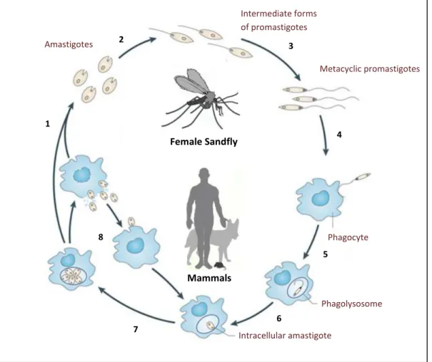
Leishmania, as well as other members of the Trypanosomatidae family, have a digenetic life cycle during which they suffer major morphological and biochemical modifications (Figure 1.1) that are associated to two distinct life stages: i) the promastigote, an elongated, extracellular single flagellated form that resides inside the alimentary tract of the vector, and ii) the amastigote, a non-motile aflagellated round shaped form that thrives inside macrophages of the host.
When an insect vector feeds on the blood of an infected host it ingests amastigote-containing macrophages and initiates the infection cycle. Inside the insect digestive tract, amastigotes are released from macrophages and immediately begin to differentiate into promastigostes. On the subsequent 6-10 days, significant morphological and biochemical changes occur, ultimately culminating in the migration of the promastigote infective form (metacyclic promastigotes) into the thoracic midgut and proboscis of the sandfly. By this time, if the infected vector feeds on a mammalian host, it will deposit several thousands of metacyclic promastigotes into its skin [35]. Parasite inoculation immediately triggers an immune response with the concomitant recruitment of phagocytic cells, like neutrophils, dendritic cells and macrophages, to the site of inoculation. Leishmania can subsequently be phagocytized by any of these cells, through a receptor-mediated phagocytosis mechanism. The role of neutrophils and dendritic cells during Leishmania invasion is still disputed. However, it is within mononuclear phagocytes that the parasite replicates and persists [36,37]. Inside these cells, parasites reside in phagolysosomes (the outcome of the fusion of a phagosome with a lysosome), where they revert into amastigotes and multiply by binary fission. Massive multiplication in these acidic vacuoles eventually leads to macrophage burst, after which parasites can be phagocytized again by other macrophages, in this way perpetuating the amastigote cycle inside the host [38].
Introduction
10
Figure 1.1. The Leishmania life cycle. During a blood meal, the female sandfly ingests macrophages containing amastigotes that are released in the insect midgut (1). Amastigotes differentiate into promastigotes (2) and these go through a series of differentiation phases that culminate in metacyclic infective forms (3). The infected sandfly feeds on a mammalian host, injecting metacyclic promastigotes into the host skin (4), where they are internalized by phagocytic cells (5). Parasite-containing phagosomes fuse with lysosomes, forming phagolysosomes, wherein promastigotes revert into amastigotes (6) and begin to replicate by binary fission (7), leading to the burst of phagocytes. Released amastigotes can be taken up by other phagocytes, in this way perpetuating the amastigote stage inside the mammalian host (8). Adapted from Kaye et al. [39].
3. Quest for new chemotherapies against leishmaniasis
3.1. Strategies for the discovery of new drugs
The unsatisfactory traits of current therapies against leishmaniasis, together with disease dissemination and the increasing reports of drug-resistant parasites, renders the development of new leishmanicidals a mandatory issue. The urgency to find new drugs arises as a major concern to researchers all around the world and efforts to increase the drug pipeline has been made in the last few years, mostly at the academic level. Two main strategies have been applied to pursuit this goal. One employs high-throughput drug screening trials, sometimes using compounds already established in the market, in order to find antiparasitic drugs. Selected compounds can then be exploited to understand their specific mode of action and, if necessary
2
3
1
4
8
5
6 7
Female Sandfly
Mammals
Intermediate forms of promastigotes
Metacyclic promastigotes
Phagocyte
Phagolysosome
Introduction
11
and possible, considered to be chemically modified to increase target specificity [40]. The other strategy, known as rational drug design, consists in identifying molecular targets that are essential to parasite survival, so that their depletion or inactivation leads to parasite death. In this context, a deep knowledge of the parasite biochemistry is crucial to find a good candidate that can be inhibited by drug-like compounds inside the macrophages of the mammalian host. This approach employs several interdisciplinary techniques, from molecular/cell biology to biochemistry and computer sciences, to assess the likelihood of a molecule to be used as drug target. The appraisement of a certain gene candidate to serve as a drug target includes its validation both at the genetic and the chemical level, as detailed next.
Genetic validation consists in evaluating the essentiality of a given gene during the life cycle of the parasite, resorting to common molecular and genetic techniques. Abrogation of gene expression is pursued and associated phenotypic changes evaluated. In the case of most
Leishmania spp., where interference RNA or inducible expression systems are either absent or still deficient, this is usually achieved by a classical gene knockout strategy. This consists in replacing the gene of interest by other that confers resistance to a toxic drug. Since Leishmania
are diploid organisms, elimination of both alleles requires two rounds of homologous recombination with different drug resistance markers. Following this approach, there are two possible outcomes: i) parasites grow normally throughout their complete life cycle, with no apparent phenotypic alterations, indicating that the target gene is not essential for parasite survival, hence not suitable as drug target; ii) elimination of both gene alleles is not compatible with parasite life, indicating that the gene may be further exploited for therapeutic purposes [41].
Chemical validation is a complement of the genetic validation. It consists of using a known inhibitor of the target molecule to verify whether its inactivation affects parasite viability. This approach addresses important “druggability” issues, namely the possibility of blocking the target molecule with low effective concentrations (EC50) of drug like-compounds
in the cell context to impair parasite survival.
In short, following a rational drug design strategy, a gene to be regarded as a valid target for drugs has to fulfill the requirements of essentiality, exclusiveness and susceptibility to inhibition in the medically relevant life stage of the parasite.
3.2. The trypanothione system as a potential target for drugs
Introduction
12
largely accomplished by the trypanosomatids’ unique thiol, N1,N8-bis(glutathionyl)spermidine, also known as trypanothione [T(SH)2]. This molecule is the major low molecular mass thiol in
trypanosomatids andplays a pivotal role in many essential metabolic pathways [reviewed in 42]. Trypanothione was first described in 1985 by Alan Fairlamb and co-workers in C. fasciculata, a member of the trypanosomatid family that is noninfectious to mammals [43]. It is a small dithiol, formed by one spermidine molecule (a cationic polyamine) with two GSH molecules covalently bound to its terminal amino groups. Originally considered as a co-substrate of GR, the actual role of T(SH)2 was only elucidated upon the finding that
typanosomatids do not possesses GR and that, instead, harbor a trypanothione reductase (TR) enzyme. It is now well established that in trypanosomatids the T(SH)2/TR system replaces most
of the functions played by the GSH/GR redox couple in mammalian cells. Even though GSH is also present in trypanosomatids, it is thought to play a minor role in the redox metabolism of parasites, as all enzymes that in mammals depend on GSH are either absent in these organisms (e.g. GR and selenium-containing GSH peroxidases) or preferentially utilize T(SH)2 as reducing
substrate [44,45].
When compared with GSH, T(SH)2 is a more reactive, positively charged molecule
capable of spontaneously reducing different biological disulfides [46]. This higher reactivity is not attributed to its redox potential (-242 mV), which does not significantly differ from that of GSH (-230 to -250 mV) [47]. Rather, it is the pKa value of the thiol groups of trypanothione
(7.4) that contributes to its increased reactivity relative to that of GSH (pKa of 8.7-9.2) [48].
This is explained by the fact that thiol-disulfide exchange reactions are highly favored when the pKa value of the thiol is proximal to the pH value of the surrounding medium. Moreover, the
dithiol nature of T(SH)2 allows the formation of an intramolecular disulfide bond that is
kinetically favored relative to intermolecular disulfide bonds, as occurs in oxidized GSH (GSSG) [49].
Trypanothione is directly or indirectly involved in many metabolic processes (Figure 1.2), from redox state regulation (by maintaining thiol redox homeostasis) to elimination of toxic compounds (including detoxification of reactive oxygen species and extrusion of toxic compounds via a trypanothione-S-transferase) [50-52] and DNA replication [by providing reducing equivalents to ribonucleotide reductase and to the universal minicircle sequence binding protein (UMSBP)] [53,54]. Many of these T(SH)2-dependent pathways have been
genetically validated as drug targets in the past [42]. Accordingly, inhibition of trypanothione biosynthesis should disturb many of these vital metabolic routes, eventually leading to parasite death. This, added to the unique character of this thiol, renders the T(SH)2 biosynthesis pathway
Introduction
13
3 3
4
5
6
7
8
9 10
GSP Spermidine
TRYS or
GSPS
GSH, Mg-ATP
GSH, Mg-ATP TRYS
1
1 2
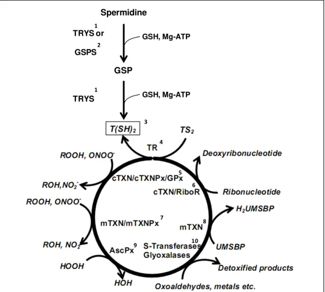
Figure 1.2. The trypanothione system.
1. De novo synthesis of trypanothione [T(SH2)] is achieved by the ATP-dependent conjugation of two
GSH molecules to spermidine. This reaction is catalyzed by trypanothione synthetase (TRYS), which in T. brucei was proven to be essential for parasite survival [55,56].
2. The first GSH conjugation can also be catalyzed by the GSPS enzyme to produce N1 or N8 -mono(glutathionyl)spermidine (GSP). This compound have been suggested to replace some of the T(SH)2
functions [57-59].
3. Trypanothione is the major thiol of trypanosomatids and is essential to many vital pathways. It delivers reducing equivalents to the enzymes of this system.
4. Trypanothione reductase (TR) is a flavin-containing disulfide reductase responsible for maintaining T(SH)2 in the reduced state. This enzyme was shown to be essential for the survival of several
trypanosomatids and is one of the most exploited drug targets in these organisms [60].
5. Trypanothione reduces cytosolic tryparedoxins (cTXN) [61], which provide the reducing equivalents to the peroxiredoxin-type tryparedoxin peroxidases (cTXNPx) [62,63] and to non-selenium glutathione peroxidase-like enzyme (cGPx) [64], the enzymes responsible for detoxification of a broad spectrum of peroxides. cTXN was shown to be essential to trypanosomatids survival [65-67].
6. Trypanothione directly, or via cTXN, reduces ribonucleotide reductase (RiboR), which is involved in DNA synthesis and repair, therefore implicating T(SH)2 in cell division [53].
7. A mitochondrial tryparedoxin (mTXN) provides reducing equivalents to the mitochondrial TXNPx (mTXNPX) and GPx (mGPx), which is responsible for elimination of reactive oxygen species [50].
8. Trypanothione, via mTXN, reduces the universal minicircle sequence binding protein (UMSBP), which is indispensable for the replication of kDNA [54].
9. Trypanothione reduces ascorbate, which serves as substrate of ascrobate peroxidase (AscPX) [68,69].
10. Trypanothione is involved in detoxification of xenobiotics via the activity of an S-transferase [51,52], oxoaldehydes via the glyoxalase system and also in detoxification of heavy metals [70,71].
Introduction
14
3.3. Trypanothione biosynthesis
As mentioned above, T(SH2) is formed by one spermidine and two GSH molecules.
The metabolic pathways of these two components are similar to those of mammalian cells. In the case of GSH, this small thiol is synthesized by the sequential conjugation of cysteine, glutamic acid and glycine residues, involving two distinct enzymes, namely glutamate cysteine ligase (GCL) and glutathione synthetase. As for spermidine, its biosynthesis starts with L
-arginine conversion to L-ornithine by arginase (ARG), followed by decarboxylation by ornithine decarboxilase (ODC) to yield putresine. In parallel, S-adenosylmethionine (AdoMet) is also decarboxylated by the S-adenosylmethionine decarboxylase (AdoMetDC) to originate decarboxylated AdoMet (dAdoMet). Finally, putresine and dAdoMet are used to generate spermidine in a reaction catalyzed by spermidine synthetase (SPDS) [reviewed in 72].
In trypanosomatids, spermidine and GSH pathways intercept to generate T(SH)2. The
enzyme responsible for the biosynthesis of this molecule is trypanothione synthetase (TRYS), a monomeric enzyme with a molecular weight ranging from 71 kDa in T. brucei to 75 kDa in C. fasciculata [73-76]. This enzyme comprises two distinct catalytic domains, a C-terminal synthetase domain displaying an ATP-grasp family fold, common to C:N ligases [77,78], and a
N-terminal amidase domain belonging to the cysteine, histidine-dependent
amidohydrolase/peptidases (CHAP) superfamily [79]. Trypanothionesynthesis is achieved by two sequential ATP-dependent conjugations of GSH molecules to spermidine, via a mono(glutathionyl)spermidine intermediate. Each conjugation step spends one ATP molecule to form a peptide bond between the carboxyl group of the glycine residue of GSH and the N1 or the
N8 amino group of spermidine, releasing ADP and orthophosphate, trough a concerted substitution mechanism [76]. Importantly, the amidase domain of TRYS is capable of hydrolyzing T(SH)2 to restore GSH and spermidine by a nucleophilic attack mechanism, again
forming mono(glutathionyl)spermidine as intermediate. How these opposing functions are regulated to avoid cells to enter in a non-productive ATP consuming futile-cycle is still unknown. However, some suggestions have been made that point for an active role of this enzyme in the regulation of free polyamine and GSH levels under certain growth conditions [80].
Trypanothione synthetase is present in all members of the Trypanosomatidae family and was validated as a drug target in T. brucei [55,56]. However, TRYS exhibits differences within this family and sometimes even between species of the same genus. Illustrating this, T. brucei
synthesizes T(SH)2 as described above with no evidence of other substrate affinity, whereas T.
Introduction
15
L. infantum and T. cruzi, GSPS is capable of catalyzing half of the synthesis reaction of TRYS, originating the N1/N8-mono(glutathionyl)spermidine (GSP) intermediate as a final product [74,80,81].
3.4. Mono(glutathionyl)spermidine biosynthesis
Glutathionylspermidine synthetase, catalyzes the formation of
mono(glutathionyl)spermidine by the ATP-consuming conjugation of one molecule of
spermidine with GSH, through a mechanism similar to that of TRYS [82]. Like TRYS, GSPS
possesses C-terminal synthetase and N-terminal amidase domains, which are capable of
synthesizing mono(glutathionyl)spermidine and hydrolyzing both
mono(glutathionyl)spermidine and T(SH)2 to GSH and spermidine [74,80,83,84]. This enzyme
was first found in E. coli as a 138 kDa homodimer [85,86] and latter in C. fasciculata as a monomeric 80 kDa protein [80]. The enzyme product is found in all parasites of the Trypanosomatidae family, even in species lacking GSPS. This occurrence is attributed to the fact that TRYS can generate mono(glutathionyl)spermidine either as a product of incomplete T(SH)2 biosynthesis or as a hydrolysis product of T(SH)2 [74-76,80]. Although the functional
relevance of mono(glutathionyl)spermidine is not completely elucidated, it has been attributed a role in cell growth modulation, by regulating polyamines availability [83,87]. Additionally, there are some evidences that it can be a substrate for TR and can substitute T(SH)2 in some
metabolic functions, namely peroxide and ribonucleotide reduction [57-59].
3.5. Targeting the trypanothione system
Targeting trypanothione biosynthesis is regarded as a good strategy to fight trypanosomatids, not only because many T(SH)2-dependent enzymes have been shown essential
for parasite survival, as it is also known that many of the currently used trypanocidal drugs exert their effect by interfering (directly or indirectly) with the trypanothione system. This is the case of eflornithine (DFMO), used to treat sleeping sickness, which inhibits ODC in the early steps of spermidine synthesis [88]. Also trivalent arsenicals react directly with T(SH)2, TR or
tryparedoxin (TXN) leading to the death of parasites [59]. In addition, drugs used to treat
Chagas’ disease, like nifurtimox or benznidazole, are known to kill parasites by inducing oxidative stress that has to be counteracted by the trypanothione-dependent peroxidase system [89,90].
Introduction
16
accomplished. This fact has been attributed to the need of high inhibition levels of TR activity (95%) to impair parasite survival [93,94]. As for TRYS, its inhibition has already been shown to impair parasite viability at reasonable drug concentrations. Several classes of compounds have been discovered that inhibit TRYS of different trypanosomatids. Recently, paullones, a class of compounds used to treat cancer, was discovered to have a potent effect as inhibitor of TRYS [95]. Paullones are 7,12-dihydroindolo[3,2-d][1]benzazepin-6(5H)-ones, a class of ATP analogues known to primarily inhibit certain protein kinases, like cyclin-dependent kinases and glycogen synthetase kinase-3. Paullones synthesis was described for the first time in 1992 and since then has been applied in several medical studies from research in neurosciences to apoptosis, diabetes, cancer, embryonic development and anti-parasitic studies [96]. Some compounds of this family, namely the group of N5-substituted 2-(6-oxo-6,7-dihydro-5H -benzo[2,3]azepino[4,5-b]indol-5-yl)-acetamides, have proven to be good inhibitors of recombinant TRYS of C. fasciculata (IC50=30 nM) and killed T. brucei in culture with an LC50 near 2 μM (M. Comini, personal communication). Paullones interact with the ATP-binding domain of TRYS, causing irreversible inhibition, which is not counteracted by physiological concentrations of ATP or any other substrate [95].
4. Aims of the work
The general aim of this study was to validate the enzymatic pathway for T(SH)2
biosynthesis as a drug target in L. infantum. As mentioned before, L. infantum harbors a TRYS enzyme, which catalyzes both conjugation steps required for T(SH)2 synthesis, as well as GSPS,
the protein that drives mono(glutathionyl)spermidine formation. So far, the essential role of
TRYS for parasite survival has only been demonstrated for T. brucei, a trypanosomatid that lacks
GSPS. In L. infantum, however, it cannot be taken for granted that TRYS is also essential, as mono(glutathionyl)spermidine, may replace some of the metabolic functions of T(SH)2. Having
this in mind, the specific goal of this work was to address the essential role of both TRYS and
Chapter 2
Results
19 1. Analysis of TRYS and GSPS amino acid sequences
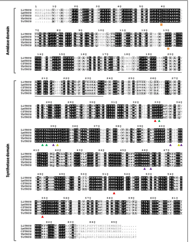
The L. infantum genome harbors a single copy of the TRYS coding sequence, which is predicted to encode a protein (LiTRYS) with 652 amino acids and a molecular mass of 74.23 kDa. Figure 2.1 shows the predicted amino acid sequence of LiTRYS and compares it to TRYS proteins of other trypanosomatids. LiTRYS exhibits 95.2% and 62.1% identity to the formerly characterized enzymes of L. major and T. brucei, respectively. Moreover, it shows 78.5% and 60.5 % identity to TRYSof C. fasciculata and T. cruzi,the species of trypanosomatids that, like
L. infantum, harbor a GSPS ORF. Trypanothione synthetase of L. infantum shares with previously established TRYS a synthetase and amidase domains, and preserves the residues that were identified as being important for substrate interaction and/or binding (Figure 2.1) [82,84,97].
Results
20
Figure 2.1. Alignment of the predicted amino acid TRYS sequences of different trypanosomatids.
Protein sequence of TRYSof L. infantum (LiTRYS, accession number A4I2Z3), L. major (LmTRYS, accession number AJ311570), C. fasciculata (CfTRYS, accession number AF006615), T. brucei (TbTRYS, accession number Q586P2) and T. cruzi (TcTRYS, accession number AF311782) are represented. The amidase and synthetase domains are indicated on the left side. Conserved and similar residues are highlighted in black background and black frames, respectively. Residues of the linker region between domains are underlined. Residues involved in substrates interaction and/or binding are indicated by arrow heads: ▲- ATP, ▲- Mg2+, ▲- GSH and ▲-spermidine. Residues involved in amidase activity are indicated with ■.
A
m
id
a
se
d
o
m
a
in
S
y
n
th
et
a
se
d
o
m
a
Results
21
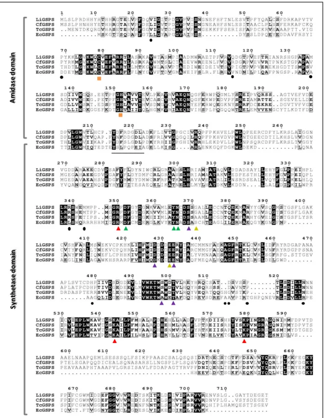
Figure 2.2. Alignment of the predicted amino acid GSPS sequences for different organisms. Protein sequence of GSPS of L. infantum (LiGSPS, accession number A4I1T8), C. fasciculata (CfGSPS, accession number U66520), T. cruzi (TcGSPS, accession number AY828997) and E. coli (EcGSPS, accession number U23148) are represented. The amidase and synthetase domains are indicated on the left side. Conserved and similar residues are highlighted in black background and black frames, respectively. Residues of the linker region between domains are underlined. Residues involved in substrates interaction and/or binding are indicated by arrow heads: ▲- ATP, ▲- Mg2+, ▲- GSH and ▲-spermidine. Residues involved in amidase activity are indicated with ■. Residues involved in EcGSPS monomer-monomer
interaction are indicated by ●.
A
m
id
a
se
d
o
m
a
in
S
y
n
th
e
ta
se
d
o
m
a
Results
22 2. Expression of TRYS along the L. infantum life cycle
Expression of TRYS was investigated by western blot throughout the life cycle of L. infantum, including promastigotes and axenically grown amastigotes. Results, depicted in Figure 2.3, show that the enzyme is expressed as a peptide of approximately 75.5 kDa in both life cycle stages of the parasite. During promastigote growth expression of TRYS gradually increases from the early logarithmic phase to the stationary phase.
Figure 2.3. Expression of TRYS during the L. infantum life cycle. Western blot analysis of total protein extracts of L. infantum, incubated with an antibody directed against the L. major TRYS. Protein extracts from early logarithmic (day 1 of culture or D1) to stationary (day 5 of culture or D5) promastigotes were analyzed, as well as from axenically grown amastigotes (A). Ponceau staining of the western blot membrane is shown as a control.
3. Genetic validation of TRYS
Essentiality of TRYS was addressed following a gene replacement strategy. Since L. infantum is a diploid organism, two rounds of gene targeting were required to target both TRYS
alleles. Accordingly, two TRYS replacement constructs were generated. To that end, part of the non-coding sequences flanking the TRYS ORF were cloned into two different plasmids, carrying either the hygromycin B phosphotransferase gene (HYG), or the phleomycin hydrolase gene (PHLEO), which confer resistance to hygromycin B and phleomycin, respectively (Figure 2.4). Prior to transfection, these constructs were linearized (by HindIII/SacI double restriction) and the replacement cassettes separated from the plasmids backbone.
D1 D3 D4 D5
Anti-Lm TryS 75.5 kDa
Ponceau
D2 A
Results
23
Figure 2.4. Schematic representation of TRYS replacement constructs. The constructs used to target
TRYS (pGL345-TRYSKØ and pGL726-TRYSKØ) are represented. Hygromycin B phosphotransferase
(HYG), phleomycin hydrolase (PHLEO), beta-lactamase (AMP) ORFs and 5’, 3’ non-coding sequences
(5’NCS and 3’NCS) flanking TRYS ORF are represented in boxes. Restriction sites used to assemble the constructs are also shown.
3.1. Generation of heterozygous trys::HYG/TRYS L. infantum parasites
The first TRYS allele was targeted with the HYG replacement cassette. Following the transfection procedure, hygromycin-resistant promastigotes were selected and individual clones tested by PCR using primers P8 and P9 (see Table 3.1 in the “Material & Methods” section). Primer P8 is a sense primer that anneals in the gene upstream of TRYS and was designed to diagnose for the correct integration of disruption constructs in the TRYS locus, whereas P9 is an anti-sense primer designed to anneal at the beginning of HYG ORF (Figure 2.5 A). Analysis of three clones testing positive for the correct integration of the HYG cassette in the TRYS locus is shown in Figure 2.5 B. These heterozygous trys::HYG/TRYS mutants (hereafter designated as
trys+/-) presented no differences regarding their morphology (not shown) and growth rate (Figure 2.5 C) when compared with wildtype parasites, indicating that one TRYS copy is sufficient to sustain promastigote growth in vitro.
HYG
AMP
3’ NCS 5’ NCS
BamHI SacI
SpeI
HindIII
pGL345-TRYSKØ
681 bp 841 bp
AMP
PHLEO 3’ NCS
5’ NCS
pGL726-TRYSKØ
681 bp 841 bp
BamHI SacI
SpeI
Results
24 A
B C
Figure 2.5. Generation of L. infantum heterozygous trys+/- promastigotes. (A) Representation of the L. infantum TRYS locus with its LinJ.27.1780 upstream gene in trys+/- parasites. Both i) wildtype and ii) HYG-targeted alleles are shown. The 5’ and 3’ NCS used to construct the HYG replacement cassette are represented in grey boxes. Primers P8 and P9 used in the PCR diagnosis for the correct integration of the HYG cassette in the TRYS locus are indicated by arrowheads. The number in between arrowheads refers to the expected size of the corresponding PCR product. (B) PCR analysis of genomic DNA from three hygromycin-resistant mutants (clones 1, 2 and 6) and wildtype parasites, using primers P8 and P9. (C)
Growth curves of wildtype (WT) and trys+/- (clone 1) promastigotes. Parasites were seeded at 106 at day 0 and counted daily with a hemocytometer. Data represent the mean and standard deviation of two independent growth curves.
3.2. Attempts to generate L. infantum TRYS null mutants
In order to replace the second TRYS allele, the trys+/- clone 1 was selected for transfection with the PHLEO replacement cassette. Following the transfection procedure, parasites resistant to both hygromycin and phleomycin were selected and three individual clones analyzed by PCR to diagnose for different events, namely (see Figure 2.6 A): i) integration of the PHLEO cassette into the TRYS locus; ii) presence of the PHLEO,HYG and TRYS ORFs; iii)
maintenance of the HYG cassette in its correct position and iv) configuration of the endogenous
TRYS locus. The results, depicted in Figure 2.6 B, show that none of the clones tested positive for the correct integration of the PHLEO cassette in the TRYSlocus. Still, the PHLEO ORF was
TRYS
1stallele
LinJ.27.1780 5’ NCS 3’ NCS
HYG LinJ.27.1780
5’ NCS 3’ NCS
P8 P9 1000 bp 1000 bp C lo n e 1 C lo n e 2 C lo n e 6 W il d ty p e
HYG integration
0 2 4 6
Results
25 1000 bp 1050 bp 1000 bp C lo n e 1 C -C lo n e 3 C lo n e 5 1950 bp 380 bpPHLEO integration (P8/P10)
HYG ORF (P11/P12)
PHLEO ORF (P13/P14)
HYG integration (P8/P9)
TRYS locus (P8/P7)
TRYS ORF (P5/P6)
1000 bp
amplified, at least in clones 3 and 5. The HYG ORF was kept in its correct location in the TRYS locus during the second transfection procedure. Importantly, all three clones retained the TRYS ORF, in its original locus (clones 3 and 5) or possibly in other position of the genome (clone 1). The failure to eliminate both gene alleles of TRYS is per se suggestive that this may be an essential gene [31].
A
B
Figure 2.6. Leishmania infantum parasites obtained upon the second round transfection with the
PHLEO replacement cassette. (A) Representation of the L. infantum TRYS locus with its LinJ.27.1780 upstream gene in i) the wildtype allele and as expected upon the recombination events with the ii) HYG and iii) PHLEO integration cassetes. The 5’ and 3’ NCS used to construct the HYG and PHLEO replacement cassettes are represented in grey boxes. Primers used in the PCR diagnosis for the different integration events in the TRYS locus, as well as to amplify the TRYS, HYG and PHLEO ORFs are indicated by arrowheads. Numbers in between arrowheads refer to the expected size of the corresponding PCR products. (B) PCR analysis of genomic DNA from three hygromycin- and phleomycin-resistant mutants (clones 1, 3 and 5). Primers used in the PCR reactions are indicated in parenthesis. The same reactions were carried out in the absence of DNA to serve as negative control (C -).
3.3. Generation of L. infantum TRYS null mutants requires previous complementation with
episomic TRYS
Following two sequential rounds of transfection we were not able to eliminate both
TRYS alleles, suggesting that this gene might be essential to promastigote survival. To rule out the possibility that technical limitations could be hindering the isolation of TRYS-depleted
TRYS LinJ.27.1780 5’ NCS
3’ NCS P27 P8 P6 P5 1000 bp 1950 bp HYG LinJ.27.1780
5’ NCS 3’ NCS
P9 P8 P12 P11 1000 bp 1050 bp
LinJ.27.1780 5’ NCS PHLEO 3’ NCS
Results
26
EpisomicTRYS (P25/P6)
HYG integration (P8/P9)
C lo n e 3 C lo n e 8 C lo n e 1 0 W il d t y p e C+ 600bp 2900 bp 1000 bp 2500 bp
pTEX R1 (P5/P10) pTEX R2 (P23/P26)
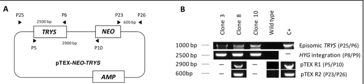
TRYS NEO AMP pTEX-NEO-TRYS 2900 bp P10 P5 P26 P23 P6 P25 600 bp 2500 bp
parasites, we followed another experimental approach. This consisted in introducing an episomal copy of the TRYS gene into trys+/- mutants prior to transfection with the PHLEO
replacement cassette. To that end the trys+/- clone 1 was transfected with the trypanosomatidal expression vector pTEX-NEO carrying the TRYS ORF (Figure 2.7 A). This vector carries the neomycin phosphotransferase gene (NEO), which confers resistence to geneticin. Following the transfection procedure, three hygromycin- and geneticin-resistant clones were confirmed by PCR (Figure 2.7 B) to carry an episomal copy of TRYS (P25/P6), as well as to preserve the HYG ORF in its the correct location in the TRYS locus (P8/P9). One of the clones (clone 8) was further tested for the integrity of the episome using different combinations of primers (P5/P10 and P23/P26). The results, depicted in Figure 2.7 B, indicated that the pTEX-NEO-TRYS
episome was intact in this clone. These trys+/- parasites carrying an extrachromosomal copy of
TRYS were subsequently used for the second round of targeting of the TRYS locus with the
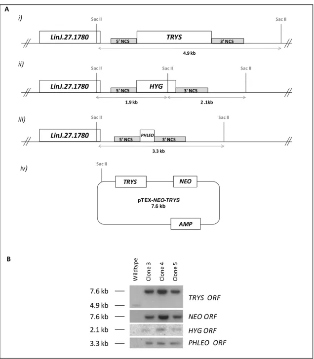
PHLEO cassette. The outcome of this transfection was the isolation of parasites resistant to hygromycin, geneticin and phleomycin. Southern blot analysis of three of these clones confirmed the successful replacement of both TRYS alleles. Illustrating this, the 4.9 kb band, which corresponds to the chromosomal TRYS copy in SacII-digested DNA (Figure 2.8 A), was not detected in any of these mutants (Figure 2.8 B). Instead, a 7.6 kb band, matching the episomal TRYS copy, was recognized by the TRYS probe. This 7.6 kb fragment also hybridized to the NEO probe, further confirming the presence of the TRYS episome. Integrity of both
HYG-and PHLEO-targeted alleles was verified using the corresponding radiolabeled-ORFs as probes. As shown in Figure 2.8 B, the HYG probe labeled one band of approximately 2 kb, which likely contains the 1.9 and 2.1 kb fragments that result from SacII restriction of the HYG allele (Figure 2.8 A). As for the PHLEO probe, it detected a 3.3 kb band, as expected for the correct integration of the PHLEO cassette into the TRYS locus. These Δtrys::HYG/Δtrys::PHLEO
[pTEX-NEO-TRYS] mutants will be from here on be designated as trys-/-/+TRYS.
Figure 2.7. Analysis of pTEX-NEO-TRYS complemented trys+/- mutants. (A) Schematic representation of the pTEX-NEO-TRYS plasmid. Trypanothione synthetase (TRYS), neomycin phosphotransferase (NEO) and beta-lactamase (AMP) ORFs are represented in boxes. Primers used in the PCR analysis in (B) are indicated by arrowheads. Numbers in between arrowheads refer to the expected size of the corresponding PCR products. (B) PCR analysis of genomic DNA from three independent pTEX-NEO-TRYS complemented trys+/- mutants (clones 3, 8 and 10) and wildtype promastigotes. Purified pTEX-NEO-TRYS plasmid was used as template to serve as positive control in some of the reactions (C +). Primers used in the PCR reactions are indicated in parenthesis and their location is represented in (A), except for primers P8 and P9, whose binding site is depicted in Figure 2.6.
Results
27 TRYS NEO AMP pTEX-NEO-TRYS 7.6 kb Sac II 4.9 kb 7.6 kbTRYS ORF
W il d ty p e C lo n e 3 C lo n e 4 C lo n e 5
7.6 kb NEO ORF
2.1 kb HYG ORF
3.3 kb PHLEO ORF
A
B
Figure 2.8. Southern blot analysis of trys-/-/+TRYS mutants. (A) Representation of the L. infantum TRYS locus with its LinJ.27.1780 upstream gene i) before and after TRYS replecement by the ii) HYG and iii) PHLEO cassettes. iv) Also depicted is the scheme of the pTEX-NEO-TRYS episome. Sizes of the predicted SacII-restriction fragments are indicated. (B) Southern blot of SacII-digested genomic DNA of wildtype and three trys-/-/+TRYS mutants (clones 3, 4 and 5) hybridized with radiolabeled NEO, TRYS, HYG and PHLEO ORFs.
3.4 Maintenance of the pTEX-NEO-TRYS episome in the absence of drug pressure confirms
gene essentiality in promastigotes
Leishmania episomes are highly unstable and are usually lost over time unless they provide an advantage to the parasite. The pTEX-NEO vector used to introduce the TRYS
episomal copy is no exception to this and, as such, it is lost when parasites are cultured in the TRYS
LinJ.27.1780
5’ NCS 3’ NCS
Sac II Sac II
4.9 kb
LinJ.27.1780
Sac II Sac II
5’ NCS PHLEO 3’ NCS
3.3 kb
LinJ.27.1780
Sac II Sac II
HYG
3’ NCS 5’ NCS
1.9 kb 2 .1kb
Sac II
i)
ii)
iii)