Genotype-Specific Interaction of Latent TGF
β
Binding Protein 4 with TGF
β
Kay-Marie Lamar1, Tamari Miller1, Lisa Dellefave-Castillo2, Elizabeth M. McNally1,2*
1Department of Human Genetics, The University of Chicago, Chicago, Illinois, United States of America, 2Center for Genetic Medicine, Northwestern University Feinberg School of Medicine, Chicago, Illinois, United States of America
Abstract
Latent TGFβbinding proteins are extracellular matrix proteins that bind latent TGFβto form the large latent complex. Nonsynonymous polymorphisms inLTBP4, a member of the latent TGFβbinding protein gene family, have been linked to several human diseases, underscor-ing the importance of TGFβregulation for a range of phenotypes. Because of strong linkage disequilibrium across theLTBP4gene, humans have two mainLTBP4alleles that differ at four amino acid positions, referred to as IAAM and VTTT for the encoded residues. VTTT is considered the“risk”allele and associates with increased intracellular TGFβsignaling and more deleterious phenotypes in muscular dystrophy and other diseases. We now evaluated
LTBP4nsSNPs in dilated cardiomyopathy, a distinct disorder associated with TGFβ signal-ing. We stratified based on self-identified ethnicity and found that theLTBP4VTTT allele is associated with increased risk of dilated cardiomyopathy in European Americans extending the diseases that associate withLTBP4genotype. However, the association ofLTBP4
SNPs with dilated cardiomyopathy was not observed in African Americans. To elucidate the mechanism by whichLTBP4genotype exerts this differential effect, TGFβ’s association with LTBP4 protein was examined. LTBP4 protein with the IAAM residues bound more latent TGFβcompared to the LTBP4 VTTT protein. Together these data provide support thatLTBP4genotype exerts its effect through differential avidity for TGFβaccounting for the differences in TGFβsignaling attributed to these two alleles.
Introduction
Latent TGFβbinding protein 4 (LTBP4) is part of a family of extracellular proteins including LTBPs 1–3 as well as the fibrillins [1,2]. Members of this family are characterized by the pres-ence of multiple epidermal growth factor-like repeats, and conserved 8-cysteine domains. LTBP4is expressed at high levels in the heart, skeletal and smooth muscle but also shows lower level expression in other tissues [1,2]. Latent TGFβis held in an inactive state in the extracellu-lar matrix as part of a extracellu-large latent complex (LLC) consisting of TGFβ, its latency associated peptide and LTBP. The regulation of TGFβis tightly controlled, and in order to become active, TGFβmust be free of both latency associated peptide and LTBP. Proteolysis of LTBP or force-OPEN ACCESS
Citation:Lamar K-M, Miller T, Dellefave-Castillo L, McNally EM (2016) Genotype-Specific Interaction of Latent TGFβBinding Protein 4 with TGFβ. PLoS ONE 11(2): e0150358. doi:10.1371/journal. pone.0150358
Editor:Nanette H Bishopric, University of Miami School of Medicine, UNITED STATES
Received:September 10, 2015
Accepted:February 12, 2016
Published:February 26, 2016
Copyright:© 2016 Lamar et al. This is an open access article distributed under the terms of the Creative Commons Attribution License, which permits unrestricted use, distribution, and reproduction in any medium, provided the original author and source are credited.
Data Availability Statement:All relevant data are within the paper and its Supporting Information files.
Funding:This work was supported by the National Institutes of Health, NS047726, HL128075.
Competing Interests:The authors have declared that no competing interests exist.
induced release of TGFβby LTBP results in liberation of the active TGFβdimer, engagement of cell surface receptors and induction of intracellular downstream signaling [3,4]. In addition to regulating the release of TGFβ, LTBP also participates in the assembly and secretion of TGFβ[5,6].
TGFβis a multifunctional molecule that regulates growth, development, and response to injury. Three TGFβisoforms, TGFβ1, 2 and 3, are highly conserved, with between 70–80% identity in their active domain. Despite high similarity, the TGFβisoforms have different spa-tiotemporal expression during development, as well as during wound healing [7,8]. In wound healing, these TGFβfamily members have been implicated in inflammation, proliferation, and tissue remodeling [9]. TGFβfamily members also directly regulate matrix deposition and fibro-sis by stimulating production of components such as fibronectin and collagen and simulta-neously downregulating matrix-degrading proteases [10–12]. Excessive fibrosis and TGFβ
signaling are found in a number of chronic pathological processes including muscular dystro-phy, liver cirrhosis, and idiopathic pulmonary fibrosis [13,14]. In these disorders, increased or
“hyper-TGFβ”signaling leads to accumulated matrix-associated proteins, scarring and fibrosis. TGFβalso undergoes auto-induction, which further amplifies its effects [8].
Non-synonymous single nucleotide polymorphisms (SNPs) inLTBP4have been associated with pathogenicity in several distinct human disorders. In humans with Duchenne Muscular Dystrophy (DMD),LTBP4genotype has been associated with prolonged ambulation in multi-ple cohorts [15–17]. In chronic obstructive pulmonary disease,LTBP4SNPs have been linked to improved exercise capacity, including increased six-minute walk test distance and greater maximum work capacity [18]. SNPs inLTBP4have also been associated with reduced expan-sion of abdominal aortic aneurysm, and less aggressive tumors in colorectal cancer [18–20].
Dilated cardiomyopathy (DCM) is genetically heterogeneous and is often characterized by fibrosis and abnormal TGFβsignaling [21,22]. Polymorphisms inTGFB1have been associated with heart failure caused by DCM, and TGFβis upregulated in the plasma and myocardium of DCM patients [23–26]. In order to assess whetherLTBP4contributes to DCM disease risk, we now genotypedLTBP4polymorphisms in cases and controls and found an overabundance of risk alleles in European American DCM subjects. To assess the biological effects of the two most commonLTBP4alleles in the human genome, we co-expressed LTBP4 protein along with TGFβ. We found that LTBP4 protein expressed with the protective four amino acids, IAAM, associated with more TGFβcompared to LTBP4 expressing the deleterious residues VTTT. In this model, decreased affinity ofLTBP4for latent TGFβaccounts for the increased TGFβand TGFβsignaling seen with the VTTT allele. Together these findings provide a molec-ular mechanism by whichLTBP4modifies chronic fibrotic disorders.
Results
LTBP4
SNPs are in linkage disequilibrium and associate with disease
phenotypes linked to TGF
β
signaling
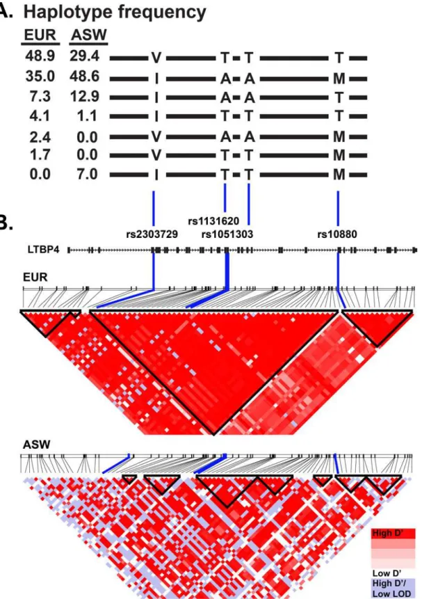
There are several common nonsynonymous SNPs in the humanLTBP4gene, and these include rs2303729, rs1131620, rs1051303, and rs10880 [27,28]. These SNPs are in strong linkage dis-equilibrium forming two major haplotypes (Fig 1). One haplotype encodes the amino acids Valine, Threonine, Threonine, and Threonine, referred to as VTTT, and the other encodes Iso-leucine, Alanine, Alanine, and Methionine, referred to as IAAM. The frequency of the VTTT haplotype in Europeans is 49%, and the IAAM allele frequency is 35%, while minor haplotypes, deviating at one or more of the SNPs contribute to the remaining 16% (Fig 1).
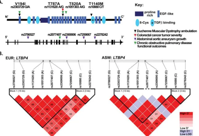
SNPs that constitute theLTBP4haplotype, rs10880 was the most significantly associated with prolonged ambulation [16]. In addition to DMD,LTBP4SNPs have also been associated with other disease phenotypes (Fig 2A). The nsSNP 1131620 (Thr787Ala) was associated with more aggressive tumors in colorectal cancer [20]. An enhanced growth rate of abdominal aortic aneurysms was linked to several intronic SNPs inLTBP4including rs3786527, rs2369006, rs7259067, rs2278242, and a borderline association with the presence of abdominal aortic aneurysms was found for synonymous SNP rs2077407 [19]. These intronic and synonymous SNPs are in high linkage disequilibrium with the VTTT haplotype (Fig 2B). In a separate study, reduced work capacity, lower exercise tolerance, and shorter 6-minute walk test distance in chronic obstructive pulmonary disease were associated with SNPs rs2303729, rs1131620, rs1051303, and rs2077407 [18]. In each of these cases, theLTBP4haplotype that specifies the VTTT residues was considered the risk allele for the phenotype of interest, and correspond-ingly the IAAM haplotype was considered the protective allele. Fibroblasts with the VTTT allele were found to have enhanced TGFβsignaling, while the IAAM allele was associated with reduced TGFβsignaling [16]. Together these data support that increased TGFβsignaling asso-ciates with increased disease burden in these disorders.
Fig 2. Single nucleotide polymorphisms (SNPs) inLTBP4modify many human diseases. A.The protein schematic of human LTPB4 protein, with arrowheads indicating the position of the non-synonymous SNPs (nsSNPs) that have been reported to associated with disease severity [15–20]. TheLTBP4
genomic locus is shown below with the vertical black bars representing exonic regions and horizontal black line representing intronic portions. Arrowheads indicate intronic and synonymous SNPs inLTBP4that modify human disease.B.Linkage disequilibrium map from Haploview of nineLTBP4SNPs previously associated with disease phenotypes in chronic obstructive pulmonary disease, colorectal cancer, abdominal aortic aneurysm, and Duchenne muscular dystrophy. This map was created using data from the 1000 Genomes EUR (European American) population or the ASW population (African Americans from Southwestern United States). Pair-wise D’values are shown in the boxes. Colors are based on the standard D’/LOD option in the Haploview software.
The
LTBP4
VTTT allele is associated with DCM in European Americans
Aberrant TGFβsignaling is also a feature of DCM, a disorder frequently associated with fibro-sis [21,22]. In order to identify genes that are co-expressed withLTBP4,LTBP4was evaluated using the Co-Regulation Database (CORD) [29].LTBP4co-regulated genes were highly expressed in the heart (p = 2.4x10⁻¹²) and over representative for the DCM KEGG pathway (p = 0.0007), positioningLTBP4in the etiology of DCM (S1–S3Tables). Furthermore, mice lacking the short isoform ofLtbp4(Ltbp4S-/-) develop cardiomyopathy with biventricular dila-tation [30]. In order to assess the effect ofLTBP4genotype on DCM risk, we genotypedLTBP4 SNPs in DCM subjects and unaffected controls. We genotypedLTBP4SNPs in African Ameri-can (AA) and European AmeriAmeri-can (EUR) DCM patients and compared each group to ethnic-ity-matched controls. Disease severity was comparable between the two cohorts of DCM patients. The left ventricular ejection fraction (LVEF) was 27.07±10.03 in AA DCM and 28.01 ±6.94 in EUR DCM (p = 0.49), and the age at which these measurements were made was com-parable 42.06±1.50 years versus 44.87±1.60 years (p = 0.228).
The frequency of the VTTT allele was increased in DCM patients compared to controls in EUR DCM (Table 1). The G allele of rs2303729 was found at a frequency of 0.712 in EUR DCM patients compared to 0.514 in healthy controls (OR = 2.259, p<0.0001). Similarly, the A allele of rs1131620 and the A allele of rs1051303, were found at frequencies of 0.720 in EUR DCM patients and 0.564 or 0.553 in controls, respectively (OR = 2.004, p = 0.0004; OR = 2.090, p = 0.0002). Finally, the C allele of rs10880 was found at a frequency of 0.721 in EUR DCM patients and 0.609 in ethnicity-matched controls (OR = 1.653, p = 0.0176). In AA with DCM, no association was observed betweenLTBP4nsSNPs and DCM status (range of p values from 0.200 to 0.996, depending on SNP,Table 2).
The frequency ofLTBP4SNPs varies with ethnicity and indeed was observed to vary slightly even among African American control cohorts. Potential admixture differences between sub-jects and controls led us to genotype two additional control DNA sets from AA populations as well as examine public databases. Adult controls, composed of adults 20–60 years of age from the Chicago area, were genotyped for rs1051303 and rs1131620. Pediatric controls from the same geographic area were genotyped for rs1051303 and rs1131620, and rs10880. No signifi-cant differences in allele frequency between African American DCM patients and controls were observed (Table 2). The frequencies in these local populations were compared with public
Table 1. LTBP4nsSNPs are associated with DCM in European Americans.
SNP Reporting frequency of“Risk”Allele (VTTT)
rs2303729 (G) rs1131620 (A) rs1051303 (A) rs10880 (C)
Population n V194I T787A T820A T1140M
DCM Patients (EUR) 122 .712 .72 .72 .721
1000 Genomes EUR 1006 .536 .548 .548 .608
Coriell Controls 200 .515 .551 .551 .613
NHLBI Exome Sequencing Project (ESP) 8600 .546 .578 .582
-Exome Aggregation Consortium (ExAC) (EUR) 73354 .514 .564 .553
-Controls combined†
.514 .564 .553 .609
Odds Ratio (95% CI) 2.259 (1.530–3.334) 2.004 (1.349–2.978) 2.090 (1.407–3.107) 1.653 (1.087–2.511)
χ2p-value <0.0001 0.0004 0.0002 0.0176
DCM = Dilated cardiomyopathy, EUR = 1000 Genomes European super population, CI = confidence interval.
†ExAC and Coriell controls were combined for rs2303729, rs1131620, and rs1051303. 1000 Genomes EUR and Coriell controls were combined for rs10880. Rs10880 has low sequencing coverage in exome sequencing in ESP and ExAC.
databases ofLTBP4SNP frequencies including the NHLBI Exome Sequencing Project, which includes 4300 EUR and 2300 AA individuals. The first threeLTBP4nsSNPS were included in this analysis since their depth of sequencing was adequate to reliably interpret genotype. SNP rs10880 was excluded in this analysis since this region is not adequately covered by exome sequencing. Taking these data into account yields similarLTBP4SNP frequencies as seen in the local control cohorts and confirms the association ofLTBP4SNPs with DCM was only observed for EUR DCM subjects.
LTBP4 IAAM interacts with TGF
β
1 more than LTBP4 VTTT
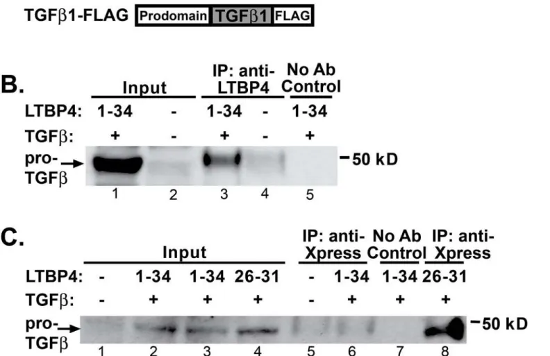
Latent TGFβbinding proteins regulate the availability of the small latent complex of TGFβ, and increased TGFβis associated with fibrosis in multiple diseases, including muscular dystro-phy [13,31]. To assess LTBP4’s interaction with latent TGFβ, co-immunoprecipitation experi-ments were carried out. Human embryonic kidney (HEK) 293T cells were co-transfected with constructs expressing the long isoform of LTBP4 and latent TGFβ1 (Fig 3A). Cells were lysed, and proteins were separated and then immunoprecipitated using an antibody to LTBP4 demonstrat-ing that TGFβ1 associates with LTBP4 (Fig 3B, lane 3). A control experiment without the LTBP4 antibody showed that the interaction with TGFβwas dependent on LTBP4. In order to refine the region of LTBP4 responsible for binding TGFβ1, amino- and carboxy-terminal fragments of LTBP4 were individually expressed and tested. The carboxy-terminal fragment encoded by exons 26–31 ofLTBP4includes the 3rdand 4th8-cysteine domains and two epidermal growth factor-like repeats (Ex26-31) (Fig 3A). This fragment of LTBP4 showed robust interaction with TGFβ(Fig 3C, lane 8). The faint 50 kDa band present in the untransfected lane (lane 5) corresponds to low level cross reactivity to the antibody 50 kDa heavy chain used for immunoprecipitation. Cells transfected with full length LTBP4 and TGFβ1 show only the heavy chain contamination (lane 6), but this negative result is attributed to the observation that full length LTBP4 is not efficiently
Table 2. LTBP4SNPs have similar frequency in African American DCM subjects and controls.
SNP Reporting frequency of“Risk”Allele (VTTT)
rs2303729 (G) rs1131620 (A) rs1051303 (A) rs10880 (C)
Population n V194I T787A T820A T1140M
DCM Patients (African American) 280 .304 .432 .432 .496
1000 Genomes ASW 122 .303 .385 .385 .434
Coriell Controls 200 .38 .439 .439 .54
Cord Blood Controls 142 - .433 .431 .537
TRIDOM Controls 396 - .435 .438
-NHLBI Exome Sequencing Project (ESP) 4406 .340 .437 .432
-Exome Aggregation Consortium (ExAC)
AFR§ 10406 .325 .426 .417
-Controls combined†
.341 .435 .432 .511
Odds Ratio (95% CI) 0.8424 (0.6478–
1.096)
0.9872 (0.7743– 1.259)
1.001 (0.7847– 1.276)
0.9715 (0.7184– 1.314)
χ2p-value 0.2002 0.9172 0.9960 0.8509
DCM = Dilated cardiomyopathy, ASW = African Americans from Southwestern United States §ExAC AFR-Includes Africans and African Americans and was not included in the p-value calculation.
†
Coriell controls, Cord Blood controls, TRIDOM controls, 1000 Genomes ASW, and ESP controls were combined for rs2303729, rs1131620, and rs1051303. 1000 Genomes ASW, Coriell controls, Cord Blood Controls, and TRIDOM controls were combined for rs10880. Rs10880 has low sequencing coverage in exome sequencing in ESP and ExAC.
pulled down with the anti-Xpress antibody. The amino-terminal region of LTBP4 did not produce a detectable association with TGFβ1 (data not shown).
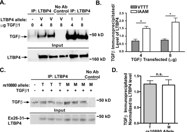
Fibroblasts with the IAAM allele ofLTBP4have lower levels of phosphorylated SMAD than cell lines carrying the VTTT allele [16]. In order to understand these differences in TGFβ sig-naling, LTBP4 proteins expressing these two haplotypes, IAAM and VTTT, were tested for binding to latent TGFβ. HEK293T cells were co-transfected with two different concentrations of plasmid expressing latent TGFβ1 and a construct expressing either the VTTT or IAAM allele of LTBP4 (V and I, respectively,Fig 4). Transfections and immunoprecipitations were done in
Fig 3. TGFβ1 binds the carboxy terminus of LTBP4. A.HEK293T cells were transfected with human constructs to express LTBP4 and TGFβ1.B. Immunoprecipitation was performed from cell lysates by immunoprecipitating with an anti-LTBP4 antibody followed by immunoblotting with anti-FLAG antibody, which detected an interaction between LTBP4 and pro-TGFβ1. The immunoblot shows that full length LTBP4 binds TGFβ1.C. Co-immunoprecipitation was performed on cell lysates by immunoprecipitating with anti-Xpress antibodies to the epitope tag on LTBP4 followed by immunoblotting with anti-FLAG antibody detecting TGFβ1. The carboxy terminal fragment of LTBP4 associated with TGFβ1. Controls were performed without adding immunoprecipitating antibody.
triplicate. Cell lysates were immunoprecipitated using an antibody to LTBP4 and immuno-blotted using an antibody to the FLAG tag on TGFβ1 (Fig 4A). The amount of TGFβ1 immu-noprecipitated was quantified and normalized to the amount of LTBP4 expression (Fig 4B). Cells lines expressing the IAAM protective allele of LTBP4 immunoprecipitated more TGFβ1 than LTBP4 with the VTTT allele when transfected with either 4μg TGFβ1 (p = 0.0106) or 8μg TGFβ1 (p = 0.0146). These data are consistent with a model in which the VTTT allele associ-ates less tightly to latent TGFβ, leading to increased available latent TGFβand therefore increased TGFβsignaling. The rs10880 SNP falls near the TGFβbinding domain of LTBP4, therefore we tested whether this SNP alone would result in differential binding of TGFβ. The exon 26–31 portion of LTBP4 was mutated to include the reference allele of rs10880 (encoding Threonine or T) or the protective allele (encoding Methionine or M). These constructs were transfected into HEK293T cells along with TGFβ1. Cell lysates were immunoprecipitated using an antibody to the Xpress tag on LTBP4, followed by immunoblotting for the FLAG tag on TGFβ1 (Fig 4C). When normalized to LTBP4 protein levels, no significant difference was seen (Fig 4D). This finding does not exclude the possibility that this variant mediates an important role in the interaction, as any single amino acid substitution may need to act in concert with the remaining protein to see this effect.
Fig 4. LTBP4-IAAM binds more TGFβ1 than LTBP4-VTTT. A.HEK293T cells were co-transfected with constructs expressing pro-TGFβ1 and either LTBP4-VTTT (V) or LTBP4-IAAM (I). Cell lysates were immunoprecipitated using an antibody to LTBP4 and immunoblotted using an antibody to the FLAG tag on TGFβ1. Both LTBP4-IAAM and LTBP4-VTTT bound pro-TGFβ1.B.The amount of TGFβ1 immunoprecipitated was quantified and normalized to the amount of LTBP4 expression (experiments performed in triplicate). Cells lines carrying the IAAM protective allele of LTBP4 immunoprecipitate more TGFβ1 than cell lines carrying the VTTT allele, (p = 0.0015).C.The carboxy terminal fragment of LTBP4 that contains rs10880 encoding either the“M”or“T”residue was expressed (Ex26-31) and tested for its interaction with TGFβ1.D.No significant difference was detected between the reference threonine (T)-containing and the protective methionine (M)-containing fragment of LTBP4 in its ability to interact with TGFβ1.
LTBP4 binds TGF
β
1, TGF
β
2, and TGF
β
3
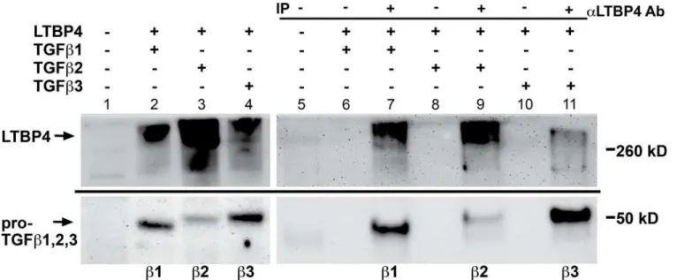
TGFβ’s isoforms TGFβ1, 2 and 3, are expressed in distinct spatiotemporal patterns [32,33]. Because of the close sequence relationship among TGFβs 1, 2 and 3, we tested each for its abil-ity to interact with LTBP4. HEK293T cells were co-transfected with LTBP4 and plasmids expressing TGFβ1, TGFβ2 or TGFβ3, and immunoprecipitations were carried out using an antibody against LTBP4 followed by immunoblotting for the FLAG-epitope tag on TGFβ1, 2 or 3 (Fig 5C). All three of the TGFβisoforms demonstrated a clear interaction with LTBP4. TGFβ2 expression was lower than that of TGFβ1 and TGFβ3, which accounts for the weaker binding result of this isoform (lanes 3,9).
Discussion
LTBP4 is a broadly expressed protein but one that shows higher level of basal expression in car-diac, skeletal and smooth muscle [1,2]. A mouse model deficient in the short isoform ofLtbp4 displays early lethality from pulmonary fibrosis, cardiomyopathy and colon cancer, indicating the importance for regulating TGFβin the heart and during development [30].LTBP4nsSNPs have been linked to a series of diseases including DMD. The two most commonLTBP4alleles found in the human population differ in four amino acids along the length of the LTBP4 pro-tein. The protectiveLTBP4haplotype correlates with milder disease in DMD, better exercise performance in chronic lung disease, smaller abdominal aneurysms and less invasive colorectal cancer [16,18–20]. Although deletion ofLtbp4in the mouse suggests that LTBP4’s activity is important in the heart,LTBP4SNPs had not previously been associated with cardiomyopathy in humans. Although replication studies are needed, a recent study did identify an association betweenLTBP4SNPs and DCM in the setting of DMD. Specifically it was found that steroid-treated DMD patients carrying the rs10880 allele encoding a threonine have a significantly increased incidence of DCM compared to patients homozygous for the protective allele, which encodes a methionine [34]. These findings, coupled with the studies in animal models, suggest a role for LTBP4 in the heart.
Fig 5. LTBP4 binds TGFβ1, 2, and 3 isoforms.HEK293T cells were co-transfected with LTBP4 and either TGFβ1, TGFβ2, or TGFβ3. Cell lysates were immunoprecipitated with an antibody to LTBP4 and immunoblotted with an antibody to the FLAG tag on TGFβ1, TGFβ2, and TGFβ3. The immunoblot shows that all three of the TGFβisoforms associate with LTBP4.
Each of these studies examining association betweenLTBP4SNPs and disease phenotypes was carried out in primarily European American populations, including non-Hispanic Euro-pean Americans from the United States, Sweden, the United Kingdom, New Zealand, and Aus-tralia. One report associatingLTBP4SNPs and ambulation loss in DMD, finding thatLTBP4 genotype was directly associated with age at loss of ambulation [15]. However, this association was found only when the population was stratified to include exclusively non-Hispanic Euro-pean Americans [15]. In this study, the number of non-EuroEuro-pean American patients was too small to make any conclusions about the effect ofLTBP4on age at loss of ambulation in other eth-nicities. Despite using a larger non-European American population, we did not find evidence for an association betweenLTBP4SNPs and DCM in African Americans. However, theLTBP4locus differs between European Americans and African Americans in its degree of linkage disequilib-rium. Specifically, there are fewer crossover events among European Americans and a greater per-centage of the European American alleles are VTTT or IAAM versus the deviating genotypes like ITTT, IAAT, and others. These studies, together with the varying linkage disequilibrium across ethnicities, suggest thatLTBP4genotype effects are more pronounced with preservations of the VTTT/IAAM diplotypes. While it is possibly that the risk alleles are in linkage disequilibrium with other, as yet unidentified genetic variants that explain the phenotypic results, extensive sequencing and examination of the LTBP4 allele has not identified additional nsSNPs that account for this result. African Americans and European Americans differ in their allele frequen-cies at many sites across the genome, and it is plausible that another missense SNP could affect TGFβbinding or signaling and confound the effect of the VTTT allele in African Americans.
Cells with theLTBP4“IAAM”allele display reduced TGFβsignaling compared to cells car-rying the“VTTT”allele, despite similar levels of LTBP4 protein [16]. The mechanism underly-ing this decreased TGFβsignaling is explained by the protective LTBP4 IAAM sequence binding TGFβmore tightly than cells expressing the risk-associated VTTT LTBP4. Increased sequestration of latent TGFβby matrix-bound LTBP4 results in less release of latent TGFβand therefore less TGFβavailable to bind TGFβreceptors. Reduced TGFβ, whether produced genetically or pharmacologically, has been shown to reduce fibrosis in muscular dystrophy [35–37]. This effect can also be reproduced through a dominant negative TGFβreceptor or through reduction of the canonical TGFβsignaling pathways [38–40]. Noncanonical TGFβ sig-naling follows this same pattern, with inhibition of noncanonical TGFβsignaling also resulting in an improved muscular dystrophy phenotype [41].
Little is known about the distinct biological roles of the three TGFβisoforms and their role in disease, although TGFβ1 is the most studied. In DCM, TGFβ1 is upregulated in the plasma and myocardium, however many studies have not examined the expression of TGFβ2 and TGFβ3 [25,26]. In an unbiased microarray study, TGFβ2 was found to be significantly increased in DCM samples compared to controls [42]. The differential roles of TGFβisoforms have been studied in several other forms of heart disease and cardiac remodeling. Regulatory variants inTGFB3have been associated with arrhythmogenic right ventricular cardiomyopa-thy [43]. In a rat model of left ventricular hypertrophy all three TGFβisoforms were induced upon cardiac remodeling, howeverTGFB2expression level increased earlier and was more robust than the other two isoforms [44]. Similarly, in a rat model of myocardial infarction, all three TGFβisoforms increase in the left ventricle after infarct, howeverTGFB2had the largest fold increase, suggesting that TGFβ2 plays an important role in myocardial remodeling [45]. Notably, this current study examined non-ischemic DCM. TGFβ1, TGFβ2 and, TGFβ3 are co-regulated during wound healing [46]. The varying levels of TGFβisoform expression, both of which interact with LTBP4, are likely critical during injury and repair.
parenchyma itself. The association ofLTBP4loss of function alleles with early onset pulmonary defects likely supports regulation of TGFβfamily members in multiple cell types, especially those critical during development, such as BMPs [47]. The postnatal role of LTBPs, including LTBP4, may be quite distinct and the molecular interactions in differentiated cell types may account for the effect in chronic lung disease, abdominal aneurysms and in colorectal cancer [18–20]. In all of these disorders, it is theLTBP4allele associated with hyper-TGFβsignaling and reduced latent TGFβsequestration that is the risk or deleterious allele. These effects may be explained by the interaction of LTBPs with multiple TGFβfamily members.
Materials and Methods
Patient populations and controls
Written informed consent was obtained from all human subjects or their parents in the case of minors under the approval of the University of Chicago’s Institutional Review Board. Peripheral blood was collected from unrelated subjects with nonischemic DCM at the University of Chicago Medical Center. Criteria for inclusion included: 1) ejection fraction<45% or 2) left ventricular (LV) end diastolic diameter>117% of the predicted value, and 3) absence of significant epicardial coro-nary artery disease, and/or hypertensive, systemic, pericardial, or congenital disease to account for DCM [48,49]. The cohort of 201 subjects included 61 European American and 140 African Ameri-can DCM subjects, as self-identified. Genomic DNA samples were extracted using the Gentra Pure-gene Blood Kit (Qiagen #158422). European American and African American control panel DNA was obtained from Coriell (catalog #s HD100CAU and HD100AA-2, respectively). African Ameri-can control DNA was also obtained through the University of Chicago TRIDOM (Translational Research Initiative in the Department of Medicine) biobank program; samples were selected based on self-identified ethnicity and those with heart disease were excluded. Control DNA from African Americans born at the University of Chicago was extracted from human umbilical cord blood. Echocardiographic data was gathered from a single reading laboratory over 15 years (1999–2014) from 169 subjects. The age at which the echocardiogram was performed was recorded. Ninety-three studies did not list a precise LVEF, classifying LV function as mildly, mildly-moderately, mod-erately, moderately-severely, or severely reduced. For these studies, the LVEF was translated as 45%, 40%, 35%, 30% and 25%, respectively, based on the reading practices of the laboratory. Six individu-als had echocardiography post heart transplant, and therefore were classified as severely reduced LVEF. One individual was estimated as severely reduced LVEF from autopsy records.
Genotyping
Genomic DNA was amplified by polymerase chain reaction (PCR) for direct sequencing of fragments encompassing SNPs rs2303729, rs1131620, rs1051303, and rs10880 (primers are listed inS4 Table). PCR fragments were purified using ExoSAP-IT reagent (Affymetrix). PCR products were then sequenced with traditional Sanger sequencing and SNP calls were made by analysis of individual chromatograms. DCM patients were separated based on self-identified ethnicity (European American or African American) and allele frequencies were compared with those of ethnicity-matched controls. A chi-square test was used for each SNP to determine whether there was a significant difference in allele frequency between DCM patients and con-trols. GraphPad Prism was used for statistical analysis.
Linkage disequilibrium analyses
ASW population (n = 61) to represent African Americans [27,50]. EUR includes CEU (Utah residents with Northern and Western European ancestry, TSI (Toscani in Italy), FIN (Finnish in Finland), GBR (British in England and Scotland), and IBS (Iberian population in Spain). ASW is composed of Americans of African ancestry in the Southwestern United States. The 1000 Genomes VCF to Ped converter was used to obtain data files and locus information files for input into Haploview. SNPs with a population frequency over five percent were included in the map. Linkage disequilibrium blocks were designated using the confidence interval method [51]. The color scheme of the linkage disequilibrium map was generated using the standard D’/ LOD option in the Haploview software.
Expression constructs
Full length humanLTBP4(NM_001042544.1) andTGFβ1(NM_000660.3) cDNA clones were purchased from Origene (catalog numbers SC311430 and SC119746, respectively).TGFβ2 (NM_003238.3) andTGFβ3(NM_003239.2) were amplified by polymerase chain reaction using cDNA from human skin fibroblasts (GM03348, Coriell Cell Repositories) as a template. The FLAG epitope tag (DYKDDDDK) was engineered on the 3’end ofTGFβ1,TGFβ2, and TGFβ3and a Kozak consensus sequence (CACC) was added immediately upstream of the initi-ation codon. The following primers were used for PCR of TGFβ2 and TGFβ3: TGFβ2 Forward: 5’CACCATGCACTACTGTGTGCTGAGCGCT 3’; TGFβ2 Reverse: 5’TCACTTGTCATCGT CATCCTTGTAATCGCTGCATTTGCAAGACTT 3’; TGFβ3 Forward: 5’CACCATGAAGAT GCACTTGCAAAGGGCT 3’; TGFβ3 Reverse: 5’TCACTTGTCATCGTCATCCTTGTAATC GCTACATTTACAAGACTT 3’. The Xpress epitope (DLYDDDDK) was added onto the 5’ /N-terminus ofLTBP4.
Cell culture
HEK293-T cells were obtained from ATCC (catalog number CRL-11268). Cells were grown in Dulbecco's Modified Eagle Medium (DMEM) supplemented with 10% fetal bovine serum (Invitrogen, lot #1420768) and 1% penicillin/streptomycin (Invitrogen) in 5% CO2.
Transfections
HEK293-T cells were plated at 1.5x10⁶cells per well in 6-well plates the day prior to transfec-tion. Media was changed to Opti-MEM Reduced Serum Media one hour prior to transfectransfec-tion. Transfections were performed using FuGENE HD transfection reagent (Promega) at a DNA: FuGENE ratio of 1:3. Opti-MEM was replaced with serum-rich media 24 hours post-transfec-tion and cells were harvested 3 days post-transfecpost-transfec-tion.
Co-immunoprecipitation
Immunoblotting
Prior to SDS-PAGE, 5X Laemmli buffer containingβ-mercaptoethanol was added to cell lysates and samples were boiled for five minutes. Fiftyμg of protein input (cell lysate pre-IP) and the entire co-IP lysate were separated on a 4–20% polyacrylamide gel (Pierce) and trans-ferred to PVDF Immobilon-P membrane (Millipore). Blocking and antibody incubations were done using StartingBlock T20 (TBS) blocking buffer (Pierce). The membrane was immuno-blotted with primary antibody at 1:4000 (anti-FLAG, anti-LTBP4) or 1:500 (anti-Xpress). Mouse monoclonal anti-FLAG was commercially available (Sigma #F1804). Rabbit polyclonal anti-LTBP4 and rabbit polyclonal anti-Xpress antibodies were generated by Pocono Rabbit Farm and Laboratory (details above). Goat anti-mouse and goat anti-rabbit secondary antibod-ies conjugated to horseradish peroxidase (Jackson Immunoresearch #115-035-003 and #111-035-003) were used at a dilution of 1:8000. Clarity ECL substrate (Bio-Rad) was applied to membranes and membranes were visualized using a Biospectrum Imager (UVP). ImageJ gel analysis tools were used for densitometry analysis. To quantify the amount of immunoprecipi-tation, immunoprecipitations were performed in triplicate and a student’s t-test was used to test whether the amount of TGFβimmunoprecipitated was significantly different depending on LTBP4 genotype. Prism software (GraphPad) was used for statistical analyses. Uncropped gels are shown inS1 Fig.
Supporting Information
S1 Fig. Uncropped gels for Figs3,4and5. (PDF)
S1 Table.LTBP4coordinately expressed genes (top 25). (PDF)
S2 Table. KEGG pathways ofLTBP4coordinately expressed genes. (PDF)
S3 Table. Tissue expression ofLTBP4coordinately expressed genes. (PDF)
S4 Table. PCR primers used for genotyping. (PDF)
Acknowledgments
We would like to thank Dr. Graeme Bell for generously providing the African American con-trol DNA samples from umbilical cord blood.
Author Contributions
Conceived and designed the experiments: KML LDC EMM. Performed the experiments: KML TM. Analyzed the data: KML EMM LDC. Contributed reagents/materials/analysis tools: LDC. Wrote the paper: KML EMM.
References
2. Saharinen J, Taipale J, Monni O, Keski-Oja J. Identification and characterization of a new latent trans-forming growth factor-beta-binding protein, LTBP-4. J Biol Chem. 1998; 273(29):18459–69. Epub 1998/07/11. PMID:9660815.
3. Shi M, Zhu J, Wang R, Chen X, Mi L, Walz T, et al. Latent TGF-beta structure and activation. Nature. 2011; 474(7351):343–9. doi:10.1038/nature10152PMID:21677751.
4. Annes JP, Munger JS, Rifkin DB. Making sense of latent TGFbeta activation. J Cell Sci. 2003; 116(Pt 2):217–24. PMID:12482908.
5. Miyazono K, Olofsson A, Colosetti P, Heldin CH. A role of the latent TGF-beta 1-binding protein in the assembly and secretion of TGF-beta 1. EMBO J. 1991; 10(5):1091–101. Epub 1991/05/01. PMID: 2022183; PubMed Central PMCID: PMC452762.
6. Ge G, Greenspan DS. BMP1 controls TGFbeta1 activation via cleavage of latent TGFbeta-binding pro-tein. The Journal of cell biology. 2006; 175(1):111–20. Epub 2006/10/04. doi:10.1083/jcb.200606058 PMID:17015622; PubMed Central PMCID: PMCPmc2064503.
7. Levine JH, Moses HL, Gold LI, Nanney LB. Spatial and temporal patterns of immunoreactive transform-ing growth factor beta 1, beta 2, and beta 3 durtransform-ing excisional wound repair. The American journal of pathology. 1993; 143(2):368–80. PMID:8342593; PubMed Central PMCID: PMCPMC1887040. 8. Bascom CC, Wolfshohl JR, Coffey RJ, Madisen L, Webb NR, Purchio AR, et al. Complex regulation of
transforming growth factor beta 1, beta 2, and beta 3 mRNA expression in mouse fibroblasts and kerati-nocytes by transforming growth factors beta 1 and beta 2. Mol Cell Biol. 1989; 9(12):5508–15. PMID: 2586525; PubMed Central PMCID: PMCPMC363721.
9. Barrientos S, Stojadinovic O, Golinko MS, Brem H, Tomic-Canic M. Growth factors and cytokines in wound healing. Wound repair and regeneration: official publication of the Wound Healing Society [and] the European Tissue Repair Society. 2008; 16(5):585–601. Epub 2009/01/09. doi: 10.1111/j.1524-475X.2008.00410.xPMID:19128254.
10. Mustoe TA, Pierce GF, Thomason A, Gramates P, Sporn MB, Deuel TF. Accelerated healing of inci-sional wounds in rats induced by transforming growth factor-beta. Science (New York, NY). 1987; 237 (4820):1333–6. Epub 1987/09/11. PMID:2442813.
11. Schultz GS, Wysocki A. Interactions between extracellular matrix and growth factors in wound healing. Wound repair and regeneration: official publication of the Wound Healing Society [and] the European Tissue Repair Society. 2009; 17(2):153–62. Epub 2009/03/27. doi:10.1111/j.1524-475X.2009.00466.x PMID:19320882.
12. Wynn TA. Common and unique mechanisms regulate fibrosis in various fibroproliferative diseases. The Journal of clinical investigation. 2007; 117(3):524–9. Epub 2007/03/03. doi:10.1172/jci31487 PMID:17332879; PubMed Central PMCID: PMCPmc1804380.
13. Border WA, Noble NA. Transforming growth factor beta in tissue fibrosis. N Engl J Med. 1994; 331 (19):1286–92. doi:10.1056/nejm199411103311907PMID:7935686.
14. Duffield JS, Lupher M, Thannickal VJ, Wynn TA. Host responses in tissue repair and fibrosis. Annual review of pathology. 2013; 8:241–76. Epub 2012/10/25. doi:10.1146/annurev-pathol-020712-163930 PMID:23092186; PubMed Central PMCID: PMCPmc3789589.
15. Bello L, Kesari A, Gordish-Dressman H, Cnaan A, Morgenroth LP, Punetha J, et al. Genetic modifiers of ambulation in the CINRG duchenne natural history study. Annals of neurology. 2015. Epub 2015/02/ 03. doi:10.1002/ana.24370PMID:25641372.
16. Flanigan KM, Ceco E, Lamar KM, Kaminoh Y, Dunn DM, Mendell JR, et al. LTBP4 genotype predicts age of ambulatory loss in Duchenne muscular dystrophy. Annals of neurology. 2013; 73(4):481–8. Epub 2013/02/27. doi:10.1002/ana.23819PMID:23440719.
17. van den Bergen JC, Hiller M, Bohringer S, Vijfhuizen L, Ginjaar HB, Chaouch A, et al. Validation of genetic modifiers for Duchenne muscular dystrophy: a multicentre study assessing SPP1 and LTBP4 variants. Journal of neurology, neurosurgery, and psychiatry. 2014. Epub 2014/12/06. doi:10.1136/ jnnp-2014-308409PMID:25476005.
18. Hersh CP, Demeo DL, Lazarus R, Celedon JC, Raby BA, Benditt JO, et al. Genetic association analy-sis of functional impairment in chronic obstructive pulmonary disease. Am J Respir Crit Care Med. 2006; 173(9):977–84. Epub 2006/02/04. 200509-1452OC [pii] doi:10.1164/rccm.200509-1452OC PMID:16456143; PubMed Central PMCID: PMC2662917.
20. Forsti A, Li X, Wagner K, Tavelin B, Enquist K, Palmqvist R, et al. Polymorphisms in the transforming growth factor beta 1 pathway in relation to colorectal cancer progression. Genes Chromosomes Can-cer. 2010; 49(3):270–81. Epub 2009/12/10. doi:10.1002/gcc.20738PMID:19998449.
21. Brooks A, Schinde V, Bateman AC, Gallagher PJ. Interstitial fibrosis in the dilated non-ischaemic myo-cardium. Heart. 2003; 89(10):1255–6. Epub 2003/09/17. PMID:12975439; PubMed Central PMCID: PMCPmc1767879.
22. Sanderson J, Lai K, Shum I, Wei S, Chow L. Transforming growth factor-β(1) expression in dilated car-diomyopathy. Heart. 2001; 86(6):701–8. doi:10.1136/heart.86.6.701PMID:PMC1729995.
23. Aharinejad S, Krenn K, Paulus P, Schafer R, Zuckermann A, Grimm M, et al. Differential role of TGF-beta1/bFGF and ET-1 in graft fibrosis in heart failure patients. American journal of transplantation: offi-cial journal of the American Society of Transplantation and the American Society of Transplant Sur-geons. 2005; 5(9):2185–92. Epub 2005/08/13. doi:10.1111/j.1600-6143.2005.01006.xPMID: 16095497.
24. Holweg CT, Baan CC, Niesters HG, Vantrimpont PJ, Mulder PG, Maat AP, et al. TGF-beta1 gene poly-morphisms in patients with end-stage heart failure. The Journal of heart and lung transplantation: the official publication of the International Society for Heart Transplantation. 2001; 20(9):979–84. Epub 2001/09/15. PMID:11557193.
25. Khan R, Sheppard R. Fibrosis in heart disease: understanding the role of transforming growth factor-β
(1) in cardiomyopathy, valvular disease and arrhythmia. Immunology. 2006; 118(1):10–24. doi:10. 1111/j.1365-2567.2006.02336.xPMID:PMC1782267.
26. Khan S, Joyce J, Margulies KB, Tsuda T. Enhanced bioactive myocardial transforming growth factor-beta in advanced human heart failure. Circulation journal: official journal of the Japanese Circulation Society. 2014; 78(11):2711–8. Epub 2014/10/10. PMID:25298166.
27. Abecasis GR, Auton A, Brooks LD, DePristo MA, Durbin RM, Handsaker RE, et al. An integrated map of genetic variation from 1,092 human genomes. Nature. 2012; 491(7422):56–65. doi:10.1038/ nature11632PMID:23128226; PubMed Central PMCID: PMCPMC3498066.
28. Karolchik D, Barber GP, Casper J, Clawson H, Cline MS, Diekhans M, et al. The UCSC Genome Browser database: 2014 update. Nucleic Acids Res. 2014; 42(Database issue):D764–70. doi:10.1093/ nar/gkt1168PMID:24270787; PubMed Central PMCID: PMCPMC3964947.
29. Fahrenbach JP, Andrade J, McNally EM. The CO-Regulation Database (CORD): A Tool to Identify Coordinately Expressed Genes. PLoS ONE. 2014; 9(3):e90408. doi:10.1371/journal.pone.0090408 PMID:24599084
30. Sterner-Kock A, Thorey IS, Koli K, Wempe F, Otte J, Bangsow T, et al. Disruption of the gene encoding the latent transforming growth factor-beta binding protein 4 (LTBP-4) causes abnormal lung develop-ment, cardiomyopathy, and colorectal cancer. Genes Dev. 2002; 16(17):2264–73. Epub 2002/09/05. doi:10.1101/gad.229102PMID:12208849; PubMed Central PMCID: PMC186672.
31. Biernacka A, Dobaczewski M, Frangogiannis NG. TGF-beta signaling in fibrosis. Growth Factors. 2011; 29(5):196–202. doi:10.3109/08977194.2011.595714PMID:21740331.
32. Roberts AB, Kim SJ, Kondaiah P, Jakowlew SB, Denhez F, Glick AB, et al. Transcriptional control of expression of the TGF-betas. Annals of the New York Academy of Sciences. 1990; 593:43–50. Epub 1990/01/01. PMID:2197962.
33. Roberts AB, Sporn MB. Differential expression of the TGF-beta isoforms in embryogenesis suggests specific roles in developing and adult tissues. Molecular reproduction and development. 1992; 32 (2):91–8. Epub 1992/06/01. doi:10.1002/mrd.1080320203PMID:1637557.
34. Barp A, Bello L, Politano L, Melacini P, Calore C, Polo A, et al. Genetic Modifiers of Duchenne Muscular Dystrophy and Dilated Cardiomyopathy. PLoS One. 2015; 10(10):e0141240. Epub 2015/10/30. doi: 10.1371/journal.pone.0141240PMID:26513582; PubMed Central PMCID: PMCPmc4626372. 35. Cohn RD, van Erp C, Habashi JP, Soleimani AA, Klein EC, Lisi MT, et al. Angiotensin II type 1 receptor
blockade attenuates TGF-beta-induced failure of muscle regeneration in multiple myopathic states. Nature medicine. 2007; 13(2):204–10. Epub 2007/01/24. doi:10.1038/nm1536PMID:17237794; PubMed Central PMCID: PMCPmc3138130.
36. Nelson CA, Hunter RB, Quigley LA, Girgenrath S, Weber WD, McCullough JA, et al. Inhibiting TGF-beta activity improves respiratory function in mdx mice. The American journal of pathology. 2011; 178 (6):2611–21. Epub 2011/06/07. doi:10.1016/j.ajpath.2011.02.024PMID:21641384; PubMed Central PMCID: PMCPmc3124227.
38. Accornero F, Kanisicak O, Tjondrokoesoemo A, Attia AC, McNally EM, Molkentin JD. Myofiber-specific inhibition of TGFbeta signaling protects skeletal muscle from injury and dystrophic disease in mice. Human molecular genetics. 2014; 23(25):6903–15. Epub 2014/08/12. doi:10.1093/hmg/ddu413PMID: 25106553; PubMed Central PMCID: PMCPmc4271062.
39. Goldstein JA, Kelly SM, LoPresti PP, Heydemann A, Earley JU, Ferguson EL, et al. SMAD signaling drives heart and muscle dysfunction in a Drosophila model of muscular dystrophy. Human molecular genetics. 2011; 20(5):894–904. Epub 2010/12/09. doi:10.1093/hmg/ddq528PMID:21138941; PubMed Central PMCID: PMCPmc3033181.
40. Goldstein JA, Bogdanovich S, Beiriger A, Wren LM, Rossi AE, Gao QQ, et al. Excess SMAD signaling contributes to heart and muscle dysfunction in muscular dystrophy. Human molecular genetics. 2014; 23(25):6722–31. Epub 2014/07/30. doi:10.1093/hmg/ddu390PMID:25070948; PubMed Central PMCID: PMCPmc4303797.
41. Wissing ER, Boyer JG, Kwong JQ, Sargent MA, Karch J, McNally EM, et al. P38alpha MAPK underlies muscular dystrophy and myofiber death through a Bax-dependent mechanism. Human molecular genetics. 2014; 23(20):5452–63. Epub 2014/05/31. doi:10.1093/hmg/ddu270PMID:24876160; PubMed Central PMCID: PMCPmc4184390.
42. Barth AS, Kuner R, Buness A, Ruschhaupt M, Merk S, Zwermann L, et al. Identification of a Common Gene Expression Signature in Dilated Cardiomyopathy Across Independent Microarray Studies. Jour-nal of the American College of Cardiology. 2006; 48(8):1610–7. doi:http://dx.doi.org/10.1016/j.jacc. 2006.07.026PMID:17045896
43. Beffagna G, Occhi G, Nava A, Vitiello L, Ditadi A, Basso C, et al. Regulatory mutations in transforming growth factor-beta3 gene cause arrhythmogenic right ventricular cardiomyopathy type 1. Cardiovascu-lar research. 2005; 65(2):366–73. Epub 2005/01/11. doi:10.1016/j.cardiores.2004.10.005PMID: 15639475.
44. Briest W, Homagk L, Rassler B, Ziegelhoffer-Mihalovicova B, Meier H, Tannapfel A, et al. Norepineph-rine-induced changes in cardiac transforming growth factor-beta isoform expression pattern of female and male rats. Hypertension. 2004; 44(4):410–8. Epub 2004/08/25. doi:10.1161/01.HYP.0000141414. 87026.4dPMID:15326086.
45. Deten A, Hölzl A, Leicht M, Barth W, Zimmer H-G. Changes in Extracellular Matrix and in Transforming Growth Factor Beta Isoforms After Coronary Artery Ligation in Rats. Journal of Molecular and Cellular Cardiology. 2001; 33(6):1191–207. doi:http://dx.doi.org/10.1006/jmcc.2001.1383PMID:11444923 46. O'Kane S, Ferguson MW. Transforming growth factor beta s and wound healing. Int J Biochem Cell
Biol. 1997; 29(1):63–78. Epub 1997/01/01. S1357272596001203 [pii]. PMID:9076942.
47. Koli K, Wempe F, Sterner-Kock A, Kantola A, Komor M, Hofmann WK, et al. Disruption of LTBP-4 func-tion reduces TGF-beta activafunc-tion and enhances BMP-4 signaling in the lung. The Journal of cell biol-ogy. 2004; 167(1):123–33. Epub 2004/10/07. doi:10.1083/jcb.200403067PMID:15466481; PubMed Central PMCID: PMCPmc2172518.
48. Hershberger RE, Lindenfeld J, Mestroni L, Seidman CE, Taylor MR, Towbin JA. Genetic evaluation of cardiomyopathy—a Heart Failure Society of America practice guideline. Journal of cardiac failure. 2009; 15(2):83–97. Epub 2009/03/04. doi:10.1016/j.cardfail.2009.01.006PMID:19254666.
49. Mestroni L, Maisch B, McKenna WJ, Schwartz K, Charron P, Rocco C, et al. Guidelines for the study of familial dilated cardiomyopathies. Collaborative Research Group of the European Human and Capital Mobility Project on Familial Dilated Cardiomyopathy. European heart journal. 1999; 20(2):93–102. Epub 1999/04/01. PMID:10099905.
50. Barrett JC, Fry B, Maller J, Daly MJ. Haploview: analysis and visualization of LD and haplotype maps. Bioinformatics (Oxford, England). 2005; 21(2):263–5. Epub 2004/08/07. doi:10.1093/bioinformatics/ bth457PMID:15297300.
51. Gabriel SB, Schaffner SF, Nguyen H, Moore JM, Roy J, Blumenstiel B, et al. The structure of haplotype blocks in the human genome. Science (New York, NY). 2002; 296(5576):2225–9. Epub 2002/05/25. doi:10.1126/science.1069424PMID:12029063.