Maritime Antarctica Soils Studied by
Mossbauer Spectroscopy and Other Methods
E. Kuzmann
, L. A. Schuch
1, V. K. Garg, P. A. de Souza Junior
2,
E. M. Guimar~aes
3, A. C. de Oliveira and A. Vertes
4Institute of Physics, University of Braslia, 70910-900 Braslia, DF, Brazil
1Department of Physics, Federal University of Santa Maria, Santa Maria, RS, Brazil 2Department of Physics, Federal University of Espirito Santo, Vitoria, ES, Brazil
3Department of Mineralogy and Petrology, University of Braslia, Braslia, Brazil 4Department of Nuclear Chemistry, Eotvos University, Budapest, Hungary
Received 12 September, 1997
Soil samples from the King George Island, Antarctica, have been studied by57Fe Mossbauer
spectroscopy, X-ray diractometry, radiometry, neutron activation analysis and chemical analytical methods. X-ray diractometry measurements have identied soils containing dif-ferent volume ratios of quartz, feldspar, chlorite as well as hematite. The dierence in the phase composition and in the iron distribution among the crystallographic sites of iron-bearing minerals (chlorite, magnetite and hematite) of samples from two dierent depths was derived from the complex Mossbauer spectra. The dierences in the mineral composi-tion, iron distribucomposi-tion, concentration of water soluble salts, pH and radioactivity of certain radionuclides indicate the occurrence of chemical weathering of minerals.
I Introduction
In Antarctica the geological and mineralogical processes are strongly inuenced by the presence of ice. Less than 2% of the continental area is ice free and is accessible for the geological and mineralogical researches. This area is distributed around the periphery of Antarctica. In these areas weathering and soil formation on the glacial deposits and exposed rocks have begun. Based on cli-mate and moisture availability, the soils of Antarctica fall into the following three major soil zones [1], the dry valleys and bare ground on the Trans Antarctic Mountains, the oases of coastal greater Antarctica, and the maritime Antarctic Peninsula. The northern part of the Antarctic Peninsula and the associated islands have a cold moist climate that makes possible the soil formation in the maritime Antarctic region to be dif-ferent from that which occurs elsewhere in Antarctica
[2-4], The chemical weathering can also be expected to be more active here than in the deserts of the north polar region. Soil studies can contribute to the knowl-edge of chronology of glacial events by correlating the weathering stages of soils in the Antarctica. A unique feature of Antarctic soils is the diversity of the nature and distribution of salts. Studies of soil sequences may reveal evidence of past variation in climatic conditions and of changes in global atmospheric circulation [1]. The objective of the present work was to investigate soil samples, from the King George Island of the South Shetland Islands in Antarctica.
II Experimental and basic
ana-lytical results
The soil samples SOLO1 and SOLO2 were collected near the Brazilian Antarctic Station Comandante
raz (62 05" latitude 58 24' longitude) in the Keller
Peninsula in the King George Island (Fig. 1) of the South Shetland Islands of Antarctica in 1991-1992. The location of the soil samples is depicted in the map of Fig. 2. Samples of SOLO1 and SOLO2 were collected from an area of 10 cm x 20 cm. Samples of SOLO1 were obtained from depths of O -5 cm, while samples of SOLO2 were collected from depths of 5 to 10 cm. The thickness of the soil layer on the rock is typically not more than 15 cm here. The mineralogical and strati-graphical characterization of the rock surrounding is similar to reported ones [5-7] Crustal structure [8], geol-ogy [9-17] and environmental parameters [18-21] of the region have been reported, wherein some of the results of the previous Brazilian missions are also summarized. The samples were analyzed by granulometry, and the results are tabulated in Table 1. The result of the neu-tron activation analysis is shown in Table 2. Table 3-4 show the analytical data of the soil samples obtained
by wet chemical method. Conventional gamma spec-troscopy was used to determine the radioactivity of ra-dionuclides in the soils. The result of this analysis is shown in Table 5. X-ray diractograms of soil sam-ples were recorded either by means of a computer con-trolled RIGAKU diractometer using CuKO radiation and a monocromator or by the help of a computer con-trolled DRON-3 diractometer using CuK radiation
and a-lter.
57Fe Mossbauer spectra of soil samples
were recorded by conventional Mossbauer spectrome-ters (WISSEL and KFKI) using a scintillation detec-tor in transmission geometry with a57Co/Rh (109Bq)
source both at room temperature and temperature of liquid nitrogen (78K). Isomer shifts are given relative to the-Fe. The evaluation of Mossbauer spectra was
per-formed by a least-square tting MOSSWINN [22] pro-gram, and the quadrupole splitting distributions were obtained by modied Hesse-Rubartsch methods.
Figure1. MapofSouthShetlandIslands.
Table 1 - Granulometric results of soils collected in the Antarctica in 1991-92. pH Granulometric composition of soils (%)
SAMPLE 2-0.20 mm 0.20-0.05 mm 0.05-0.02 mm <0.02 mm
Table 2 - Analytical data of neutron activation analysis of soils collected from Antarctica in 1991-92. Sample Elements Solo 1 Solo 2
Sc (ppm) 21.30.9 25.00.3
Cr (ppm) 21.61.1 23.71.4
Fe(%) 5.8 0.3 5.4 0.2
Co (ppm) 22.20.6 23.71.6
Zn (ppm) 73.82.5 80.54.2
As (ppm) 6.60.5 9.00.7
Rb (ppm) 43.93.0 45.33.7
Sr (ppm) ND ND Sb (ppm) 46030 59030
La (ppm) 25.22.1 25.71.3
Ce (ppm) 50.21.5 53.52.7
Nd (ppm) 30.22.7 38.74.1
Sm (ppm) 5.270.54 6.090.34
Eu (ppm) 1.610.06 1.500.06
Tb (ppm) 0.700.06 0.770.04
Yb (ppm) 2.70.2 2.60.3
Lu (ppm) 0.40O.03 0.400.02
Hf(ppm) 5.7O.2 5.80.2
Th (ppm) 6.80.2 7.780.07
U (ppm) 1.90.1 2.00.5
Br (ppm) 0.990.18 0.300.02
Cs (ppm) ND ND
Table 3 - Analytical data of soils collected in the Antarctica in 1991-92.
Sample P(ppm) Mg++ (%) Ca++(%) K+(%) Na+(%) C organic(%) N C/N
Solo 1 167 6.1 2.1 1.1 1.88 0.95 0.08 12 Solo 2 167 7.4 2.9 1.12 2.68 0.33 0.03 11
Table 4 - Analytical data of soluble salts of soils collected in the Antarctica in 1991-92. Sample Soluble salts ionic ratio relative to potassium
Ca++ Mg++ Na+ K+
Solo 1 19 10 10 1 Solo 2 14 10 2 1
Table 5 - Determination of-radiating radionuclides in soils collected in 1991-92, on the King George Island of
South Shetland Islands of Antarctica. Material Radionuclides
Cs-137 Ra-226 Ra-228 K-40 BqKg,1
Solo 1 2:940:32 20:51:3 22:51:8 38321
X-ray diractograms of soil samples are depicted in Fig. 3, and the reections of quartz, feldspar, chlorite or smectite and hematite can be identied. The lattice spacing of chlorite in SOLO1 is dierent from that of SOLO2. This indicates that the chlorite in SOLO1 contains signicantly more iron than the chlorite in SOLO2. Table 6 shows the normalized integrated inten-sities of peaks belonging to the identied main phases, giving the estimated occurrence of the main minerals of Antarctic soil samples.
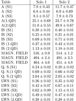
Room temperature Mossbauer spectra of soil sam-ples SOLO1 and SOLO2 are depicted in Fig. 4. These spectra are very complex in nature. The envelop of these spectra shows peaks that lie from -8 mm/s to 8.5 mm/s. This can be considered as a superposition of magnetically split sextets and a number of paramag-netic subspectra. Several attempts were made to nd the optimum deconvolution of these spectra into sub-spectra. In these cases the spectra recorded at 300K and 77K were evaluated for the same components of minerals under the corresponding constrains. It was found that the magnetically split part can be very well understood by the decomposition of room temperature spectra into 3 sextets (S1, S2 and S3) whose Mossbauer parameters are in Table 7. Sextet S1 is attributed to Fe in the hematite,-Fe
2O3. Since the isomer shift, ,
quadrupole splitting, E
Q and the internal magnetic
eld, H, are typically characteristic of the hematite [23-27] and the isomer shift and the hyperne eld exhibit the expected temperature dependence at 77K.
Figure 3. Details of X-ray diractograms of soil samples collected in King George Island in Antarctica in the years 1991-92.
Figure 4. Room temperature Mossbauer spectra of soil sam-ples. (The middle part o the spectra is depicted as decom-pesed only into a doublet of Fe3+and a doublet o Fe2+)
:
Table 6 - Result of X-ray diractometry, (normalized integrated intensities of peaks belonging to the identied main phases).
Table 7 - Mossbauer parameters of soils collected from the Antarctica in 1991-1992. Table Solo 1 Solo 2
A (S1) 7:80:43 11:70:47
A (S2) 6:80:44 6:90:69
A (S3) 8:50:57 7:90:73
A(1.QD) 25:10:68 23:70:76
A(2.QD) 51:80:55 49:80:67
IS (S1) 0:380:01 0:400:01
IS (S2) 0:250:01 0:310:01
IS (S3) 0:690:01 0:710:01
IS (1.QD) 0:370:01 0:430:01
IS (2.QD) 1:130:01 1:180:01
MAGN. FIELD 516:0:9 514:1:2
MAGN. FIELD 494:2:4 491:3:5
MAGN. FIELD 464:4:6 451:4:8
Q. S.(S1) 0:240:02 0:200:02
Q. S.(1 QD) 0:690:02 0:660:02
Q. S.(2 QD) 2:640:02 2:650:02
LWS (S1) 0:260:02 0:350:02
LWS (S2) 0:420:07 0:670:11
LWS (S3) 0:620:09 1:150:15
LWS (1.QD) 0:490:02 0:470:02
LWS (2.QD) 0:380:01 0:380:01
Based upon their Mossbauer parameters hematite [23-27], sextet S2 corresponds to the Fe substituted to the tetrahedral in A site, and the sextet S3 reects the Fe incorporated into the octahedral B site in magnetite, Fe3O4. The occurrence of these Fe microenvironments
in hematite and in both cation sites of magnetite are represented by the relative area of these subspectra (Ta-ble 8). Since the paramagnetic part of the spectrum is complex and may contain many subspectra, and the absorption percentage can be dierent, therefore, the conventional spectrum analysis alone cannot lead us to an unambiguous solution. Namely, in most cases of iron bearing paramagnetic minerals, the decomposition of the Mossbauer spectra is generally performed by tting a few quadrupole split subspectra of Lorentzian lines in the frame of an appropriately chosen model containing the constraints between the Mossbauer parameters of subspectra and the dierent iron sites. In the model it is considered that the site assignment of the Mossbauer subspectra corresponds well to the distinct cation po-sitions at dierent crystallographic sites. The result of the evaluation procedure strongly depends, rst of all, on the correct knowledge of the number of iron po-sitions that can be distinguished by Mossbauer
spec-troscopy. Because of dierent microenvironments of the same site (e.g. the crystallographic site with dierent cation neighbors) the number of subspectra can be dif-ferent from the exact number of crystallographic iron sites which can also be distinguished by the Mossbauer method. The presence of numerous micro-environments can make the conventional evaluation unreliable. For such a case we can use the calculation of quadrupole splitting distribution derived from the Mossbauer spec-trum. The correspondence can be given between the peaks of the distribution curve and the dierent micro-environments. With the help of this method we can dis-tinguish the dierent micro-environments more reliably compared to the conventional spectrum evaluation.
Fig. 5. shows the distribution curves of Fe2+ and
Fe3+ obtained for the SOLO1 and SOLO2 samples
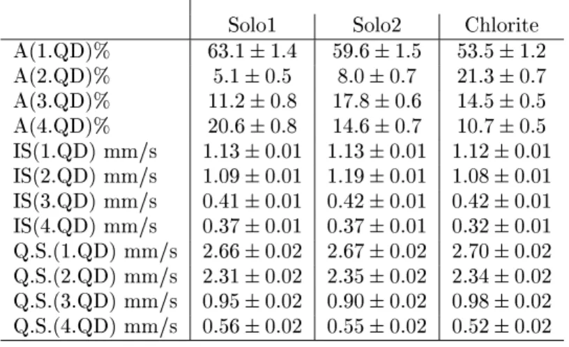
Table 8 - Mossbauer parameters of paramagnetic spectral parts of Antarctic soils and of a pure chlorite sample Solo1 Solo2 Chlorite
A(1.QD)% 63:11:4 59:61:5 53:51:2
A(2.QD)% 5:10:5 8:00:7 21:30:7
A(3.QD)% 11:20:8 17:80:6 14:50:5
A(4.QD)% 20:60:8 14:60:7 10:70:5
IS(1.QD) mm/s 1:130:01 1:130:01 1:120:01
IS(2.QD) mm/s 1:090:01 1:190:01 1:080:01
IS(3.QD) mm/s 0:410:01 0:420:01 0:420:01
IS(4.QD) mm/s 0:370:01 0:370:01 0:320:01
Q.S.(1.QD) mm/s 2:660:02 2:670:02 2:700:02
Q.S.(2.QD) mm/s 2:310:02 2:350:02 2:340:02
Q.S.(3.QD) mm/s 0:950:02 0:900:02 0:980:02
Q.S.(4.QD) mm/s 0:560:02 0:550:02 0:520:02
Figure 5. Paramagnetic part of Mossbauer spectra (on the left side) and quadrupol splitting distributions (on the right site) of soil sample SOLO2 (a, d), chlorite (b,e) and soil sample SOLO1 (C, f).
Based on the results of the quadrupole splitting dis-tribution method, four doublets of Lorentzian lines were used for the conventional decomposition of the obtained Mossbauer spectra of soil samples. By this way, all paramagnetic part of the Mossbauer spectrum could be decomposed into four doublets (D1, D2, D3 and D4); the corresponding parameters are tabulated in Table 9. By comparing the above data, corresponding paramag-netic mineral of the soil samples was determined to be chlorite in both soil samples, (the Mossbauer parame-ters are in fairly good agreement with those observed with a pure chlorite sample as well as with those
re-ported in the literature [24,28]. This conclusion is also in agreement with the X-ray diractometry, wherein ex-cept hematite, only chlorite could be identied as iron-bearing mineral.
(kaoli-nite, smectite) can not be entirely excluded apart from chlorite. Because our results can be well understood and because iron prefers to accumulate in the chlorite [27] the presence of other silicate phases is neglected. The four doublets in the chlorite Mossbauer spectrum are assigned as follow [23]: D1 represents Fe2+ at M2
site, D2 as reects Fe2+at M1 site, D3 belongs to Fe3+
at M1 site and D4 represents Fe3+ at M2 site.
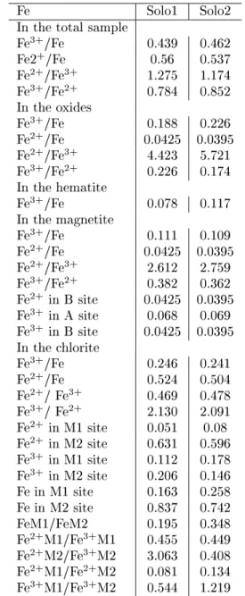
Table 9 - Distribution of iron in Antarctic soils Fe Solo1 Solo2 In the total sample
Fe3+/Fe 0.439 0.462
Fe2+/Fe 0.56 0.537
Fe2+/Fe3+ 1.275 1.174
Fe3+/Fe2+ 0.784 0.852
In the oxides
Fe3+/Fe 0.188 0.226
Fe2+/Fe 0.0425 0.0395
Fe2+/Fe3+ 4.423 5.721
Fe3+/Fe2+ 0.226 0.174
In the hematite
Fe3+/Fe 0.078 0.117
In the magnetite
Fe3+/Fe 0.111 0.109
Fe2+/Fe 0.0425 0.0395
Fe2+/Fe3+ 2.612 2.759
Fe3+/Fe2+ 0.382 0.362
Fe2+ in B site 0.0425 0.0395
Fe3+ in A site 0.068 0.069
Fe3+ in B site 0.0425 0.0395
In the chlorite
Fe3+/Fe 0.246 0.241
Fe2+/Fe 0.524 0.504
Fe2+/ Fe3+ 0.469 0.478
Fe3+/ Fe2+ 2.130 2.091
Fe2+ in M1 site 0.051 0.08
Fe2+ in M2 site 0.631 0.596
Fe3+ in M1 site 0.112 0.178
Fe3+ in M2 site 0.206 0.146
Fe in M1 site 0.163 0.258 Fe in M2 site 0.837 0.742 FeM1/FeM2 0.195 0.348 Fe2+M1/Fe3+M1 0.455 0.449
Fe2+M2/Fe3+M2 3.063 0.408
Fe2+M1/Fe2+M2 0.081 0.134
Fe3+M1/Fe3+M2 0.544 1.219
The dierence in the characteristic isomer shift and the quadrupole splitting of doublets reects dierences in the microenvironments of M1 and M2 sites due to the
dierence in the cation distribution in the soil samples. This is in good correlation with changes found in the lattice parameters of the chlorites in these soils by the help of X-ray diractometry. Also, signicant dier-ences between the relative areas of doublets of SOLO1 and SOLO2 (Table 8) have been found. These reect dierent site occupation of iron ions in the two soil samples. The iron distribution data derived from the Mossbauer parameters are shown in Table 9.
Considering the summarized analytical result of Mossbauer spectroscopy (Table 10), it can be concluded that the hematite content is signicantly higher in sam-ple SOLO2 than in samsam-ple SOLO1. The dierence in the phase composition, and the dierence of iron dis-tribution between the dierent crystallographic sites in the minerals of these soil samples reect the weathering.
Table 10. Distribution of iron in the iron bearing phases derived from the Mossbauer analysis.
Material Oxides Silicates sample Fe2O3% Fe3O4 % Chlorite %
Solo1 7:80:4 15:31:01 76:91:2
Solo2 11:70:5 14:81:4 7351:4
In desert varnish layers originated from the Upper Taylor Valley of Antarctica weathering has also been observed by Mossbauer spectroscopy [29], however, the iron distribution in the cold desert samples was com-pletely dierent from the samples of the present investi-gation, that is, no hematite, magnetite or chlorite could be identied in the desert varnish layer. The iron con-tent of the our soil samples (Table 2) is much higher than that (0.56-2.5 wt%) found in the cold desert soils in Antarctica [30]
in our soil samples indicate the chemical weathering of
the local rock minerals according to the decomposition process Antarctica [39].
c
d
Upper Cretaceous-Lower Tertiary volcanic rocks are below the investigated soils [5-7]. Pyroxene andesite (which is the dominant lava), contains phenocrysts of plagioclase, augite, hypersthene and opaque ore as well as varying amounts of ferromagnesian minerals [5-7]. The original ferromagnesian minerals have been al-tered or completely destroyed, but from the shape of the phenocryst [5] concluded that the original rocks included olivine basalts, hypersthene-augite-andesites and augite-andesites [5]. Apart from the eect of plu-tonic intrusions [5], characterized by epidote and pyrite mineralization accompanied by widespread albitization of plagioclase, the alterations of these rocks can be as-sociated with weathering.
It has been suggested [5] that pyrite mineralization has taken place in the Keller peninsula. Presence of hematite in our soil samples is in agreement with this suggestion; because, a part of the mineralized pyrite has transformed to a part of hematite after chemical weathering processes.
The correlation of Na/K ratio (Table 4) with the feldspar content (Table 6) can also indicate that a large part of these cations arises from mineral weathering [1]. The pH of the upper layer of soil is 5.1 (Table 1) which increases to 7.0 in the lower soil layer which is much lower than that found in the soils in the McMurdo Oasis (pH = 8) or in Inexpressible Island (pH = 9) or Edisto Inlet (pH = 9)[31] where the marine inuence is relatively strong. Even in the xerous and ultaxer-ous soils from Shackleton Glacier the pH was found to be between 6.0 and 6.5, which was associated with the eect of acidic salt derived from upper atmospheric cir-culation [31]. The low, acidic pH value of soil in our case indicate that the upper atmospheric circulation is an essential source of weathering in the King George
Is-land, even by taking into consideration some variations of meteorological parameters?[19]. However, some in-uence can also be attributed to the organic matter con-tent (Table 3), which decreases at the lower soil layer, where the pH is neutral. The dierence in the pH value is a indication of dierent chemical weathering condi-tions. The higher pH value gave a favorable condition for the oxidation processes at the lower layers.
The eect of atmospheric circulation is also sup-ported by the result of radiometry measurements on the soil samples. Namely, the Cs-137 activity of SOLO1 sample came from the Cs on the surface originated from the atmospheric circulation [32].
IV Conclusions
Soils from the Keller Peninsula, King George Island, Antarctica, were investigated by57Fe Mossbauer
spec-troscopy, X-ray diractometry, radiometry, neutron activation analysis and chemical analytical methods. Quartz, feldspar, chlorite, hematite and magnetite were identied as constituent minerals in the soil samples. The iron distribution among the crystallographic sites of chlorite, magnetite and hematite has been deter-mined. At dierent depths soils had signicant dif-ferences in the mineral composition, in the iron dis-tribution among the crystallographic site of chlorite, in pH, in the occurrence of water soluble salts, and in the specic radioactivity of Cs and K radionuclides. These results indicates chemical weathering of minerals in this maritime part of Antarctica.
Acknowledgments
Kuz-mann thanks the Conselho Nacional de Desenvolvi-mento Cientco e Tecnologico for a foreign visiting re-search fellowship (project CNPQ VKG 520414/96-9).
References
[1] G.G.C. Claridge, I. B. Campbell, Physical Geography, Soils, In: Antarctica, Key Environments, Pergamon Press, N.Y. Oxford, Toronto, Sydney, Paris, Frankfurt, pp 62-71 (1984).
[2] R.M.G. O'Brien, J.C.C. Romans, L. Robertson, Bull. Br. Antarc, Surv.47, 1 (1979).
[3] K. R. Everett, Rep. Inst. Polar. Stud.58, 14 (1976).
[4] K. Hall, Br Antarc Survey Bull.79, 17 (1988).
[5] D. D. Hawkes, Sci. Repts. Falkl. Isl. Dep. Survey,26,
1 (1961).
[6] C. M. Barton, Sci Repts Br Antarc Survey, 44, 1
(1965).
[7] J. L. Smellie, R. J. Pankhurst, M. R. Thompson, R. E. S. Davies, Sci. Repts. Br. Antarc. Survey,87, 1 (1984).
[8] W. A. Ascroft, Sci. Repts. Br. Antarc. Survey, 66, 1
(1972).
[9] P. E. Baker, I. McReath, M. R. Harvey, M. J. Rool-bol, T. J. Davies, Sci. Repts. Br. Antarc. Survey,78, 1
(1975).
[10] K. Birkenmajer, Stud. Geol. Polon. 64, 7 (1980).
[11] P. C. Kang, M. S. Jing, Petrology and Geologic Struc-tures of the Barton Peninsula, King George Island, Antarctica, In (Antarctic Science: Geology and Biol-ogy (Eds. H. T. Huh, B.K. Park, S.H. Lee), Korean Ocean Research, Seoul, 121-135 (1989).
[12] R. A. J. Trouw, A. Ribeiro, F. V. P. Paciullo, Anais da Academia Brasileira Ci^encias, Suppl.58, 157 (1986).
[13] E. Valenzuela, F. Herve, Geology of Byers Penin-sula, Livingston Island, South Shetland Islan eds, In:
Antarctica Geology and Geophysics (R.J. Adie Ed.), Universitatsforlaget, Oslo, p.83-91 (1972).
[14] H. C. Fensterseifer, E. Soliani Jr, M. A. F. Hansen, F. L. Trojan, Ser. Scient. INACH,38, 29 (1988).
[15] F. L. Trojan, H. C. Fensterseifer, M. A. F. Hansewn, Ser. Scient. INACH,40, 9 (1990).
[16] M. A. Parada, J. P. Orsini, A. Hurtado, l. Garido, A. Sina, Ser. Scient. INACH,36, 9 (1987).
[17] P. R. Dos Santos, A. C. Rocha Campos, R. Trompette, A. Uhlein, M. Gipp, J. C. Simoes, Preliminary report, Pesq. Antarct. Bras.2, 87 (1990).
[18] Y. Ikeda, L. B. De Miranda, M. Iwai, V. V. Furtado, P. L. Cacciari, Ann. Acad. Sci. Br. Suppl. 117-135 (1986). [19] I. Fonseca, A. Cavalcanti, Ann. Acad. Sci. Br. Suppl.
172-179 (1986).
[20] E. B. Pereira, D. J. R. Nordeman, M. B. A. Vasconcelos Ann. Acad. Sci. Br. Suppl. 182-186 (1986).
[21] A. M. Paviglione, Y. Ikeda, P. L. Cacciari, Ann. Acad. Sci. Br. Suppl. 150-156 (1986).
[22] Z. Klencsar, E. Kuzmann, A. Vertes, J. Radional. Nucl. Chem. Lett.210, 105. (1996).
[23] E. Kuzmann, S. Nagy, A. Vertes, T. G. Weiszburg, V. K. Garg, Geological and Mineralogical Application of Mossbauer eect, inNuclear Methods in Geology(eds. A. Vertes, S. Nagy, K. Suvegh), Plenum Press, NY. 1998.
[24] J. G. Stevens, H. Pollak, Lhi. Zhe, V. E. Stevens, R. M. White, J. L. Gibson, Mineral: Data, Mossbauer Ef-fect Data Center, Univ. North Carolina, Asheville, NC 28814 (1982).
[25] E. Murad, Hyp. Int.47, 33 (1989).
[26] E. Kuzmann, S. Nagy, A. Vertes, Analytical Applica-tion of Mossbauer Spectroscopy, In: Nuclear Methods in Chemical Analysis (Ed Z. Alfassi) N.Y. (1994). [27] E. Murad, J. H. Johnston, Iron oxydes and
oxyhydrox-ides, In Mossbauer Spectroscopy Applied to Inorganic Chemistry(Ed. G.J. Long), Vol. 2. Plenum, NY. Lon-don pp 507-583 (1984).
[28] S. Mitra,Applied Mossbauer Spectroscopy, Physics and Chemistry of Earth Vol. 18, Pergamon, Oxford, N.Y. (1992). p. 197.
[29] J. H. Johnston, C. M. Cardile, Chem Geol 45, 73
(1984).
[30] J. R. Keys, K. Williams, Geochim. Cosmichim. Acta
45, 2299 (1981).
[31] G. G. C. Claridge, l. B. Campbell, Soil Sci.,123, 377
(1977).


