http://dx.doi.org/10.1590/S1806-37132015000004247
Formation of multiple pulmonary nodules
during treatment with leflunomide*
Formação de múltiplos nódulos pulmonares durante tratamento com leflunomida
Gilberto Toshikawa Yoshikawa1, George Alberto da Silva Dias1, Satomi Fujihara1,
Luigi Ferreira e Silva2, Lorena de Britto Pereira Cruz3, Hellen Thais Fuzii4,
Roberta Vilela Lopes Koyama1
Abstract
Pulmonary involvement is one of the extra-articular manifestations of rheumatoid arthritis and can be due to the disease itself or secondary to the medications used in order to treat it. We report the case of a 60-year-old woman who had been diagnosed with rheumatoid arthritis and developed multiple pulmonary nodules during treatment with leflunomide.
Keywords: Arthritis, rheumatoid; Immunosuppressive agents; Rheumatoid nodule; Lung.
1. Senior Assistant Professor. Federal University of Pará, Belém, Brazil. 2. Medical Student. Federal University of Pará, Belém, Brazil. 3. Junior Assistant Professor. Federal University of Pará, Belém, Brazil. 4. Adjunct Professor II. Federal University of Pará, Belém, Brazil. *Study carried out at the Federal University of Pará, Belém, Brazil.
Correspondence to: Gilberto Yoshikawa. Avenida Senador Lemos, 443, Edifício Village Executive, sala 908/909, CEP 66050-000, Belém, PA, Brasil.
Tel. 55 91 3241-7905. E-mail: [email protected] Financial support: None.
Submitted: 8 May 2014. Accepted, after review: 24 November 2014.
Introduction
Rheumatoid arthritis (RA) is an autoimmune disease of unknown etiology that is characterized by symmetric polyarthritis and can lead to joint deformity and destruction.(1,2) When RA involves
other organs besides the joints, there is an increase in morbidity and severity, and life expectancy can be reduced by 5 to 10 years.(1)
Pulmonary involvement in RA was first described by Ellman and Ball,(3) who reported
diffuse pulmonary fibrosis in three patients with RA. Since then, the association between pulmonary involvement and RA has been described by several authors. The risk factors for pulmonary involvement include middle age, male gender, severe erosive arthritis, high titers of rheumatoid factor, subcutaneous nodules, smoking, genetic predisposition (HLA-DRB1), and other extra-articular manifestations of RA.(4,5)
Pulmonary involvement is a severe complication of RA and can manifest as upper airway disease, interstitial lung disease, pleural effusion, bronchiolitis obliterans, fibrosing alveolitis, pulmonary rheumatoid nodules, bronchiectasis,
Caplan syndrome, pulmonary hemorrhage, organizing pneumonia, vasculitis, and pulmonary infections.(6-9)
Case report
A 60-year-old White female librarian, who had been born and raised in the city of Belém, Brazil, presented with an approximately 10-year-history of RA, as defined on the basis of the 2010 American College of Rheumatology/European League Against Rheumatism classification criteria for RA. At the time of diagnosis, she was started on methotrexate, the dose of which was progressively increased to 15 mg/week. Subsequently, methotrexate was discontinued because of gastrointestinal intolerance, and, at that time, it was decided to start the patient on leflunomide (20 mg/day) and deflazacort (12 mg/day). During the treatment period, the patient had joint improvement and, on her own initiative, reduced her leflunomide dose from 20 mg/day to 20 mg/every other day.
A chest X-ray performed 8 years previously to screen the patient for fractures after chest trauma
J Bras Pneumol. 2015;41(3):281-284
Yoshikawa GT, Dias GAS, Fujihara S, Silva LF, Cruz LBP, Fuzii HT, et al.
http://dx.doi.org/10.1590/S1806-37132015000004247
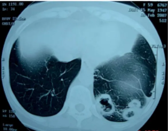
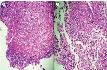
and fluconazole. After bronchoscopy showed negative BAL fluid cultures, prednisone (40 mg/ day) was commenced in an attempt to stabilize her condition. However, despite the therapeutic approach used, another CT scan of the chest revealed increased pulmonary nodules (Figure 2). At that point, the patient was referred to the city of São Paulo, Brazil, for evaluation. A lung biopsy (Figure 3) by video-assisted thoracoscopy showed a chronic inflammatory lesion, with an exudative center, adjacent to the lung parenchyma (disrupted by lymphocytic vasculitis), as well as a central cavitation filled with fibrinoleukocytic exudate and a lymphocytic infiltrate surrounded by granulation tissue. The results of AFB and fungal testing were negative, as was the result of neoplastic cell testing. In view of this result, which ruled out neoplastic and infectious disease, it was decided to discontinue leflunomide. Systemic corticosteroid therapy was continued, and azathioprine (1 mg/kg/day) was commenced. Six months after leflunomide was discontinued, the pulmonary nodules disappeared. At this writing, the patient was free of disease activity, was taking abatacept (500 mg/month), and had been off systemic corticosteroid therapy for over a year.
Discussion
Pulmonary rheumatoid nodules are extra-articular manifestations of RA.(2,4) The prevalence
of these nodules is variable: they are detected on chest X-ray in only 1% of RA patients, whereas they had shown a pulmonary nodule in the right lung
base. A chest X-ray performed before the patient was started on leflunomide had been normal. On that basis, the patient sought a pulmonologist, who requested further investigation. The results of the ancillary tests were as follows: blood workup, no changes; ESR, 65 mm/h; levels of transaminase and nitrogenous compounds, normal; antinuclear factor, negative; PCR, 6.2 mg/L; serology for viral hepatitis, negative; rheumatoid factor, positive (443 IU/mL); perinuclear and cytoplasmic antineutrophil cytoplasmic antibody (p-ANCA and c-ANCA), negative on several occasions; bronchoscopy results, normal; BAL microscopy, negative for bacteria; BAL cultures, negative; and BAL cytology, low cellularity and autolytic pattern. A CT scan of the chest showed multiple cavitary pulmonary nodules predominantly in the left lung base (Figure 1). A biopsy of a peripheral nodule revealed an acute suppurative inflammatory process with necrosis. At the time, it was decided that the patient should undergo clinical and radiological follow-up.
After a five-month follow-up of the pulmonary nodules, the patient developed dyspnea on exertion accompanied by dry cough, but no fever. Another bronchoscopy revealed laryngitis and a nodule on the right vocal fold. The tracheobronchial tree was endoscopically normal; BAL was negative for AFB, BAL microscopy revealed gram-negative bacilli (Klebsiella pneumoniae and Pseudomonas fluorescens were isolated by using an automated culture system), BAL cultures for mycobacteria and fungi were negative, and BAL cytology revealed no neoplastic cells. At that time, the patient was started on antibiotic therapy with clindamycin
Figure 1 - CT scan of the chest showing cavitary
nodular opacities.
Figure 2 - CT scan of the chest showing a nodular
opacity adjacent to the pleural surface, located in the right lower lobe.
J Bras Pneumol. 2015;41(3):281-284
Formation of multiple pulmonary nodules during treatment with leflunomide
http://dx.doi.org/10.1590/S1806-37132015000004247
rheumatoid nodules can appear during treatment with leflunomide; and there have been reports of peripheral and/or pulmonary rheumatoid nodules in patients undergoing treatment with leflunomide.(8,15,16)
References
1. Zhu H, Deng FY, Mo XB, Qiu YH, Lei SF. Pharmacogenetics and pharmacogenomics for rheumatoid arthritis responsiveness to methotrexate treatment: the 2013 update. Pharmacogenomics. 2014;15(4):551-66. http:// dx.doi.org/10.2217/pgs.14.25
2. Atzeni F, Boiardi L, Sallì S, Benucci M, Sarzi-Puttini P. Lung involvement and drug-induced lung disease in patients with rheumatoid arthritis. Expert Rev Clin Immunol. 2013;9(7):649-57. http://dx.doi.org/10.158 6/1744666X.2013.811173
3. Ellman P, Ball RE. Rheumatoid disease with joint and pulmonary manifestations. Br Med J. 1948;2(4583):816-20. http://dx.doi.org/10.1136/bmj.2.4583.816 4. Chanin K, Vallejo-Manzur F, Sternbach GL, Fromm
R, Varon J. Pulmonary manifestations of rheumatoid arthritis. Hosp Physician. 2001;37(7):23-8.
5. van Ede A, den Broeder A, Wagenaar M, van Riel P, Creemers MC. Etanercept-related extensive pulmonary nodulosis in a patient with rheumatoid arthritis. J Rheumatol. 2007;34(7):1590-2.
6. Karadag F, Polatli M, Senturk T, Kacar F, Sen S, Cildag O. Cavitary necrobiotic nodule imitating malignant lung disease in a patient without articular manifestations of rheumatoid arthritis. J Clin Rheumatol. 2003;9(4):246-52. http://dx.doi.org/10.1097/01.rhu.0000081260.50171.bf 7. Dawson JK, Graham DR, Lynch MP. Lung disease in
patients with rheumatoid arthritis. CPD Rheumatol. 2002;3(2):38-42.
8. Gauhar UA, Gaffo AL, Alarcón GS. Pulmonary manifestations of rheumatoid arthritis. Semin Respir
are identified on chest HRCT in up to 20-22% and are detected by open lung biopsy in 32%.(7,10-12)
Pulmonary rheumatoid nodules can be single or multiple and range from a few millimeters to 7 cm in diameter. They are mostly asymptomatic or produce few symptoms, although they can cause cough and bloodstained sputum, and tend to involve both lungs. They occur at the periphery of the lung, just beneath the pleura, and can cavitate in approximately one third of cases, causing hemoptysis, bronchopleural fistula, spontaneous pneumothorax, secondary infection, and abscess.(4,10,13)
The appearance of pulmonary nodules in RA patients is a diagnostic problem, and the possibilities of malignancy and tuberculosis should be ruled out.(10,13) Nodules due to lung
cancer are characteristically greater than 10 mm in diameter and have irregular borders. Metastases can also appear as multiple nodules in the lungs; however, no primary cancer was found in the case reported here.(11,12,14)
Other diagnostic possibilities include mycobacterial and fungal infections, which can manifest as nodules; however, the absence of systemic symptoms is rare.(11)
A relevant aspect in the case reported here is that the pulmonary nodules were likely related to the use of leflunomide, since there are several arguments in favor of this hypothesis: the patient in question had a long-term history of RA and had shown no signs of pulmonary involvement prior to treatment with leflunomide; pulmonary
A B
Figure 3 - Lung biopsy. In A, fibrin and collagen deposition (red arrow) surrounded
by cell debris (dark arrow), with an area of necrosis. In B, inflammatory infiltrate with multinucleated cells (dark arrow) and central necrosis.
J Bras Pneumol. 2015;41(3):281-284
Yoshikawa GT, Dias GAS, Fujihara S, Silva LF, Cruz LBP, Fuzii HT, et al.
http://dx.doi.org/10.1590/S1806-37132015000004247
53-year-old woman with arthritis and pulmonary nodules. N Engl J Med. 2001;344(13):997-1004. http://dx.doi. org/10.1056/NEJM200103293441308
13. Kairalla RA. Manifestações pulmonares das doenças do tecido conectivo (DTC). In: Zamboni M, Pereira CAC, editors. Pneumologia - Diagnóstico e Tratamento. vol.1. São Paulo: Editora Atheneu; 2006. p. 235-44. 14. Alušík Š, Fanta J, Eis V, Mandys V, Pavlicek J. Formation
of rheumatoid pulmonary nodules during the leflunomide treatment. Case Rep Clin Pract Rev. 2006;7(2):139-42. 15. Yachoui R, Ward C, Kreidy M. A rheumatoid nodule in
an unusual location: mediastinal lymph node. BMJ Case Rep. 2013;2013. pii: bcr2013009516.
16. Rozin A, Yigla M, Guralnik L, Keidar Z, Vlodavsky E, Rozenbaum M, et al. Rheumatoid lung nodulosis and osteopathy associated with leflunomide therapy. Clin Rheumatol. 2006;25(3):384-8. http://dx.doi.org/10.1007/ s10067-005-0024-1
Crit Care Med. 2007;28(4):430-40. http://dx.doi. org/10.1055/s-2007-985664
9. Rosa DJ, Paula EA, Bonfante HLM, Bonfante HL, Areal CE, Baião GS, et al. Accelerated nodulosis in rheumatoid arthritis during Leflunomide therapy [Article in Portuguese]. Rev Bras Reumatol. 2007;47(3):228-31. http://dx.doi. org/10.1590/S0482-50042007000300015
10. Burke GW, Carrington CB, Grinnan R. Pulmonary nodules and rheumatoid factor in the absence of arthritis. Chest. 1977;72(4):538-40. http://dx.doi.org/10.1378/ chest.72.4.538
11. Espinoza-Poblano E, Betancourt-Hernández L, Canizales-Cobos M, Careaga-Reyna G, Esparza-Pantoja J. Nódulos pulmonares necrobióticos en ausencia de artritis reumatoide. Reporte de un caso. Neumol Cir Torax. 2000;59(4):109-11.
12. Case records of the Massachusetts General Hospital. Weekly clinicopathological exercises. Case 10-2001. A
J Bras Pneumol. 2015;41(3):281-284