Abstract
Objective: To compare a once-daily long-acting β2 agonist (indacaterol 150 µg) with a once-daily long-acting anticholinergic (tiotropium 5 µg) in terms of their effects on exercise endurance (limit of tolerance, Tlim) in patients with moderate COPD. Secondary endpoints were their effects on lung hyperinlation, exercise-related dyspnea, and daily-life dyspnea. Methods: This was a randomized, single-blind, crossover pilot study involving 20 patients (mean age, 60.9 ± 10.0 years; mean FEV1, 69 ± 7% of predicted). Spirometric parameters, Transition Dyspnea Index scores, Tlim, and exertional dyspnea were compared after three weeks of each treatment (with a one-week washout period between treatments). Results: Nineteen patients completed the study (one having been excluded because of COPD exacerbation). Improvement in Tlim from baseline tended to be greater after treatment with tiotropium than after treatment with indacaterol (96 ± 163 s vs. 8 ± 82 s; p = 0.06). Tlim signiicantly improved from baseline after treatment with tiotropium (having increased from 396 ± 319 s to 493 ± 347 s; p = 0.010) but not after treatment with indacaterol (having increased from 393 ± 246 to 401 ± 254 s; p = 0.678). There were no differences between the two treatments regarding improvements in Borg dyspnea scores and lung hyperinlation at “isotime” and peak exercise. There were also no signiicant differences between treatments regarding Transition Dyspnea Index scores (1.5 ± 2.1 vs. 0.9 ± 2.3; p = 0.39). Conclusions: In patients with moderate COPD, tiotropium tends to improve Tlim in comparison with indacaterol. No signiicant differences were observed between the two treatments regarding their effects on lung hyperinlation, exercise-related dyspnea, and daily-life dyspnea. Future studies, including a larger number of patients, are required in order to conirm our indings and explore mechanistic explanations.
(ClinicalTrials.gov identiier: NCT01693003 [http://www.clinicaltrials.gov/])
Keywords: Pulmonary disease, chronic obstructive; Exercise; Bronchodilator agents.
Effects of indacaterol versus tiotropium on
exercise tolerance in patients with moderate
COPD: a pilot randomized crossover study
Danilo Cortozi Berton1, Álvaro Huber dos Santos2, Ivo Bohn Jr.2,Rodrigo Quevedo de Lima2, Vanderléia Breda2, Paulo José Zimermann Teixeira2,3,4
Correspondence to:
Paulo José Zimermann Teixeira. Pavilhão Pereira Filho, Santa Casa de Misericórdia de Porto Alegre, UFCSPA, Avenida Independência, 155, CEP 93510-250, Porto Alegre, RS, Brasil.
Tel.: 55 51 3346-9513. Fax: 55 51 3346-9513. E-mail: [email protected] Financial support: This study received inancial support from Novartis.
INTRODUCTION
Bronchodilators have consistently been shown to result in long-term improvements in clinical outcomes
(symptoms, exercise capacity, and airlow limitation) and
are currently considered the therapeutic mainstay for patients with COPD.(1) According to current guidelines, all
symptomatic patients with COPD should be prescribed a short-acting bronchodilator to be used on an as-needed basis. A long-acting bronchodilator should be added and used regularly if symptoms are inadequately controlled with short-acting bronchodilator therapy or if patients are at an increased risk for poor outcomes, such as frequent exacerbations and disease that is more severe.(1,2)
Until recently, a long-acting anticholinergic (LAMA) was preferred over a long-acting β2 agonist (LABA) because
most of the effects of once-daily LAMAs appeared to be superior to those of twice-daily LABAs.(3-8) The advent of once-daily LABAs (ultra-LABAs) changed that, studies
comparing once-daily LAMAs with once-daily LABAs
having demonstrated the clinical beneits of the latter. (9,10)
However, no studies have compared once-daily LABAs with once-daily LAMAs regarding clinical outcomes during exercise, including exercise tolerance, dyspnea, and
dynamic hyperinlation. Therefore, we conducted a pilot study aimed at comparing a once-daily LABA (indacaterol) with a once-daily LAMA (tiotropium) in terms of their effects
on exercise tolerance in patients with moderate COPD. Indacaterol and tiotropium were also compared in terms
of their effects on lung hyperinlation, exercise-related
dyspnea, and daily-life dyspnea.
METHODS
This was a phase IV, randomized, single-blind (i.e.,
with single-blind masking of outcome assessors),
placebo-controlled, two-period, crossover pilot study conducted at a single center specializing in respiratory
care (ClinicalTrials.gov identiier: NCT01693003).(11)
The study protocol was approved by the local research ethics committee.
1. Programa de Pós-Graduação em Pneumologia, Faculdade de Medicina, Universidade Federal do Rio Grande do Sul – UFRGS – Porto Alegre (RS) Brasil.
2. Universidade Federal de Ciências da Saúde de Porto Alegre – UFCSPA – Porto Alegre (RS) Brasil.
3. Universidade Feevale, Novo Hamburgo (RS) Brasil.
4. Pavilhão Pereira Filho, Santa Casa de Misericórdia de Porto Alegre, Porto Alegre (RS) Brasil.
Submitted: 6 January 2016.
Accepted: 31 May 2016.
Patients were randomly assigned to receive three weeks of treatment with 150 µg of inhaled indacaterol (Onbrize® Breezhaler®; Novartis, Basel, Switzerland) delivered via a capsule-based dry powder inhaler (DPI),
followed by another three weeks of treatment with 5 µg
of inhaled tiotropium (Spiriva® Respimat®; Boehringer Ingelheim, Ingelheim, Germany) delivered via a soft mist inhaler (SMI), with a one-week washout period
between the two treatment periods; or three weeks
of treatment with 5 µg of inhaled tiotropium (Spiriva® Respimat®; Boehringer Ingelheim) delivered via an SMI, followed by another three weeks of treatment with
150 µg of inhaled indacaterol (Onbrize® Breezhaler®; Novartis) delivered via a capsule-based DPI, with a
one-week washout period between the two treatment
periods (Figure 1). After a screening visit (on day 7), all
long-acting bronchodilators were discontinued. Patients were allowed to use short-acting bronchodilators, being instructed to use two puffs every 4 h as rescue medication. They were also allowed to use inhaled corticosteroids, provided that the dose, schedule, and formulation remained unchanged.
At the baseline visit, patients underwent clinical evaluation, pulmonary function testing, and incremental symptom-limited cardiopulmonary exercise testing
(CPET). At visits 1 through 4, patients underwent constant-rate CPET to the limit of tolerance (Tlim), at
~80% of the maximum load reached during incremental CPET. Activity-related breathlessness was assessed at
baseline with the Baseline Dyspnea Index (BDI), and
changes in daily breathlessness were assessed with
the Transition Dyspnea Index (TDI),(12) being recorded at the end of each treatment period (Figure 1).
Patients
Patients presenting with stable COPD (FEV1/FVC <
0.7 and 50% < post-bronchodilator FEV1 < 80% of
predicted) and a long smoking history (> 20 pack-years) were enrolled. The exclusion criteria were as follows:
cardiovascular or neuromuscular disease potentially affecting exercise tolerance; recent exacerbation (in
the last month); long-term oxygen therapy or resting SaO2 < 90%; and treatment with oral corticosteroids.
Procedures
All spirometric tests were performed with a calibrated pneumotachograph (Vmax29®; SensorMedics, Yorba Linda, CA, USA). Spirometric variables were measured
at the baseline visit (before and 20 min after inhalation
of 400 µg of albuterol via a metered dose inhaler);
at visits 1 and 3 (after a one-week long-acting
bron-chodilator washout period and before CPET); and at
visits 2 and 4 (2 h after administration of the study
medications and before CPET). A constant-volume
body plethysmograph (Vmax Autobox®; SensorMedics)
was used in order to measure RV, functional residual
capacity, and TLC. Single-breath DLCO was measured using a Vmax System (SensorMedics). All pulmonary
function tests were performed in accordance with international standards.(13-15) The variables obtained
were expressed as absolute and percent predicted values.(16-18)
All exercise tests were performed on an elec-tromagnetically braked cycle ergometer (Corival;
Lode, Groningen, the Netherlands), with the use of
a computer-based breath-by-breath CPET system (Vmax29®; SensorMedics). HR was determined from the R-R interval of a 12-lead electrocardiogram, and SaO2
was measured by pulse oximetry. All CPET variables were presented as 20-s averages. Participants rated their shortness of breath and leg effort using the 0-10 Borg scale(19) every 2 min. During incremental
CPET, the workload was increased every 1 min from a baseline of 2 min of loadless pedaling at a rate of 5-10 W/min to Tlim. Incremental load increases were highest in patients with FEV1 > 1 L. Constant-rate
CPET was performed with loadless pedaling for 2 min
at a pedaling frequency of 60 ± 5 rpm, immediately
followed by loaded pedaling at ~80% of the maximum workload achieved during incremental CPET. Assuming that resting TLC remains constant during exercise, we
considered that changes in inspiratory capacity (IC) relected changes in end-expiratory lung volume, i.e., end-expiratory lung volume = TLC − IC.(20) IC
maneuvers were performed every 2 min. Exercise responses were compared at peak exercise and at “isotime”, i.e., the longest exercise duration common to all constant-rate cardiopulmonary exercise tests performed by a given individual.
The BDI and TDI were used in order to measure
daily-life dyspnea, and both have three domains: 1)
functional impairment, which determines the impact of breathlessness on the ability to carry out activities;
2) magnitude of task, which determines the type of task that causes breathlessness; and 3) magnitude of
effort, which establishes the level of effort that results in breathlessness. The BDI domain scores range from
0 (very severe impairment) to 4 (no impairment) and
are summed to determine the total score, which can range from 0 to 12. The TDI domain scores range from
−3 (major deterioration) to +3 (major improvement).
The sum of all domains yields the total score, which
can range from −9 to +9.(12) The minimal clinically
important difference for the TDI score is 1.(21) Safety
Safety assessments included adverse events and
serious adverse events at the end of each treatment period. HR correction of the QT interval was performed using Bazett’s correction.
Statistical analysis
Data are reported as mean ± SD or median (range),
except where otherwise indicated. Generalized estimating equations were used in order to test for
signiicant differences between treatments at different
visits and time points. Paired t-tests were used in order to compare TDI scores after each treatment and calculate the sample size required to detect a
regarding improvement in exercise tolerance (with a
type II error of 20%). The chi-square test was used
in order to compare categorical data. Differences were
considered signiicant if p < 0.05.
RESULTS
Of the 69 patients who were screened, 20 were randomized. Of those, 19 (95%) completed the
study. One patient (in the group of patients assigned
to receive indacaterol irst) was excluded because of COPD exacerbation (during treatment with indacaterol).
The baseline demographic, anthropometric, and clinical characteristics of the patients studied are
described in Table 1. A Consolidated Standards of Reporting Trials (CONSORT) low diagram of the study
is shown in Figure 2.
Effects on spirometric variables and daily-life dyspnea
After three weeks of treatment, FEV1 was signiicantly
improved from baseline in both groups (Table 2).
However, in addition to having resulted in greater improvement in FEV1, indacaterol signiicantly improved FVC when compared with tiotropium. There were
no signiicant differences between indacaterol and tiotropium regarding TDI scores (1.5 ± 2.1 vs. 0.9 ± 2.3; p = 0.39) or the proportion of patients in whom TDI scores were ≥ 1 (58% vs. 37%; p = 0.19).
Effects on exercise responses
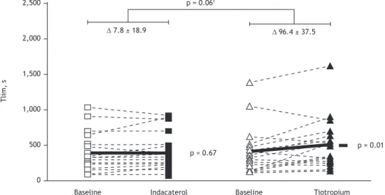
Improvement in Tlim from baseline (the primary
study outcome) tended to be greater after treatment
with tiotropium than after treatment with indacaterol
(96 ± 163 s vs. 8 ± 82 s; p = 0.06; Figure 3). Additionally, Tlim signiicantly improved from baseline
after treatment with tiotropium (having increased from
396 ± 319 s to 493 ± 347 s; p = 0.010) but not after
treatment with indacaterol (having increased from 393
± 246 s to 401 ± 254 s; p = 0.678). A sample size
of 28 was estimated to be required in order to detect
a signiicant difference in exercise tolerance between
the two treatments.
There were no differences between the two treatments regarding the magnitude of improvement in Borg
dyspnea scores (at isotime and peak exercise) or lung hyperinlation, as estimated from serial measurements of IC (at rest, isotime, and peak exercise). Lung hyperinlation was found to have improved signiicantly after treatment with bronchodilators (2.00 ± 0.33 L vs. 2.09 ± 0.31 L; p = 0.03) at all time points analyzed (i.e., at rest, isotime, and peak exercise). The same was true for exercise-related dyspnea (p = 0.067).
Safety
The overall incidence of adverse events was exactly
the same in both treatment groups (i.e., 58%), the majority of the events being mild in severity. No serious adverse events (hospitalization or death)
were reported during the study period. There was no difference between indacaterol and tiotropium in terms of their effects on the resting corrected QT
interval (445 ± 48 ms vs. 439 ± 47 ms; p > 0.05),
post-bronchodilator values being no different from
baseline values (456 ± 34 ms).
DISCUSSION
This was a pilot study designed to collect preliminary data regarding the comparative effects of indacaterol
150 µg (the lowest available dose in most countries)
and tiotropium 5 µg on exercise tolerance in patients with moderate COPD. Previous studies(22,23) have
demonstrated that indacaterol 300 µg results in
signiicant improvement in exercise tolerance and lung hyperinlation at rest and during exercise when
compared with placebo in patients with moderate to
severe COPD. Surprisingly, the present study showed that a lower dose of indacaterol (150 µg) in a subset
of patients with less severe disease did not increase
Figure 1. Flowchart of the study design. incCPET: incremental cardiopulmonary exercise testing; crCPET: constant-rate cardiopulmonary exercise testing; BDI: Baseline Dyspnea Index; and TDI: Transition Dyspnea Index.
Run-in period
7 days 21 days
Indacaterol
Indacaterol Tiotropium
Tiotropium 21 days Washout period
5-7 days
Clinical Evaluation Resting Lung Function
incCPET
• TDI • crCPET • BDI
• crCPET R
Baseline Visit
exercise tolerance from baseline. In contrast, tiotropium
5 µg signiicantly improved exercise tolerance from baseline, a inding that is consistent with those of
previous studies in which a different drug dose and delivery system were used (i.e., 18 µg of tiotropium
delivered via a DPI).(24-27)
In the present study, both drugs resulted in signiicant improvement in lung hyperinlation and exercise-related
Table 1. Baseline characteristics of the patients studied
(N = 19).a
Variable Result
Demography and anthropometry
Age, years 60.9 ± 10.0
Male/female, n/n 9/10
BMI, kg/m2 24.8 ± 3.5
Smoking history, pack-yearsb 45 (6-108) Pulmonary function
Pre-BD spirometry FEV1, L
FEV1, % of predicted
1.86 ± 0.62 67.4 ± 8.6 FVC, L
FVC, % of predicted
3.26 ± 0.83 94.1 ± 10
FEV1/FVC 57 ± 8
Post-BD spirometry FEV1, L
FEV1, % of predicted
1.89 ± 0.58 68.7 ± 7.4 FVC, L
FVC, % of predicted
3.27 ± 0.8 94.6 ± 11.2
FEV1/FVC 58 ± 8
Plethysmography IC, L
IC, % of predicted
2.15 ± 0.9 66.3 ± 20.5 TLC, L
TLC, % of predicted
5.67 ± 1.4 109.1 ± 12.7
IC/TLC 0.37 ± 0.1
RV, L
RV, % of predicted
2.36 ± 0.73 122.5 ± 33.6 DLCO, mmol/min/kPa
DLCO, % of predicted
4.4 ± 1.4 67.4 ± 18.3 Symptoms
mMRC score 2.3 ± 1.1
BDI score 8.4 ± 2.4
Peak incremental CPET
VO2, mL/min 1,083 ± 349
VO2, % of predicted 74.7 ± 16.6
VE, L 42.4 ± 14.9
VE/MVV 0.69 ± 0.17
SaO2, % 96 ± 2
HR, % of predicted 79 ± 12
VO2/HR, mL/min/bpm 8.45 ± 2.12
Borg scale, dyspnea scoreb 4 (0.5-10) Borg scale, leg effort scoreb 7 (1-10)
aValues expressed as mean ± SD, except where
otherwise indicated. bValues expressed as median
(range). BD: bronchodilator; IC: inspiratory capacity; mMRC: modiied Medical Research Council; BDI: Baseline Dyspnea Index; CPET: cardiopulmonary
exercise testing; VO2: oxygen uptake; VE: minute
ventilation; and MVV: maximal voluntary ventilation.
dyspnea, as previously described for tiotropium (18
µg delivered via a DPI)(12,19-21) and indacaterol (300 µg),(17,18) with no signiicant difference between the
two treatments. However, it is possible that our small sample size did not allow us to detect individual drug effects on the aforementioned variables or differences between the two treatments.
Although both treatments improved FEV1 from baseline, the magnitude of change was greater for
indacaterol. Similar indings have previously been
described.(10,28) With regard to clinical outcomes, a clinically relevant improvement in total TDI and Saint
George’s Respiratory Questionnaire scores is more likely to be achieved with indacaterol 150 µg than with tiotropium 18 µg in patients with moderate to severe COPD.(29) However, tiotropium has been reported to
afford greater protection against exacerbations.(30)
In the present study, indacaterol resulted in greater improvement in FEV1 than did tiotropium (Table 2). However, it did not result in improved exercise toler-ance, probably because constraints on tidal volume
expansion as a result of lung hyperinlation constitute
the main mechanism related to dyspnea and exercise
capacity, independently of the magnitude of airlow
obstruction.(20,26,31) Nevertheless, because of its small
size, our sample was probably underpowered to detect differences between the two treatments regarding this physiological variable. Therefore, other mechanisms to explain improved exercise tolerance after treatment with tiotropium should be considered and further investigated.(32) For instance, it is impossible to rule out
that our small sample size randomly included primarily
patients who were more likely to beneit from one speciic
pharmacological class of bronchodilators. Polymorphisms
of β2-adrenergic receptors can result in differences in
pharmacological responses to bronchodilators.(33,34) This
underscores the need for further, larger studies. If our
indings are conirmed, adequately powered studies
will be required in order to investigate physiological and molecular mechanistic aspects.
The present study has methodological limitations that should be noted. First, because this was an exploratory study including only a small number of patients, the results should be interpreted with caution. Our sample was possibly underpowered to detect differences in important outcomes, such as dyspnea
and lung hyperinlation, and our main indings should be conirmed in studies including a larger number of patients. Second, because the present study included
only patients with moderate COPD, the results should not be generalized to patients with mild or severe COPD. Finally, we used a low dose of indacaterol and
a full dose of tiotropium delivered via an SMI. The
dose of indacaterol used in the present study (i.e.,
150 µg) did not improve exercise tolerance as did the dose used in other studies (i.e., 300 µg).(22,23) In fact, it has been shown that indacaterol is more beneicial to resting pulmonary function at higher doses (> 200 µg) than at lower doses (of 50 µg and 100 µg); however,
Table 2. Lung function parameters at baseline and after three weeks of treatment with indacaterol or tiotropium.a
Variable Indacaterol Diff Tiotropium Diff
Baseline Post-treatment Baseline Post-treatment
FEV1, L 1.62 ± 0.12 1.82 ± 0.12* 0.20 1.69 ± 0.13 1.79 ± 0.14* 0.10
FEV1, % of predicted 56 ± 2 63 ± 2* 7† 58 ± 2 61 ± 2 3
FVC, L 2.94 ± 0.2 3.15 ± 0.17* 0.21† 3.06 ± 0.19 3.12 ± 0.2 0.06
FVC, % of predicted 80 ± 2 87 ± 2* 7† 84 ± 2 86 ± 2 2
FEV1/FVC, % 55.5 ± 2.0 57.6 ± 1.6* 2.1 55.0 ± 1.9 56.9 ± 1.9* 1.9
aData presented as mean ± SE. BD: bronchodilator; and Diff: difference between mean post-treatment values and
mean baseline values. *p < 0.05 baseline vs. post-treatment. †p < 0.05 comparison between treatment changes.
Figure 2.Consolidated Standards of Reporting Trials (CONSORT) low diagram of the study.
Excluded (n = 49)
- Failed to meet the inclusion criteria (n = 34) - Declined to participate (n = 14)
- Other reasons (n = 1) Assessed for eligibility (n = 69)
Randomized (n = 20)
Enrollment
Allocation
Follow-up
Analysis
Allocated to receive indacaterol first (n = 11) - Received allocated intervention (n = 11) - Did not receive allocated intervention (n = 0)
Allocated to receive tiotropium first (n = 9) - Received allocated intervention (n = 9) - Did not receive allocated intervention (n = 0)
Lost to follow-up (n = 1)
Reason: Discontinued treatment because of COPD exacerbation during treatment with indacaterol
Lost to follow-up (n = 0) Discontinued treatment (n = 0)
Analyzed (n = 10)
- Excluded from analysis (n = 1)
Analyzed (n = 9)
- Excluded from analysis (n = 0)
Figure 3.Individual values (dashed lines) and mean values (solid lines) of changes from baseline in the limit of tolerance (Tlim) during constant-rate cardiopulmonary exercise testing after three weeks of treatment with indacaterol (squares) or tiotropium (triangles). *p < 0.05 from baseline. †p = 0.06 for between-treatment difference.
Indacaterol
T
li
m
,
s
Tiotropium p = 0.06†
p = 0.67
p = 0.01 2,500
2,000
1,500
1,000
500
0
∆ 96.4 ± 37.5 ∆ 7.8 ± 18.9
drug result in signiicant improvement.(28) In contrast,
it has been shown that 5 µg of tiotropium delivered
via an SMI and 18 µg of the same drug delivered via
a DPI are comparable in terms of their effects on lung function(35,36) and clinical outcomes (rescue medication use, death, and exacerbation rate).(30) Given that the
doses of indacaterol approved for use in different
countries vary from 75 µg to 300 µg and that the only dose of SMI-delivered tiotropium approved for use in
COPD patients is 5 µg, we sought to compare doses that are more commonly used in clinical practice.
In conclusion, although treatment with tiotropium
at a daily dose of 5 µg resulted in a signiicant
improvement in exercise tolerance in patients with moderate COPD, treatment with indacaterol at a daily
dose of 150 µg did not. No signiicant differences
were observed between the two treatments regarding
their effects on lung hyperinlation, exercise-related
dyspnea, and daily-life dyspnea. Further studies, including a larger number of patients, are required in
order to conirm our indings and explore mechanistic
explanations.
REFERENCES
1. Vestbo J, Hurd SS, Agustí AG, Jones PW, Vogelmeier C, Anzueto A, et al. Global strategy for the diagnosis, management, and prevention
of chronic obstructive pulmonary disease: GOLD executive summary. Am J Respir Crit Care Med. 2013;187(4):347-65. http:// dx.doi.org/10.1164/rccm.201204-0596PP
2. Montes de Oca M, López Varela MV, Acuña A, Schiavi E, Rey MA, Jardim J, et al. ALAT-2014 Chronic Obstructive Pulmonary Disease
(COPD) Clinical Practice Guidelines: questions and answers. Arch Bronconeumol. 2015;51(8):403-16. http://dx.doi.org/10.1016/j.
arbres.2014.11.017
3. Donohue JF, van Noord JA, Bateman ED, Langley SJ, Lee A, Witek TJ Jr, et al. A 6-month, placebo-controlled study comparing lung function and health status changes in COPD patients treated with
tiotropium or salmeterol. Chest. 2002;122(1):47-55. http://dx.doi. org/10.1378/chest.122.1.47
4. Brusasco V, Hodder R, Miravitlles M, Korducki L, Towse L, Kesten S. Health outcomes following treatment for six months with once daily tiotropium compared with twice daily salmeterol in patients
with COPD. Thorax. 2003;58(5):399-404. Erratum in: Thorax. 2005;60(2):105. http://dx.doi.org/10.1136/thorax.58.5.399 5. Briggs DD Jr, Covelli H, Lapidus R, Bhattycharya S, Kesten S, Cassino
C. Improved daytime spirometric eficacy of tiotropium compared
with salmeterol in patients with COPD. Pulm Pharmacol Ther.
2005;18(6):397-404. http://dx.doi.org/10.1016/j.pupt.2005.02.013 6. Hodder R, Kesten S, Menjoge S, Viel K. Outcomes in COPD patients
receiving tiotropium or salmeterol plus treatment with inhaled
corticosteroids. Int J Chron Obstruct Pulmon Dis. 2007;2(2):157-67. 7. Santus P, Centanni S, Verga M, Di Marco F, Matera MG, Cazzola M.
Comparison of the acute effect of tiotropium versus a combination
therapy with single inhaler budesonide/formoterol on the degree of resting pulmonary hyperinlation. Respir Med. 2006;100(7):1277-81. http://dx.doi.org/10.1016/j.rmed.2005.10.008
8. van Noord JA, Aumann JL, Janssens E, Smeets JJ, Verhaert J, Disse B, et al. Comparison of tiotropium once daily, formoterol twice daily and both combined once daily in patients with COPD. Eur Respir J.
2005;26(2):214-22. http://dx.doi.org/10.1183/09031936.05.0014040
4
9. Rodrigo GJ, Neffen H. Comparison of indacaterol with tiotropium
or twice-daily long-acting -agonists for stable COPD: a systematic review. Chest. 2012;142(5):1104-10. http://dx.doi.org/10.1378/
chest.11-2252
10. Vogelmeier C, Ramos-Barbon D, Jack D, Piggott S, Owen R, Higgins
M, et al. Indacaterol provides 24-hour bronchodilation in COPD: a
placebo-controlled blinded comparison with tiotropium. Respir Res.
2010;11:135. http://dx.doi.org/10.1186/1465-9921-11-135 11. ClinicalTrials.gov [homepage on the Internet]. Bethesda: National
Institutes of Health [cited 2015 Jan 14]. Indacaterol Versus Tiotropium on Dynamic Hyperinlation in COPD. Available from: https://clinicaltrials.gov/ct2/show/NCT01693003
12. Mahler DA, Weinberg DH, Wells CK, Feinstein AR. The measurement of dyspnea. Contents, interobserver agreement, and physiologic
correlates of two new clinical indexes. Chest. 1984;85(6):751-8. http://dx.doi.org/10.1378/chest.85.6.751
13. Miller MR, Hankinson J, Brusasco V, Burgos F, Casaburi R, Coates A,
et al. Standardisation of spirometry. Eur Respir J. 2005;26(2):319-38. http://dx.doi.org/10.1183/09031936.05.00034805
14. Wanger J, Clausen JL, Coates A, Pedersen OF, Brusasco V, Burgos F, et al. Standardisation of the measurement of lung volumes. Eur
Respir J. 2005;26(3):511-22. http://dx.doi.org/10.1183/09031936.05
.00035005
15. Macintyre N, Crapo RO, Viegi G, Johnson DC, van der Grinten CP, Brusasco V, et al. Standardisation of the single-breath determination
of carbon monoxide uptake in the lung. Eur Respir J. 2005;26(4):720-35. http://dx.doi.org/10.1183/09031936.05.00034905
16. Pereira CA; Sato T; Rodrigues SC. New reference values for forced
spirometry in white adults in Brazil. J Bras Pneumol. 2007;33(4):397-406. http://dx.doi.org/10.1590/S1806-37132007000400008 17. Crapo RO, Morris AH, Clayton PD, Nixon CR. Lung volumes in healthy
nonsmoking adults. Bull Eur Physiopathol Respir. 1982;18(3):419-25. 18. Crapo RO, Morris AH. Standardized single breath normal values
for carbon monoxide diffusing capacity. Am Rev Respir Dis.
1981;123(2):185-9.
19. Borg GA. Psychophysical bases of perceived exertion. Med Sci
Sports Exerc. 1982;14(5):377-81
http://dx.doi.org/10.1249/00005768-198205000-00012
20. O’Donnell DE. Hyperinlation, dyspnea, and exercise intolerance
in chronic obstructive pulmonary disease. Proc Am Thorac Soc.
2006;3(2):180-4. http://dx.doi.org/10.1513/pats.200508-093DO 21. Jones PW, Beeh KM, Chapman KR, Decramer M, Mahler
DA, Wedzicha JA. Minimal clinically important differences in
pharmacological trials. Am J Respir Crit Care Med. 2014;189(3):250-5. http://dx.doi.org/10.1164/rccm.201310-1863PP
22. O’Donnell DE, Casaburi R, Vincken W, Puente-Maestu L, Swales J, Lawrence D, et al. Effect of indacaterol on exercise endurance and
lung hyperinlation in COPD. Respir Med. 2011;105(7):1030-6. http:// dx.doi.org/10.1016/j.rmed.2011.03.014
23. Beeh KM, Wagner F, Khindri S, Drollmann AF. Effect of indacaterol
on dynamic lung hyperinlation and breathlessness in hyperinlated patients with COPD. COPD. 2011;8(5):340-5. http://dx.doi.org/10.31 09/15412555.2011.594464
24. O’Donnell DE, Flüge T, Gerken F, Hamilton A, Webb K, Aguilaniu
B, et al. Effects of tiotropium on lung hyperinlation, dyspnoea and exercise tolerance in COPD. Eur Respir J. 2004;23(6):832-40. http:// dx.doi.org/10.1183/09031936.04.00116004
25. Maltais F, Hamilton A, Marciniuk D, Hernandez P, Sciurba FC, Richter K, Kesten S, O’Donnell D. Improvements in symptom-limited exercise performance over 8 h with once-daily tiotropium in patients
with COPD. Chest. 2005;128(3):1168-78. http://dx.doi.org/10.1378/
chest.128.3.1168
26. O’Donnell DE, Hamilton AL, Webb KA. Sensory-mechanical relationships during high-intensity, constant-work-rate exercise in
COPD. J Appl Physiol (1985). 2006;101(4):1025-35. http://dx.doi. org/10.1152/japplphysiol.01470.2005
27. Verkindre C, Bart F, Aguilaniu B, Fortin F, Guérin JC, Le Merre C, et
al. The effect of tiotropium on hyperinlation and exercise capacity in chronic obstructive pulmonary disease. Respiration. 2006;73(4):420-7. http://dx.doi.org/10.1159/000089655
28. Rennard S, Bantje T, Centanni S, Chanez P, Chuchalin A, D’Urzo A, et al. A dose-ranging study of indacaterol in obstructive airways disease,
with a tiotropium comparison. Respir Med. 2008;102(7):1033-44. http://dx.doi.org/10.1016/j.rmed.2008.02.001
29. Buhl R, Dunn LJ, Disdier C, Lassen C, Amos C, Henley M, et al. Blinded 12-week comparison of once-daily indacaterol and
30. Decramer ML, Chapman KR, Dahl R, Frith P, Devouassoux G, Fritscher C, et al. Once-daily indacaterol versus tiotropium for patients
with severe chronic obstructive pulmonary disease (INVIGORATE):
a randomised, blinded, parallel-group study. Lancet Respir Med.
2013;1(7):524-33. http://dx.doi.org/10.1016/S2213-2600(13)70158-9 31. O’Donnell DE, Laveneziana P, Webb K, Neder JA. Chronic obstructive
pulmonary disease: clinical integrative physiology. Clin Chest Med. 2014;35(1): 51-69. http://dx.doi.org/10.1016/j.ccm.2013.09.008 32. Trevethick M, Clarke N, Strawbridge M, Yeadon M. Inhaled
muscarinic antagonists for COPD--does an anti-inlammatory mechanism really play a role? Curr Opin Pharmacol. 2009;9(3):250-5. http://dx.doi.org/10.1016/j.coph.2009.02.003
33. Wechsler ME, Lehman E, Lazarus SC, Lemanske RF Jr, Boushey HA, Deykin A, et al. beta-Adrenergic receptor polymorphisms and
response to salmeterol. Am J Respir Crit Care Med. 2006;173(5):519-26. http://dx.doi.org/10.1164/rccm.200509-1519OC
34. Umeda N, Yoshikawa T, Kanazawa H, Hirata K, Fujimoto S.
Association of beta2-adrenoreceptor genotypes with bronchodilatory
effect of tiotropium in COPD. Respirology. 2008;13(3):346-52. http:// dx.doi.org/10.1111/j.1440-1843.2008.01259.x
35. Wise RA, Anzueto A, Cotton D, Dahl R, Devins T, Disse B, et al. Tiotropium Respimat inhaler and the risk of death in COPD. N
Engl J Med. 2013;369(16):1491-501. http://dx.doi.org/10.1056/
NEJMoa1303342
36. van Noord JA, Cornelissen PJ, Aumann JL, Platz J, Mueller A, Fogarty