227
RENAL CELL CARCINOMA IN CHILDHOOD Pediatric Urology
International Braz J Urol
Official Journal of the Brazilian Society of Urology
Vol. 30 (3): 227-229, May - June, 2004
RENAL CELL CARCINOMA IN CHILDHOOD
LUCIANO R. BARROS, SIDNEY GLINA, LUIZ F. MELLO
Section of Urology, Ipiranga Hospital, São Paulo, SP, Brazil
ABSTRACT
The renal cell carcinoma (RCC) rarely occurs in childhood. We report here 3 cases of RCC in children.
Two girls and 1 boy aged 14, 8 and 13 years old, respectively, presented with gross hema-turia as their main complaint. They underwent ultrasonography and computerized tomography, which revealed unilateral renal tumor with lymph nodal involvement in all 3 cases. They were treated with radical nephrectomy associated with regional lymphadenectomy, with histopathology of RCC. In-complete adjuvant radiotherapy was performed in 2 cases and no complementary treatment in the other one. All are disease-free in a period ranging from 9 to 77 months after diagnosis.
Radical nephrectomy associated with regional lymphadenectomy is the best treatment for RCC in childhood. The disease appears to have a less aggressive behavior in children.
Key words: kidney neoplasms; children; carcinoma, renal cell; nephrectomy Int Braz J Urol. 2004; 30: 227-9
INTRODUCTION
The incidence of renal cell carcinoma (RCC) is estimated in 0.1% to 0.3% of all tumors and 1.8% to 6.3% of all malignant renal tumors in childhood (1).
No proper therapy has been defined for chil-dren with RCC. Surgery constitutes the main treat-ment and results in cure when the tumor is localized and completely resected. The importance of radio-therapy and immunoradio-therapy is not clear and different chemotherapy regimens showed only minimal activ-ity when tested in clinical trials (1).
The authors report 3 cases of RCC in child-hood.
CASE REPORTS
Case 1
Fourteen year-old girl was seen in June 1997 with left renal colic and hematuria for 3 months. The
ultrasound (US) revealed a nodule in the upper pole of the left kidney, measuring 4.3 x 4.0 cm, with en-larged perihilar lymph nodes. The computerized to-mography (CT) confirmed the findings. She under-went radical nephrectomy with regional lym-phadenectomy in July 1997 with histopathological diagnosis of RCC in left kidney and lymph nodes in-volvement (4/4). The service of oncology initially indicated radiotherapy, which was terminated after the fourth session. The patient has been semestrally followed by US or CT and is disease-free.
Case 2
228
RENAL CELL CARCINOMA IN CHILDHOOD
involved hilar lymph nodes. Radiotherapy was ini-tially indicated by the oncology service, and termi-nated after the first session. She is disease-free.
Case 3
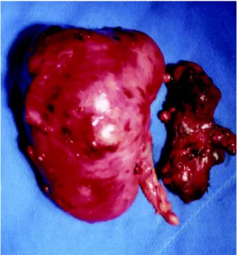
Thirteen years old boy, seen in February 2003 with hematuria for 8 months, hemoglobin of 9.3 mg/ dL and US performed 30 days before, showed a right renal tumor, which was confirmed by CT (Figure-1). He underwent radical nephrectomy with regional lym-phadenectomy (Figure-2) in March 2003, with histo-pathological diagnosis of RCC in right kidney, with predominance of papillary variant, with lymph node metastasis. He did not undergo any adjuvant therapy and is disease-free.
DISCUSSION
Recent studies showed that the RCC corre-sponds to 1.4% of renal tumors in patients under 4 years old, 15.2% between 5 and 9 years and 52.6% from 10 to 15 years old (2).
Palpable mass occurs in 38%, hematuria in 38% and abdominal pain in 50%, with the classic triad being found in only 6% of cases (3). Metastases oc-cur in lungs (40-65%), liver (35-57%), bones (10-42%) or bladder, brain or pleura (7-15%) (2). Sur-gery constitutes the main treatment (1).
Tumor staging is the most important prog-nostic factor. Overall 5-year survival is approximately 60%, with poor prognosis (9%) for stage IV (2).
Two of our patients started adjuvant radio-therapy. Since we did not find in the literature any in-centive for such procedure, we decided jointly with the service of oncology to terminate it. Considering this fact and the good survival we achieved, we can agree with the unanimous opinion expressed in the works we reviewed, that radical nephrectomy associ-ated with regional lymphadenectomy is the best treat-ment for RCC in childhood. Our results also suggest a less aggressive behavior of the disease in this age range.
REFERENCES
1. Indolfi P, Terenziani M, Casale F, Carli M, Bisogno G, Schiavetti A, et al.: Renal cell carcinoma in children: A clinicopathologic study. J Clin Oncol. 2003; 21: 530-5.
2. Uchiyama M, Iwafuchi M, Yagi M, Inuma Y, Masahiro O, Tomita Y, et al.: Treatment of childhood renal cell carcinoma with lymph node metastasis: Two cases and a review of literature. J Surg Oncol. 2000; 75: 266-9.
Figure 2 – Surgical specimen from right radical nephrectomy
and regional lymphadenectomy.
Figure 1 – Computerized tomography evidencing right renal
229
RENAL CELL CARCINOMA IN CHILDHOOD
3. Carcao MD, Taylor GP, Greenberg ML, Bernstein ML, Champagne M, Hershon L, et al.: Renal cell carcinoma
EDITORIAL COMMENT
The authors present 3 interesting cases of a rare pediatric tumor, that is renal cell carcinoma. Since the treatment with radiation therapy is not the stan-dard of care, I do not know why they treated their patients with this therapy.
The authors say that the tumor has good prog-nosis in children, and I do not believe this is what the literature says. Also, they cannot base their conclusion on their limited experience with a short term follow up
(the authors stated that because one of their patients had advanced disease and is alive, this point to better prognosis in children). It is important to note that most patients with renal cell carcinoma do not have posi-tive nodes, and that in the present series the incidence was higher than expected (even though again it is a limited experience). Also, it is important to remember that lymph node disease is known to significantly worsen the survival of patients with renal cell carci-noma.
Dr. E. Tavora Fernandes
Chief of Urology Department of Veterans Affairs Minneapolis, Minnesota, USA
in children: A different disorder from its adult coun-terpart? Med Pediatr Oncol. 1998; 31:153-8.
Received: December 12, 2003 Accepted after revision: April 4, 2004
Correspondence address:
Dr. Luciano da Rocha Barros Rua do Arraial, 209 / 24B São Paulo, SP, 04122-030, Brazil Fax: + 55 11 6169-9629