Modification of the gasless fetoscopy technique for the
treatment of large myelomeningocele: a study in sheep
Modificação da técnica de fetoscopia de suspensão para tratamento fetal de grandes
meningomieloceles: estudo em ovelhas
Denise Araujo Lapa Pedreira1, Gregório Lorenzo Acácio2, Rogério Chaccur Abou-Jamra3†, Rita de Cássia Sanches Oliveira4, Elia Garcia Caldini5, Paulo Hilário Nascimento Saldiva6
ABSTRACT
Objective: To change the gasless fetoscopy technique in order to
reduce the diameter of entry orifices in the myometrium. Methods:
Seven pregnant ewes were submitted to fetoscopy for repairing a large skin defect measuring 4.0 x 3.0 cm, created in the fetal lumbar region at the gestational age of 100 days. The defect was repaired through continuous suture of the skin with approximation of borders. Gasless fetoscopy was used for performing the suture with three orifices to allow entry of the trocar into the myometrium. A 5.0-mm optical trocar, and 3.5-5.0-mm grasping, dissecting and suturing forceps were used. After surgery, pregnancy was maintained until the animals were euthanized on the 133rd day of gestation, and the
fetuses were evaluated. Results: Seven pregnant ewes underwent surgery; the first two cases were characterized as the Pilot Group, in which the endoscopic technique was modified and caliber reduction was possible in two out of three entry orifices in the myometrium. In the five remaining cases (Study Group), the repair was successfully carried out in all the fetuses, and the mean duration of fetoscopy was 98 minutes. There was a case of maternal death attributed to intrauterine infection. Mean intrauterine permanence after surgery was 12 days. Conclusions: The technique was successfully modified, allowing reduction of the uterine orifices necessary to perform the repair of a skin defect in the fetal lumbar region through a new fetoscopy technique. The impact of this modification in repair of myelomeningocele in human fetuses should be studied.
Keywords: Fetoscopy/methods; Fetal therapies/methods;
Meningomyelocele/surgery; Sheep/abnormalities; Disease models, animal; Surgical procedures, operative; Suture techniques; Ultrasonography; Video-assisted surgery
RESUMO
Objetivo: Modificar a técnica de fetoscopia de suspensão para
redução do calibre dos orifícios de entrada no miométrio. Métodos:
Sete ovelhas grávidas foram submetidas à fetoscopia para correção de um grande defeito de pele medindo 4,0 x 3,0 cm, criado na região lombar fetal com 100 dias de gestação. O defeito era corrigido através de sutura contínua da pele, aproximando-se as suas bordas. Para realizar a sutura, foi realizada a fetoscopia de suspensão, utilizando-se três orifícios para entrada de trocarte no miométrio. O trocarte da ótica era de 5,0 mm e das pinças de apreensão, dissecção e sutura eram de 3,5 mm. Após a cirurgia, a gestação era mantida até o sacrifício, realizado no 133º dia de gestação, quando os fetos eram avaliados.
Resultados: Sete ovelhas prenhes foram operadas. Os dois primeiros
casos constituíram o Grupo Piloto, no qual a técnica endoscópica foi modificada e a redução do calibre foi possível em dois dos três orifícios de entrada no miométrio. Nos cinco demais casos (Grupo de Estudo) a correção foi realizada com sucesso em todos os fetos e o tempo médio de duração da fetoscopia foi de 98 minutos. Houve um caso de morte materna atribuído à infecção intrauterina. A média de permanência intraútero após a cirurgia foi de 12 dias. Conclusões:
A técnica foi modificada com sucesso, permitindo a redução do calibre dos orifícios uterinos necessários para realizar a correção de um defeito de pele na região lombar do feto através de uma nova técnica de fetoscopia. O impacto desta modificação na correção da meningomielocele em fetos humanos deve ser estudado.
Descritores: Fetoscopia/métodos; Terapias fetais/métodos;
Meningomielocele/cirurgia; Ovinos/anormalidades; Modelos animais de doenças; Procedimentos cirúrgicos operatórios; Técnicas de sutura; Ultrassonografia; Cirurgia vídeo-assistida
Study carried out at Centro de Experimentação e Treinamento em Cirurgia – CETEC, Instituto Israelita de Ensino e Pesquisa Albert Einstein – IIEPAE, São Paulo (SP), Brazil.
1 PhD; Fetal Medicine specialist, Department of Pathology of Faculdade de Medicina da Universidade de São Paulo – USP, São Paulo (SP), Brazil.
2 PhD; Fetal Medicine specialist, Department of Obstetrics of Faculdade de Medicina da Universidade de Taubaté – UNITAU, São Paulo (SP), Brazil.
3 †In Memoriam; Veterinarian, PhD student, Department of Pathology, Faculdade de Medicina da Universidade de São Paulo – USP, São Paulo (SP), Brazil. 4 PhD; Fetal Medicine specialist, Department of Pathology of Faculdade de Medicina da Universidade de São Paulo – USP, São Paulo (SP), Brazil. 5 Pathologist; Professor of Department of Pathology of Faculdade de Medicina da Universidade de São Paulo – USP, São Paulo (SP), Brazil.
6 Pathologist; Chairman of Department of Pathology of Faculdade de Medicina da Universidade de São Paulo – USP, São Paulo (SP), Brazil.
Corresponding author: Denise Araujo Lapa Pedreira – Rua Bagé, 163, apto 182 – Vila Mariana – CEP 04012-140 – São Paulo (SP), Brazil - Tel.: 11 5572-2033 - e-mail: wdpedreira@uol.com.br
Received on: Jul 30, 2009 – Accepted on: Dec 14, 2009
INTRODUCTION
Prenatal repair of myelomeningocele seems to improve the neurological prognosis of affected individuals and its repair has been investigated in a North American collaborative study named MOMS (Management of Myelomeningocele Study)(1). In this study, the defect repair is carried out through exposure of the fetal area to be operated with open surgery, i.e., after maternal laparotomy, followed by opening of the uterus and amniotic membranes.
Open surgery in fetuses were first performed in the end of the 1980’s for surgical treatments of fetal anomalies; however, the possibility of neurological sequelae in the operated fetuses led to discontinuation of this approach(2). Therefore, repair of fetal defects has gradually migrated to other techniques, such as endoscopic fetal surgery or fetoscopy. The fetoscopic approach was initially established for treating feto-fetal transfusion syndrome(3,4) and, recently, for the repair of congenital diaphragmatic hernia(5).
Fetoscopy is performed through a single percutaneous entry until the uterine cavity is reached(3-5). One of the main complications of this type of procedure is the premature rupture of membranes, and this risk seems to be directly related to the diameter of the instrument used. To date, there are no established techniques to close the orifice caused in the membranes by inserting the fetoscope(6), which could reduce this risk.
Our group has been studying an alternative technique for endoscopic repair of myelomeningocele in animals(7) by developing an innovative technique named gasless fetoscopy. The original technique was based on the insertion of three 5.0-mm trocars in the uterine wall for fetal surgery. However, new laparoscopy instruments with smaller diameters were developed in the last years, and we decided to try them with the purpose of reducing the risk of premature rupture of membranes in the postoperative clinical course.
OBJETIVE
To change the gasless fetoscopy technique in order to reduce the diameter of entry orifices in the myometrium.
METHODS
Seven pregnant ewes with known gestational age were brought to the laboratory at least seven days before the surgical procedure to allow their acclimatization. The animals were kept in a closed and calm environment with day/night variation.
Surgery: day 100 of gestation
The animals were submitted to feed fasting for two days, and water was allowed up to 12 hours before the surgery. Still at the pen, the animals were administered 0.2 to 0.4 mg/kg acepromazine 1% and 0.3 to 0.5 mg/kg midazolam intravenously so they were sedated during hair shaving and transportation to the operating room. Thiopental (7.5 to 10 mg/kg) was used for anesthesia, followed by endotracheal intubation and maintenance with isoflurane 2%. Intravenous enrofloxacin (5.0 mg/ kg) was used for antibiotic prophylaxis before the surgical procedure.
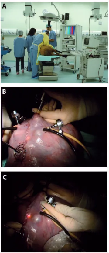
After asepsis, antisepsis and placement of surgical drapes, the maternal abdominal wall was opened and the uterus was exposed. Fetal exposure was verified by means of ultrasound and, after locating the fetal lumbar region, a 7.0-cm hysterotomy was performed to create a defect in the fetus skin (Figures 1A and B). The defect created was large enough to prevent immediate approximation of fetal skin. This defect measured 4.0 x 3.0 cm and skin and subcutaneous tissue were removed.
The fetal skin defect was left open, and the uterus was closed with continuous suture. Subsequently, three unvalved trocars were inserted, one measuring 5.0 mm and two measuring 3.5 mm, and fixed by purse-string suture (left with repair) at the site of uterine insertion.
Straight optics measuring 4.0 mm with a 30o angle was inserted (HStratner®, Brazil, Karl Storz®, Germany) through the 5.0-mm trocar for inspection of the cavity and direct visualization of the defect. A total of 100 ml of amniotic fluid was removed and stored in sterile syringes, which were maintained heated for further restitution.
The myometrium located immediately over the center of the lesion created on the fetal dorsum was grasped with a forceps. In this region, a helical forceps was inserted for distancing the uterine wall above the lesion and forming a “tent”. Myometrial “suspension” clamp, with a cutting tip, was inserted in the myometrial thickness through forceps rotation in its larger axis with divulsion of muscular fibers in its path until finally penetrating the amniotic membranes through a single orifice.
space between the dermis and subcutaneous tissue that allowed partial reapproximation of the skin borders.
A biosynthetic cellulose film (Bionext®, São Paulo, Brazil) measuring 2.0 x 1.0 cm was placed on the skin defect in the fetal lumbar region. Artificial skin (Integra®, Integra, USA) measuring 3.0 x 2.0 cm was placed over the film, below the previously dissected borders under the skin.
An attempt to approximate the skin margins on the defect midline was carried out with continuous mononylon 4-0 suture, burying the cellulose and artificial skin, which was purposely exposed in the median region of the defect. Our purpose was to impede the lesion size from allowing the reapproximation of the skin borders, thus mimicking what happens in a large myelomeningocele after birth.
After completing the fetal skin suture, the suspension clamp was removed, followed by the trocars. Upon removal of the last trocar, the stored amniotic fluid was recovered and efforts were made to remove all the air before closing the purse-string suture that remained repaired for closure. After transuterine ultrasound evaluation of fetal vitality, maternal abdominal wall closure was performed by layers, and the animals were sent back to the pen after anesthetic recovery.
Euthanasia: gestational day 133
The ewes were euthanized , assuring that they did not have suffering. To that end, the same aforementioned anesthetic protocol was used, except for the orotracheal intubation and the use of isoflurane. The thiopental dose used was increased to 20 mg/kg, assuring maternal and fetal sedation. Once sedation was assured, a bolus injection of 0.4 ml/kg KCL 19.1% was injected in the maternal bloodstream. After arrest of the maternal heart beats, the uterus wall was opened and the fetus was removed, once the stop of its heart beats was assured.
The fetuses were photographed and had their skin, subcutaneous tissue and paravertebral musculature below the operated region removed in a block for anatomopathological examination.
This study was approved by the Research Ethics Committee of Instituto Israelita Albert Einstein.
RESULTS
Seven animals were operated, and the first two animals composed the pilot group. In these cases, the utilization of the smaller diameter (2.0 and 3.0 mm) optics was tested, but it did not provide an appropriate image of the defect. The smaller the optics diameter was, the smaller the wide angle visualization and the illumination within the uterine cavity were. Therefore, it was established
Figure 1. (A) Animal preparation for endoscopic fetal surgery. Note the ultrasound equipment located beside the videolaparoscope. The procedure utilizes a sono-endoscopic approach. (B) After uterine exposure and creation of a skin defect over the fetal spine, the three trocars were placed. (C) Ultrasound is used to examine fetal vitality during the procedure.
dislodgement outside the uterine cavity and helping in the enlargement of the uterine “tent”.
that the minimum optics diameter was 4.0 mm for an appropriate visualization of the defect. However, it was possible to reduce the diameter of the two auxiliary orifices for forceps passage from 5.0 to 3.5 mm, and this was the technique used in the five remaining cases.
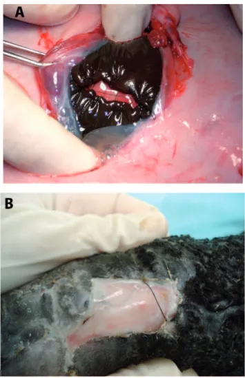
In the pilot cases, closure of the fetal skin defect was carried out through an open technique (Figure 2A) in order to avoid prolonged surgical time; however, the technique for closing the defect itself was the same in all seven cases.
In three out of four remaining cases, premature birth occurred 3, 4 and 15 days after surgery, with animal euthanasia carried out in a case of 21-day intrauterine stay. The mean intrauterine permanence after the repair was 12 days.
The suture was observed, with approximation of the borders of the skin defect in the fetal lumbar region in all cases (Figure 2A). Cellulose and artificial skin used to correct the defect were observed in all cases but one, in which none of the materials was found at birth. In this case, we believe they may have been dislodged during the premature labor that occurred four days after repair.
The artificial skin was firmly adhered to the fetal skin in the two specimens that remained for a longer time in the uterus (15 and 21 days). In the latter, there were signs of mild infection at the suture site (Figure 2B).
DISCUSSION
The animal model chosen for the study in fetal surgery has been sheep fetuses(8-10). However, there are limitations associated with this model, such as difficult evaluation of amniotic fluid loss in the postoperative period. Fetoscopy with a single entry orifice increases the risk of premature rupture of the membranes, which may be observed in 10 to 20% of cases(11). This risk varies depending on the type of approach to be performed in the uterine cavity, with greater manipulation leading to higher risk of rupture of the membranes. To date, the risks of fetoscopy with three entry orifices are unknown; however, reducing the number and diameter of the entry
Figure 2. (A) Appearance of continuous suture approximating the defect borders which was performed outdoors during the pilot study. (B) Same fetus, after birth; it is possible to observe the suture and adherence of the artificial skin to the adjacent tissues
The mean total operative time in the five cases was 224 minutes, and the duration of the endoscopic procedure was 98 minutes (Table 1 and Figure 3).
All fetuses survived at the end of surgery; however, there was a maternal death and, consequently, a fetal death 20 hours after the end of surgical procedure. This maternal death was attributed to intrauterine infection, for the membranes and amniotic fluid showed a large quantity of purulent secretion of fetid odor. This case was excluded from the other analyses.
Case
GA at surgery
(days)
Course of gestation
Intrauterine permanence
(days)
Total time (minutes)
Endoscopy time (minutes)
1 100 PD 4* 210 90
2 108 PD 15† 260 120
3 106 PD 3 260 120
4 112 MD - 200 100
5 114 S 21 190 60
Mean 108 - 12 224 98
Table 1. Operative time and duration of gestation after surgery in five of the studied cases
GA: gestational age at surgery; MD: maternal death; PP: premature delivery; S: sacrifice. * cellulose and artificial skin not found on defect; † signs of local infection.
Duration (minutes)
Animals
Total
Fetoscopy
orifices has the potential to reduce maternal/fetal risks. Likewise, the use of sealants or plugs to close the orifices in the membranes may also reduce the occurrence of membrane rupture.
In a previous study in which the gasless fetoscopy technique was developed, three 5.0-mm trocars were used to correct a defect created in the fetal skin; however, the defect size allowed complete skin closure(7) and these were the main differences between the two studies.
In the current study, premature labor was the main complication found, and it was comparable to our previous study, accounting for 50% of cases(7).
As to duration of the endoscopic surgery, the mean time in the current study was comparable to our previous study(7), which lasted 105 minutes. Taking into account that the surgical technique used for closing the defect in the present study was a little more complex than the one utilized in the previous study, we can consider that the learning curve of this fetoscopy technique is already stabilized. In the current study, artificial skin was used to cover cellulose, adding this operative time to the total endoscopy time.
In the present study, fetal mortality was much lower than that found in our previous studies, when we evaluated the surgical technique for closing a defect that was similar to myelomeningocele in the fetuses. The mortality rates found in previous studies was 38.5 and 36.1%(9,10), while in the current study there was only one death out of five operated cases. We believe that this difference is probably related to the defect and to the gestational age of its creation. In this study, no defects in the spinal cord were created, but only a deep skin defect, while in previous studies the defect involved removal of tissue in all layers: skin, paravertebral musculature, dura mater until reaching the spinal cord. We suppose that a more aggressive defect tends to lead to higher fetal mortality rates. Also, in the current study the defect was created at the time of repair surgery, while in the previous analyses the defect was created three weeks before the repair. In our opinion, the earlier the gestational age of defect creation is, the higher the fetal mortality rates associated to its creation are.
We recommend that, when the study purpose is only to modify the surgical approach and not repair the defect itself, the creation of a skin defect at the same time as the repair is an efficient way to reducing fetal mortality. This approach also aims to comply with the ethical standards that provide using the lowest number of animals as possible in a study(12).
In our caseload, we had a case of severe intrauterine infection which may have been the most probable cause of maternal death, although the possibility of amniotic embolism cannot be ruled out; we believe that potential modifications of the technique on this subject might be
necessary. This occurrence also suggests that the use of intra-amniotic antibiotic is indicated in addition to the intravenous prophylaxis that was used in all of our animals.
Gasless fetoscopy has still not been used in humans, and we suppose that any attempt to reduce its risks might be beneficial. We consider that new modifications of the technique and its training in an animal model might be carried out before performing this study in humans.
In 1999, Brunner et al.(13) used an endoscopic technique with gas injection in the cavity for repairing myelomeningocele in four human fetuses, but the lack of success led to discontinuation of this technique. In our opinion, the study of the new technique proposed by the author could have been benefited from its previous application and training in more adequate animal models.
In a recent North American review about prenatal repair of myelomeningocele, the author emphasizes the importance of the study of new techniques for repairing the defect and cites gasless fetoscopy as one of the promising techniques in this field(14). The modification presented in this study has the potential to reduce the maternal/fetal risks associated with the endoscopic fetal surgery, and this aspect must be studied.
CONCLUSIONS
Gasless fetoscopy technique with reduction of the diameter of the two entry orifices in the uterus was successfully modified.
ACKNOWLEDGEMENTS
This study was funded by Instituto de Ensino e Pesquisa da Sociedade Beneficente Israelita Albert Einstein, São Paulo (SP), Brazil.
The company Promedon, a retailer of Integra® (Plainsboro, USA), donated the artificial skin used in this study.
This paper is dedicated to the memory of the student Rogério Chaccur Abou-Jamra, who will always be present in our hearts.
REFERENCES
1. MOMS: Management of Myelomeningocele Study. [homepage on internet]. [cited 29 March 2009] Available from: http://www.spinabifidamoms.com/ 2. Bealer JF, Raisanen J, Skarsgard ED, Long SR, Wong K, Filly RA, et al. The
incidence and spectrum of neurological injury after open fetal surgery. J Pediatr Surg. 1995;30(8):1150-4.
4. Pedreira DA, Acácio GL, Drummond CL, Oliveira RC, Deustch AD, Taborda WG. Laser for the treatment of twin to twin transfusion syndrome. Acta Cir Bras. 2005;20(6):478-81.
5. Deprest J, Gratacos E, Nicolaides KH; FETO Task Group. Fetoscopic tracheal occlusion (FETO) for severe congenital diaphragmatic hernia: evolution of a technique and preliminary results. Ultrasound Obstet Gynecol. 2004; 24(2):121-6. 6. Lewi L, Liekens D, Heyns L, Poliard E, Beutels E, Deprest J, et al. In vitro
evaluation of the ability of platelet-rich plasma to seal an iatrogenic fetal membrane defect. Prenat Diagn. 2009;29(6):620-5.
7. Pedreira DAL, Oliveira RCS, Valente PR, Abou-Jamra RC, Araújo A, Saldiva PH. Gasless fetoscopy: a new approach for endoscopic closure of a lumbar skin defect in fetal sheep. Fetal Diagn Ther. 2008;23(4):293-8.
8. Meuli M, Meuli-Simmen C, Yingling CD, Hutchins GM, Hoffman KM, Harrison MR, et al. Creation of myelomeningocele in utero: a model of functional damage from spinal cord exposure in fetal sheep. J Pediatr Surg. 1995;30(7):1028-32.
9. Pedreira DAL, Sanchez e Oliveira Rde C, Valente PR, Abou-Jamra RC, Araújo A, Saldiva PHN. Validation of the ovine fetus as an experimental model for the human myelomeningocele defect. Acta Cir Bras. 2007;22(3):168-73. 10. Abou-Jamra RC, Valente PR, Araújo A, Sanchez e Oliveira Rde C, Saldiva PH,
Pedreira DA. Simplified correction of a meningomyelocele-like defect in the ovine fetus. Acta Cir Bras. 2009;24(3):239-44.
11. Peiró JL, Carreras E, Guillén G, Arévalo S, Sánchez-Durán MA, Higueras T, et al. Therapeutic indications of fetoscopy: a 5-year institutional experience. J Laparoendosc Adv Surg Tech A. 2009;19(2):229-36.
12. Schnaider TB. Ética e pesquisa. Acta Cir Bras. 2008;23(1):107-11.
13. Bruner JP, Tulipan N, Paschall RL, Boehm FH, Walsh WF, Silva SR, et al. Fetal surgery for myelomeningocele and the incidence of shunt-dependent hydrocephalus. JAMA. 1999;282(19):1819-25.

