J Bras Pneumol. 2008;34(10):881-884
881
Case Report
Case report
A retired, hypertensive 67-year-old male, a former smoker and alcoholic, was referred to our facility with complaints of progressive dyspnea, wheezing and chest pain for the last 6 months. A chest X-ray obtained 8 years previously showed right hilar enlargement. He also had a long-standing history of episodic hemoptysis that ceased 10 years prior, together with a history of recurrent respiratory infections. Upon physical examination, he presented diminished breath sounds and slight crackles at the lung bases. A second chest X-ray revealed no changes in comparison with the first. The
Introduction
The amyloidoses constitute a group of diseases in which extracellular protein folding, or rather misfolding, plays a prominent role.(1) The disease can be hereditary or
acquired and can be systemic or localized. Some patients are asymptomatic, whereas others exhibit a range of symp-toms, and, in certain cases, the disorder can even be severe and life-threatening.(2) Tracheobronchial amyloidosis is an
uncommon localized form of amyloidosis, characterized by amyloid deposits restricted to the trachea, main bronchi and segmental bronchi.(3,4)
Primary tracheobronchial amyloidosis*
,**
Amiloidose traqueobrônquica primária
José Wellington Alves dos Santos1, Ayrton Schneider Filho2, Alessandra Bertolazzi3,
Gustavo Trindade Michel4, Lauro Vinícius Schvarcz da Silva5, Carlos Renato Melo6,
Vinícius Dallagasperina Pedro7, Daniel Spilmann7, Juliana Kaczmareck Figaro7
Abstract
Tracheobronchial amyloidosis is an uncommon localized form of amyloidosis, characterized by amyloid deposits restricted to the trachea, main bronchi and segmental bronchi. We present the case of a retired 67-year-old man with long-term progressive dyspnea, wheezing and chest pain. A diagnosis of tracheobronchial amyloidosis was made after the third fiberoptic bronchoscopy and histological confirmation through Congo red staining of tissue samples.
Keywords: Amyloidosis/trachea; Bronchoscopy; Tracheal diseases/diagnosis.
Resumo
A amiloidose traqueobrônquica é uma forma pouco comum de amiloidose localizada, caracterizada por depósitos amilóides limitados à traquéia, brônquios principais e brônquios segmentares. Nós apresentamos o caso de um homem aposentado de 67 anos com dispnéia progressiva de longa data, sibilância e dor torácica. O diagnóstico de amiloidose traqueobrônquica foi realizado após três fibrobroncoscopias e confirmação histopatológica com coloração vermelho congo.
Descritores: Amiloidose/traquéia; Broncoscopia; Doenças da traquéia/diagnóstico.
* Study carried out at the Hospital Universitário de Santa Maria – HUSM, Santa Maria University Hospital – Universidade Federal de Santa Maria – UFSM, Federal University of Santa Maria – Santa Maria, Brasil.
1. Director of the Departmento de Residência Médica/Ministério da Educação –DEREM/MEC, Medical Residence Department/Ministry of Education – Brasília, Brazil. 2. Head Bronchoscopist in the Pulmonology Department of the Hospital Universitário de Santa Maria – HUSM, Santa Maria University Hospital – Universidade Federal de Santa Maria – UFSM, Federal University of Santa Maria – Santa Maria, Brasil.
3. Assistent Professor of Pulmonology at the Hospital Universitário de Santa Maria – HUSM, Santa Maria University Hospital – Universidade Federal de Santa Maria – UFSM, Federal University of Santa Maria – Santa Maria, Brasil.
4. Assistant Professor in the Pulmonology Department of the Hospital Universitário de Santa Maria – HUSM, Santa Maria University Hospital – Universidade Federal de Santa Maria – UFSM, Federal University of Santa Maria – Santa Maria, Brasil.
5. Resident in the Pulmonology Department of the Hospital Universitário de Santa Maria – HUSM, Santa Maria University Hospital – Universidade Federal de Santa Maria – UFSM, Federal University of Santa Maria – Santa Maria, Brasil.
6. Head of the Pathology Laboratory. Universidade Federal de Santa Maria – UFSM, Federal University of Santa Maria – Santa Maria, Brasil. 7. Medical Student. Universidade Federal de Santa Maria – UFSM, Federal University of Santa Maria – Santa Maria, Brasil.
Correspondence to: Vinícius Dallagasperina Pedro. Rua Benjamin Constant, 1257, apto. 302, CEP 970500-023, Bairro Centro, Santa Maria (RS) Brasil. Tel 55 55 3025-7661. E-mail: [email protected]
Financial support: None.
Submitted 26 November 2007. Accepted, after review, 18 February 2008.
882 Santos JWA, Schneider Filho A, Bertolazzi A, Michel GT, Silva LVS, Melo CR et al.
J Bras Pneumol. 2008;34(10):881-884
enomegaly (Figure 1). An induced sputum sample tested negative for bacteria, acid-fast bacilli and fungi. Bronchoalveolar lavage revealed a nonspe-cific inflammatory process.
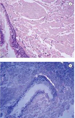
Fiberoptic bronchoscopy was performed three times during the diagnostic approach. All exami-nations revealed extrinsic compression of the right side of the trachea, nodular mucosal infiltrates distributed from the main carina to the right main and intermediary bronchus, as well as stenosis of the intermediary bronchus (Figure 2). Only during the third fiberoptic bronchoscopy was it possible to obtain a usable sample of the endobronchial infiltrates. Histopathological analysis of the tissue sample with Congo red staining demonstrated apple-green birefringence when viewed under polar-ized light (Figure 3). A diagnosis of amyloidosis was strongly suggested. Samples of periumbilical adipose tissue and bone marrow revealed no evidence of systemic disease. The results of urinalysis (for 24-h proteinuria and creatinuria) and protein profiles were within normal limits. An echocardiogram, an electrocardiogram and an abdominal ultrasound showed no abnormalities. A diagnosis of tracheo-bronchial amyloidosis was made. Six months later, the patient required only short-acting bronchodi-lators, presenting mild respiratory symptoms and normal lung function test results.
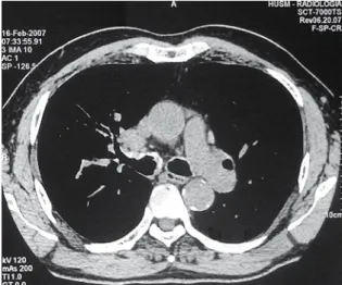
results of pulmonary function tests (spirometry and the six-minute walk test) and laboratory tests were within normal limits. A recent computed tomog-raphy scan of the chest showed partial stenosis of the intermediary and right main bronchi, together with laminar mucosal calcifications extending from the superior third of the trachea inferiorly to stenotic areas, as well as mediastinal
lymphad-Figure 1 - Computed tomography scan of the chest showing calcified wall thickening with stenosis of the intermediary and right main bronchi.
Figure 2 - Bronchoscopic views. a) Multiple nodular involvement of the right main bronchi; b) Narrowing of the intermediary lobar bronchi.
Primary tracheobronchial amyloidosis
J Bras Pneumol. 2008;34(10):881-884
883
calcifications, bronchiectasis and hilar adenop-athy.(4,11) Computed tomography and fiberoptic
bronchoscopy reveal bronchial wall thickening, irregular narrowing of the airway lumen and calci-fied nodules within the submucosa.(3,4,6) Computed
tomography scans can suggest the diagnosis, but fiberoptic bronchoscopy allows better visualization of the lesions and has the advantage of allowing the collection of samples for histopathological analysis. In most cases, the diagnosis is reached only after sequential fiberoptic bronchoscopy examinations.
During the diagnostic approach, it is essential to consider neoplastic diseases, granulomatous disease, tracheobronchopathia osteochondroplastica and relapsing polychondritis.(8,9,13,14) The long-term
clinical symptoms, together with the results of the imaging studies and fiberoptic bronchoscopy, narrow the diagnostic possibilities, tracheobronchopathia osteochondroplastica and relapsing polychon-dritis being the principal differential diagnoses.(8,13)
In the case presented here, the histopathological diagnosis was not made until the third fiberoptic bronchoscopy, which reflects the low level of suspi-cion of tracheobronchial amyloidosis on the part of pathologists and the bronchoscopists. Although the recently developed therapeutic approach known as external beam radiation therapy might become a viable option,(15) we have had no experience with this
intervention at our facility. The patient described here refused surgical intervention and was therefore treated with short-acting bronchodilators alone.
In conclusion, patients with tracheobronchial amyloidosis can present a wide variety of nonspecific symptoms and characteristic findings on imaging exams. The diagnosis is confirmed essentially through fiberoptic bronchoscopy, with appropriate (Congo red) staining of the bronchial tissue samples obtained.
References
1. Merlini G, Bellotti V. Molecular mechanisms of amyloidosis. N Engl J Med. 2003;349(6):583-96.
2. Tracheobronchial amyloidosis: a surgical disease with long-term consequences. J Thorac Cardiovasc Surg. 2004;128(5):789-92.
3. Georgiades CS, Neyman EG, Barish MA, Fishman EK. Amyloidosis: review and CT manifestations. Radiographics. 2004;24(2):405-16.
4. Berk JL, O’Regan A, Skinner M. Pulmonary and tracheobronchial amyloidosis. Semin Respir Crit Care Med. 2002;23(2):155-65.
Discussion
Tracheobronchial amyloidosis is a rare disease resulting from abnormal submucosal protein deposi-tion in the trachea and large bronchi.(1-7) It is a slowly
progressive disease that requires histopathological evidence in order to confirm the diagnosis.(2,4,5)
Tracheobronchial amyloidosis typically presents in patients between the fourth and fifth decades of life, accounting for 0.5% of symptomatic tracheo-bronchial lesions.(2,4) Common presenting symptoms
include chronic cough, dyspnea, wheezing, hemo-ptysis and recurrent pneumonia.(2-12) Cases of
tracheobronchial amyloidosis simulating difficult-to-manage asthma have been reported.(12) Half of all
patients present with a normal chest X-ray; the most common alterations are lobar atelectasis, bronchial
a
Figure 3 - a) Histopathological analysis of a tissue sample shows eosinophilic masses of amyloid material; b) Congo red staining demonstrating apple-green birefringence.
884 Santos JWA, Schneider Filho A, Bertolazzi A, Michel GT, Silva LVS, Melo CR et al.
J Bras Pneumol. 2008;34(10):881-884
11. Daniels JT, Cury JD, Diaz J. An unusual cause of postobstructive pneumonia. Chest. 2007;131(3):930-3. 12. Corrêa da Silva LM, Bellicanta J, Marques RD, Corrêa da
Silva LC. Amiloidose traqueobrônquica. J Bras Pneumol 2004;30(6):581-4.
13. Ozbay B, Dilek FH, Yalçinkaya I, Gencer M. Relapsing polychondritis. Respiration. 1998;65(3):206-7.
14. Sarodia BD, Dasgupta A, Mehta AC. Management of airway manifestations of relapsing polychondritis: case reports and review of literature. Chest. 1999;116(6):1669-75.
15. Neben-Wittich MA, Foote RL, Kalra S. External beam radiation therapy for tracheobronchial amyloidosis. Chest. 2007;132(1):262-7.
5. Rubinow A, Celli BR, Cohen AS, Rigden BG, Brody JS. Localized amyloidosis of the lower respiratory tract. Am Rev Respir Dis. 1978,;118(3):603-11.
6. Capizzi SA, Betancourt E, Prakash UB. Tracheobronchial amyloidosis. Mayo Clin Proc. 2000;75(11):1148-52. 7. Gillmore JD, Hawkins PN. Amyloidosis and the respiratory
tract. Thorax. 1999;54(5):444-51.
8. Prakash UB. Tracheobronchopathia osteochondroplastica. Semin Respir Crit Care Med. 2002;23(2):167-75.
9. Moura e Sá J, Almeida J, Amado J, Fernandes B, Caminha J, Ferraz JM. Traqueobroncopatia osteocondroplástica. Experiência de uma Unidade de Broncologia. Rev Port Pneuml. 2002;VIII:329-39