Pyoderma Gangrenosum: A Review Article
Pioderma Gangrenoso: Um Artigo de Revisão
Clóvis Luíz Konopka,1 Geórgia Andrade Padulla,2 Michele Purper Ortiz,3 Anderson Kahl Beck,4 Mariana Rechia Bittencourt,5 Diogo Chagas Dalcin6
Abstract
Pyoderma gangrenosum (PG) is a chronic dermatosis with peculiar characteristics and uncertain etiology, often diicult to diagnose. Painful skin ulcers form rapidly and progressively, most commonly in the lower limbs. Ulcerations may occur spontaneously or after various types of trauma. he time between the onset of lesions and the correct diagnosis is often long. here is no standard treatment or simple algorithm for the choice of therapy. In this study, we conducted a comprehensive review of current literature on the pathophysiology, diagnosis, and treatment of this disease through the systematic analysis of references in four databases, namely PubMed, Scielo, Medline and Lilacs.
Keywords:pyoderma gangrenosum; skin ulcer; diferential diagnosis.
Resumo
O pioderma gangrenoso (PG) é uma dermatose crônica com características peculiares e de etiologia desconhecida, muitas vezes de difícil diagnóstico. Manifesta-se através de lesões cutâneas ulceradas e dolorosas com evolução rápida e progressiva, mais comumente em membros inferiores. As ulcerações podem surgir espontaneamente ou depois de variados tipos de trauma. O período entre o início das lesões e o diagnóstico correto costuma ser prolongado. Não existe nenhum tratamento padronizado ou algoritmo simples para a escolha da terapia. Neste artigo, os autores fazem uma ampla revisão da literatura atual acerca da isiopatologia, do diagnóstico e do tratamento desta patologia através de análise sistemática das referências bibliográicas atuais nas bases de dados PubMed, Scielo, Medline e Lilacs.
Palavras-chave: pioderma gangrenoso; úlcera cutânea; diagnóstico diferencial.
A R T I C L E
1 Universidade Federal de Santa Maria - UFSM, Hospital Universitário de Santa Maria, Santa Maria, RS, Brazil. 2 Universidade Federal de Santa Maria - UFSM, Hospital de Clínicas de Porto Alegre - HCPA, Porto Alegre, RS, Brazil. 3 Universidade Federal de Santa Maria - UFSM, Hospital Universitário de Santa Maria, Santa Maria, RS, Brazil. 4 Universidade Federal de Santa Maria - UFSM, Hospital Universitário de Santa Maria, Santa Maria, RS, Brazil. 5 Universidade Federal de Santa Maria - UFSM, Hospital Universitário de Santa Maria, Santa Maria, RS, Brazil. 6 Universidade Federal de Santa Maria - UFSM, Hospital Universitário de Santa Maria, Santa Maria, RS, Brazil.
Financial support: None.
Conlicts of interest: No conlicts of interest declared concerning the publication of this article. Submitted on: 22.04.12. Accepted on: 27.11.12.
Pyoderma Gangrenosum: A Review Article
26 J Vasc Bras. 2013 Mar; 12(1):25-33
INTRODUCTION
Pyoderma gangrenosum (PG) is a rare, chronic
and often recurrent neutrophilic dermatosis. Its
etiology is uncertain, and it is often associated with
inlammatory bowel diseases (ulcerative colitis and
Crohn’s disease), malignancies, arthritis and blood
dyscrasias. It may even mimic, in certain cases,
surgical wound infections.
1This disease, irst described in 1916 by Brocq,
was later better characterized by Brusting in 1930,
2who named it PG because it was believed to be
a streptococcal infection causing skin gangrene.
Currently, its pathogenesis remains mostly uncertain,
but it has been deined that PG is not directly caused
by bacteria and is not, therefore, an infectious
pathology.
3Its incidence is estimated at 3 to 10 cases per
million people per year,
4,5and several studies in the
literature report on single cases or small patient series.
In the United States, the Mayo clinic diagnosed only
180 cases of PG in 53 years.
4,6PG affects individuals of any age, but is more
common in young adults 25 to 54 years old, and
affects women more often than men.
2It rarely affects
children (less than 4% of the cases), in which case it
is usually associated with other systemic diseases.
7Reports published in scientiic journals refer to its
development in immunodepressed patients due to
immunosuppressive therapy or chemotherapy, as
well as in cases of HIV infection.
5,6The clinical presentation of PG is variable, but
essentially characterized by multiple or single painful
ulcerative skin lesions that progress rapidly and have
a mottled, erythematous appearance.
8Lower limbs
are its most common site.
5,9This study conducted a broad review of the current
literature about the clinical and pathological aspects
of PG, its differential diagnosis and its treatment by
systematic analysis of references in the PubMed,
Scielo, Medline and Lilacs databases from 1992 to
2012 using the following keywords (in English and
Portuguese): pyoderma gangrenosum, pioderma
gangrenoso, neutrophilic dermatosis, skin ulcer and
úlcera cutânea. In addition to the electronic search,
other articles, relevant for the production of our
manuscript, were speciically selected using the
Periódicos Capes tool.
PATHOGENESIS
The initial lesion usually has the appearance of
a nodule or pustule that, as it breaks, progressively
forms an ulcer with a necrotic center and irregular
borders. Neutrophils are prominently found in the
lesions, and abnormalities in their functions, such as
defects in their chemotaxis or hyperresponsiveness,
have been reported a few times.
2Autoantibodies to
skin antigens have also been described, but there has
been no conirmation that they may be associated
with the causes of the skin lesions.
3Evidence of
disorders in immune cells is convincing,
10but
insufficient to fully explain PG pathogenesis.
Lazarus et al.
11found no response to intradermal
injection of puriied protein derived from tuberculin,
streptokinase-streptodornase and mumps antigens.
Greenberg et al.
12reported that the inhibition of
several cell immune responses was mediated by
a non-dialyzable and thermostable serum factor,
speciically in one patient under study. This factor
inhibited allogeneic and autologous lymphocytic
proliferation and the production of leukocyte
inhibitory factor.
3In an experiment, a circulatory
dermonecrotic factor found in the serum of patients
with PG induced necrosis in the skin of pigs, but its
effect in humans has not been determined.
A possible key to explaining PG etiology may
be its frequent association with systemic diseases,
which have well-known autoimmune mechanisms.
According to some authors, pathergy, which explains
the development of new lesions after local trauma
and is suggestive of an altered, exaggerated and
incontrollable inlammatory response to unspeciic
stimuli, might also be found in PG lesions.
Therefore, despite all scientific advances in
understanding PG, its pathogenesis remains uncertain.
Evidence points to immune disorders as the factors
responsible for its etiology, but such changes seem to
be detectable only in isolated patients.
13Nevertheless,
several authors classify PG as a disease of immune
origin.
CLINICAL PRESENTATION
PG usually begins as a painful deep nodule
or a hemorrhagic supericial pustule, often after
minor skin trauma.
14The nodule or pustule is
characteristically followed by the appearance of a
dark red or purplish inlammatory, painful ulcerative
lesion with irregular borders and a granular necrotic
base, usually mottled with small abscesses. A
hemorrhagic and purulent exudate is produced
inside the ulcerative lesions. The peripheral growth
of the lesions gives them a serpiginous coniguration
resulting from edge undermining and the appearance
of new hemorrhagic pustules. Supericial ulcers may
be conined to the dermis, but usually extend into
the fat-laden panniculus and the underlying fascia.
also be seen. They usually appear simultaneously
or consecutively in different parts of the body,
2with a predilection for the lower limbs, buttocks
and abdomen.
1,15Mucous membranes are usually
spared, but aphthae may appear in the oral mucosa,
often affecting the oral cavity, larynx and pharynx
massively.
Several times the lesions appear after minor
trauma, such as injections, insect bites, biopsies, or
surgical incisions,
1,16,17which suggests the occurrence
of the phenomenon known as pathergy.
18Systemic
symptoms, such as fever, arthralgia and myalgia,
may be associated.
19Extracutaneous involvement of
bones and lungs has already been described, with the
presence of neutrophilic iniltrates.
17,20PG onset may follow two distinct clinical
progression patterns:
• Sudden onset with rapid spread of lesions, clini
-cally characterized by pain, systemic toxicity, fever, hemorrhagic pustules, suppuration and borders with an inlammatory halo.
• Slow and indolent onset, which massive granula
-tion inside the ulcer, crust and hyperkeratosis in the edges, involving large areas and characterized by spontaneous retrogression in certain areas and progression in others.2
PG has four different clinical and histopathological
variants:
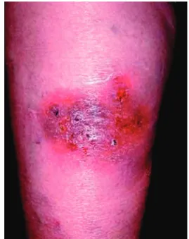
1. Vegetative – 12.5%21: Limited and not aggressive,
has supericial verrucose lesions and a non-purulent base (Figure 1), which differentiates it from the ulcerative variant. It is also called superficial granulomatous pyoderma and usually affects the trunk, head or neck. Many patients have no associ
-ated systemic diseases and respond to appropriate therapy rapidly. The differential diagnosis should include infections by mycobacteria, sporotrichosis and skin malignancies.22
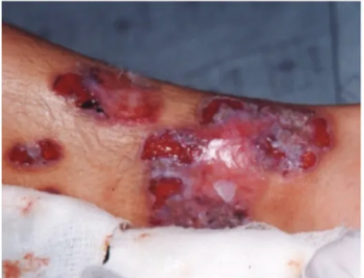
2. Bullous – 6.25%21: Associated with leukemia, its on
-set is sudden; it is more supericial and characterized by purple papules and bluish, bullous and hemor
-rhagic lesions (Figure 2). Its differential diagnosis should include acute febrile neutrophilic dermatosis (Sweet syndrome), cellulitis, bullous dermatosis and spider bites.22,23
3. Ulcerative – 81.52%21: Begins with a small pustule
surrounded by an inlammatory halo (Figure 3), is painful and progresses rapidly. Lesion resolution results in atrophic cribriform scarring.24 Malignant
pyoderma, an aggressive and potentially lethal vari
-ant of ulcerative PG, usually affects the head and neck and may be associated with systemic vasculites. The differential diagnosis should include systemic vasculites (Wegener granulomatosis, cryoglobu
-linemia, polyarteritis nodosa and antiphospholipid antibody syndrome), infections (sporotrichosis, ame
-biasis, syphilitic ulcer and ecthyma gangrenosum),1
malignancies, ischemic ulcers and insect bites.22
4. Pustular – rare: It is associated with fever and arthral
-gia, frequently associated with inlammatory bowel diseases (IBD).6,25 Pustules may either progress or
not into ulcerative lesions (Figure 4) and usually affect the extensor side of extremities. After IBD is controlled, it may retrogress without scarring, but lesions may coexist with the ulcerative variant.22
Periostomal PG in colostomies or ileostomies is a rarer variant reported almost exclusively in patients with IBD.26 The differential diagnosis should include
pustular vasculites, folliculitis and pustular eruption due to drugs and infections.22
LABORATORY FINDINGS
There are no specific laboratory findings.
Tests show leukocytosis and invariably elevated
erythrocyte sedimentation rate and protein C-reactive
levels.
4There may be anemia and low serum iron
Figure 2. Bullous PG.
Pyoderma Gangrenosum: A Review Article
28 J Vasc Bras. 2013 Mar; 12(1):25-33
levels, as well as hyper- and hypoglobulinemia.
Speciic autoantibodies are not usually found, and
circulating immunocomplexes are not detected.
HISTOPATHOLOGY
Histopathological indings are not characteristics
and may include edema, neutrophilic iniltrate, small
and medium-caliber vessel thrombosis, necrosis
and hemorrhage. Polymorphonuclear leukocyte
iniltrate is usually extremely dense, which leads to
the formation of microabscesses with liquefaction
necrosis associated with the secondary thrombosis
of venules. Neutrophils are PG markers.
3The occurrence of necrotizing vasculitis is
controversial, and some authors have described
only the presence of ibrinoid necrosis, whereas
others found lymphocytic vasculites or describe
it as indistinguishable from the lesions found in
immunocomplex diseases. Nevertheless, the current
correlation between PG and necrotizing vasculitis is
important.
2Histopathological indings also depend on the
biopsy site (edge, center or necrotic area of the
ulcer), the stage of progression and the disease variant
(vegetative, bullous, ulcerative or pustular).
6ASSOCIATION WITH SYSTEMIC DISEASES
In 50% to 70% of all cases, PG is associated
with systemic diseases, such as inlammatory bowel
diseases (idiopathic ulcerative colitis and Crohn’s
disease), rheumatoid arthritis, paraproteinemia,
multiple myeloma, leukemia, chronic active hepatitis,
Behçet’s disease, malignancies, HIV infection and
immunosuppression in transplant recipients.
8,24,25,27-29In the other cases, PG is a primary, isolated lesion
limited to the skin and is, therefore, classiied as
idiopathic.
In the case of inflammatory bowel diseases,
PG is found in about 5% of the cases of ulcerative
colitis, and in only 0% to 1.2% of the cases of
Crohn’s disease.
6,30In most patients with colitis,
bowel symptoms precede PG onset. However, in
some cases PG skin lesions are seen irst and may
persist for a long time, whereas IBD symptoms
may remain quiescent. Some reports also associate
it with psoriasis and the development of lesions in
the inguinal, axillary, perineal and penile regions.
31DIAGNOSIS
As mentioned above, there are no specific
laboratory findings or pathognomic histological
characteristics and, therefore, PG diagnosis
depends exclusively on the observation of clinical
characteristics and disease progression. However,
some criteria may be suggestive of PG, such as the
fact that ulcerative lesions are painful and progress
rapidly from onset. Other criteria should also be taken
into consideration, such as the presence of pathergy,
the association with systemic diseases (secondary
PG) and a rapid response to the administration of
corticosteroids. Histopathological examination of
biopsy material may show sterile neutrophilia and
lymphocytic vasculites,
18which may exclude other
etiologies of skin ulcers.
26,32Ferrándiz-Pulido and Briones
24recommend the
following routine to establish the diagnosis of an
ulcerative lesion
:
1. Clinical history – rapid or slow progression, no response to antibiotics, associated diseases. 2. Physical exam – necrotic ulcer, purple erythematous
edges, involvement of other organs.
3. Skin biopsy for Gram stains and culture – bacteria, fungi and mycobacteria.
4. Skin biopsy for histopathological examination (HE, PAS)*.
5. Laboratory blood tests - complete blood count, biochemical parameters, erythrocyte sedimentation rate, protein electrophoresis, clotting, anticardiolipin antibodies, antiphospholipid antibodies, ANCA* and cryoglobulins.
Figure 3. Ulcerative PG.
C
ló
vis L
uí
z K
o
n
o
p
ka
, G
eó
rg
ia A
n
dr
ad
e P
adulla e
t a
l.
29
J V
as
c B
ra
s. 2013 M
ar
; 12(1):25-33
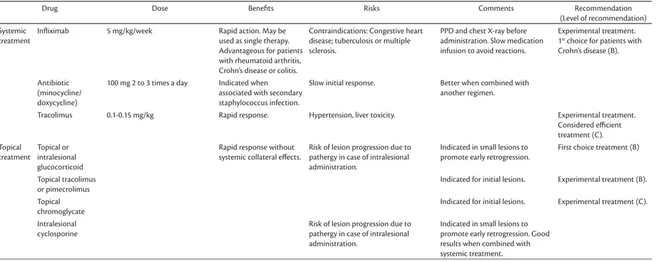
(Level of recommendation) Systemic
treatment
Glucocorticoid (prednisone or equivalent)
Moderate high dose: 0.5-1 mg/kg/day
Pulse therapy 1 g/day for 1-5 days
Rapid action in ulcer resolution Analgesic action
Long term: infections, insulin resistance, or no change.
PPD test before beginning of treatment Use for 2-4 weeks, if possible. Reduce dose gradually if use for >21 days – avoid it. Secondary adrenal insuiciency.
First choice treatment (B)
Cyclosporine 4-5 mg/kg/day Rapid response. Renal toxicity, hypertension,
hypertriglyceridemia, hypertrichosis.
Initiate directly with dose of 4-5 mg/kg/day, not less.
First choice treatment (B)
Azathioprine 100-300 mg/day Greater compliance in
case of long treatment duration.
Slow initial response, bone marrow suppression, gastrointestinal intolerance.
Evaluate levels of thiopurine methyltransferase enzyme to predict toxicity at low levels or absence.
Experimental treatment (C).
Dapsone 50-200 mg/day Better safety proile in the
long run Also used to save corticosteroids.
Slow initial response. No resolution of more severe cases.
Anemia, methemoglobinemia, neuropathy.
Periodical evaluation of blood count (white blood count reduction of up to 10% is normal).
Experimental treatment (C).
Methotrexate 10-30 mg/week Weekly administration,
better compliance in the long run.
Liver toxicity, bone marrow suppression.
Test serum for viral hepatitis before beginning of treatment.
Experimental treatment (C).
Mycophenolate mofetil
2-3 g/day Good safety proile in the
long run
Slow initial response, neutropenia. Better performance after initial
administration together with glucocorticoid due to slow initial response.
Experimental treatment (C).
halidomide 50-200 mg/day Teratogenesis, sleepiness, neuropathy,
bleeding disorders.
Night dose due to sleepiness. Experimental treatment.
Eicacy documented (C).
Colchicine 0.6 mg 2 to 3 times a day Safe drug, if tolerated. Diarrhea Begin with low dose, increase
gradually due to gastrointestinal irritation.
Cyclophosphamide 1.5-3.0 mg/kg/day Hemorrhagic cystitis, azoospermia. Keep good hydration before and
after treatment.
Experimental treatment. Consider cost-beneit (B).
Clofazimine 200 mg/day Useful in treatment of
refractory PG.
Changes color of skin an bodily luids Administrate with food or milk for
maximum absorption.
P
yo
d
er
m
a G
ang
re
n
o
sum: A R
ev
ie
w A
rt
ic
le
30
J V
as
c B
ra
s. 2013 M
ar
; 12(1):25-33
Drug Dose Beneits Risks Comments Recommendation
(Level of recommendation)
Systemic treatment
Inliximab 5 mg/kg/week Rapid action. May be
used as single therapy. Advantageous for patients with rheumatoid arthritis, Crohn’s disease or colitis.
Contraindications: Congestive heart disease; tuberculosis or multiple sclerosis.
PPD and chest X-ray before administration. Slow medication infusion to avoid reactions.
Experimental treatment. 1st choice for patients with Crohn’s disease (B).
Antibiotic (minocycline/ doxycycline)
100 mg 2 to 3 times a day Indicated when
associated with secondary staphylococcus infection.
Slow initial response. Better when combined with
another regimen.
Tracolimus 0.1-0.15 mg/kg Rapid response. Hypertension, liver toxicity. Experimental treatment.
Considered eicient treatment (C). Topical
treatment
Topical or intralesional glucocorticoid
Rapid response without systemic collateral efects.
Risk of lesion progression due to pathergy in case of intralesional administration.
Indicated in small lesions to promote early retrogression.
First choice treatment (B)
Topical tracolimus or pimecrolimus
Indicated for initial lesions. Experimental treatment (B).
Topical chromoglycate
Indicated for initial lesions. Experimental treatment (C).
Intralesional cyclosporine
Risk of lesion progression due to pathergy in case of intralesional administration.
6. Chest X-ray
7. Abdominal ultrasound.
8. If digestive symptoms are present: upper digestive tract endoscopy and colonoscopy.
9. If complete blood count shows abnormalities: bone marrow aspirate and biopsy.
*ANCA: antineutrophil cytoplasmic antibodies;
HE: hematoxylin-eosin; PAS: periodic acid Schiff
protocol.
TREATMENT
The main objective of PG treatment is to limit
tissue destruction, promote wound healing and
obtain a good esthetic result. Surgical debridement
and skin grafts should be avoided because of the
potential risk of pathergy and the consequent lesion
deterioration.
4,33Simple direct cleaning procedures and aseptic
dressing are indicated. Moreover, the administration
of medication should almost always be taken into
consideration to promote healing of existing ulcers
and prevent new lesions. In this sense, treatments
available include the use of topical or intralesional
corticosteroids,
346-mercaptopurine or azathioprine,
topical chromoglycate, dapsone, clofazimine and
cyclosporine, as seen in Table 1. Hyperbaric oxygen
therapy may be indicated for patients that do not
tolerate or do not respond to high doses of systemic
corticosteroids.
35,36Administration of systemic corticosteroids is
the most effective treatment of PG.
4,8The use of
corticosteroids may interrupt ulcerative progression
and prevent the development of new lesions.
High initial doses are necessary in most cases,
usually about 100-200 mg/day of prednisolone or
60-80 mg/day of prednisone.
5,8,37The use of sulfas may be beneicial, but response
is not uniform. Daily doses range from 4 to 6 g
and should be reduced progressively after some
clinical improvement is observed to about 0.5 to 1
g/day. These drugs may be added to treatment in
combination with corticosteroids in the initial phase
of the disease.
39Alternatively, cyclosporine, at 6-10
mg/kg/day, may produce signiicant improvement
and lesion healing in about 1 to 3 months. Some
authors reported on the use of clofazimine at
daily doses of 200-300 mg; they found that lesion
progression was interrupted in 1 to 2 months and
full healing was achieved in up to 5 months.
3Oral
thalidomide was efficient in a patient that did
not respond to high doses of corticosteroids, and
complete lesion cure was achieve after 10 weeks
of administration.
38TNF-alpha inhibitors, such as
inliximab, have been used with good results,
25,39especially for patients whose other comorbidities
also improve with the use of this drug. In the cases
of PG secondary to Crohn’s disease, this drug is the
treatment of choice.
4,25,37Surgery may play a role in the treatment of
PG secondary to IBD in unstable patients that do
not respond to medication.
6Colectomy may be
considered in cases of extensive colitis, but the
ulcerative lesions may persist after surgery.
26,40CONCLUSIONS
PG has an unpredictable and highly variable
course from its onset and during its progression.
41Some patients develop lesions of sudden onset
associated with progressive increase, followed by
pain and fever. Others have chronic lesions, with
ulcerations that progress slowly. Regardless of the
variant, most lesions are self-limiting and progress
to spontaneous healing in a short time.
6Some
patients have recurrent lesions periodically. Also,
some do not respond to the medications commonly
used, which then have to be empirically replaced
while other drugs are added to ensure PG cure
or retrogression.
4,8,42Prognosis is usually good,
especially for patients that rapidly respond to the
initial treatment regimen.
43Similarly, the lesions of
patients with secondary PG usually retrogress after
the treatment of the primary disease.
4,43REFERENCES
1. Ogon M, Wimmer C, Behensky H, Sepp NT. A surgical wound infection? Lancet. 2000;356:1652. http://dx.doi.org/10.1016/ S0140-6736(00)03161-5
2. Bennet ML, Jackson JM, Jorizzo JL, Fleischer Junior AB, White WL, Callen JP. A comparison of typical and atypical forms with an emphasis on time to remission. Case review of 86 patients from 2 institutions. Medicine. 2000;79:37-46. http://dx.doi. org/10.1097/00005792-200001000-00004
3. Fitzpatrick TB, Eisen AZ, Wolf K, Fredberg IM, Austen KFK. Dermatology in general medicine. McGraw-Hill; 1993. p.1173-79.
4. Ahronowitz I, Harp J, Shinkai K. Etiology and management of pyoderma gangrenosum: a comprehensive review. Am J Clin Dermatol. 2012;13(3):191-211. PMid:22356259. http://dx.doi. org/10.2165/11595240-000000000-00000
5. Bolognia JL, Jorizzo JL, Rapini RP. Pyoderma gangrenosum. Dermatology. 2003;1:415-418.
6. Alam M, Grossman ME, Schneiderman PI, Blume RS, Benvenisty AI. Surgical management of pyoderma gangrenosum: case report and review. Dermatol Surg. 2000; 26(11):1063-6. PMid:11096397. http://dx.doi.org/10.1046/j.1524-4725.2000.0260111063.x 7. Sarma N, Bandyopadhyay SK, Boler AK, Barman M. Progressive
Pyoderma Gangrenosum: A Review Article
32 J Vasc Bras. 2013 Mar; 12(1):25-33
8. Reichrath J, Bens G, Bonowitz A, Tilgen W. Treatment recommendations for pyoderma gangrenosum: an evidence-based review of the literature evidence-based on more than 350 patients. J Am Acad Dermatol. 2005;53:273-83. PMid:16021123. http:// dx.doi.org/10.1016/j.jaad.2004.10.006
9. Barbato MT, Bakos L , Masiero NCMS , Bolson. Per f il clinicopatológico dos pacientes com pioderma gangrenoso do Hospital de Clínicas de Porto Alegre (RS) – Brasil (2000-2006). An Bras Dermatol. 2008;83(5):431-6. http://dx.doi.org/10.1590/ S0365-05962008000500006
10. Coors EA, Von Den Driesh P. Pyoderma gangrenosum in a patient with autoimmune haemolytic anaemia and complement deficiency. Br J Dermatol. 2000;143(1):154-6. http://dx.doi. org/10.1046/j.1365-2133.2000.03606.x
11. Lazarus GS, Goldsmith LA, Rocklin RE, Pinals RS, De Buisseret JP, David JR. Pyoderma gangrenosum, altered delayed hypersensitivity and polyarthritis. Arch Dermatol. 1972;105(1):46-51. PMid:5009622. http://dx.doi.org/10.1001/archderm.1972.01620040018003
12. Greenberg SJ, Jegasothy BV, Johnson RB,Lazarus GS. Pyoderma GangrenosumOccurrence With Altered Cellular Immunity and a Circulating Serum Factor. Arch Dermatol. 1982;118(7):498-5 0 2 . P M i d : 7 0 9 2 2 7 6 . h t t p : / / d x . d o i . o r g / 1 0 . 1 0 0 1 / archderm.1982.01650190052019
13. Khandpur S, Mehta S, Reddy BS. Pyoderma gangrenosum i n t w o s i b l i n g s : a f a m i l i a l p re d is p o s i ti o n . Pe d i atr Dermatol. 2001;18:308-12. PMid:11576404. http://dx.doi. org/10.1046/j.1525-1470.2001.01936.x
14. Hadi A , Lebwohl M. Clinic al features of pyo derma gangrenosum and current diagnostic trends. J Am Acad of Dermatology. 2011;64(5):950-54(e2).
15. Wani I, Bhat IHG, Mir M, Mir M, Hassan N, Mustafa A. Pyoderma Gangrenosum of Abdominal Wall: A Case Report. Oman Med J. 2011;26(1):64–65. PMid:22043386 PMCid:3191625. http:// dx.doi.org/10.5001/omj.2011.18
16. Umezawa Y, Oyake S, Nagae T, Ishimaru S. A case of pyoderma gangrenosum on the stump of an amputated right leg. J. Dermatol. 2000;27(8):529-32. PMid:10989578.
17. Vasili E, Shkodrani E, Labinoti L, Xhaja A. A case of atypical pyoderma gangrenosum. J Dermatol Case Rep. 2010;4(1):18-21. PMid:21886741 PMCid:3157807. http://dx.doi.org/10.3315/ jdcr.2010.1044
18. Su WP, Davis MP, Weenig RH, Powell FC, Perry HO. Pyoderma gangrenosum: clinic pathologic correlation and proposed diagnostic criteria. Int J Dermatol. 2004;43:790-800. PMid:15533059. http://dx.doi.org/10.1111/j.1365-4632.2004.02128.x
19. Callen JP. Pyoderma gangrenosum. Lancet. 1998;351:581-5. http:// dx.doi.org/10.1016/S0140-6736(97)10187-8
20. Vignon-Pennamen MD, Wallach D. Neutroilic disease: a review of extra-cutaneous manifestation. Eur J Dermatol. 1995;5:449-55.
21. Conrad C, Ralph M. Pyoderma Gangrenosum. Article Review. J Dtsch Dermatol Ges. 2005;3:334-42. PMid:16372799. http:// dx.doi.org/10.1111/j.1610-0387.2005.05022.x
22. Newman B, Cescon D, Domenchini A, Siminovitch KA. CD2BP1 and CARD 15 mutations are not associated with pyoderma gangrenosum in patients with inlammatory Bowel Disease. J Invest Dermatol. 2004;122:1054-5. PMid:15102098. http://dx.doi. org/10.1111/j.0022-202X.2004.22430.x
23. Sakiyama M, Kobayashi T, Nagata Y, Fujimoto N, Satoh T, Tajima S. Bullous pyoderma gangrenosum: A case report and review of the published work. J Dermatol. 2012;39(12):1010-1015.
24. Ferrándiz-Pulido C, García-Patos V. Pioderma gangrenoso. Diagnóstico y tratamiento. Briones Piel. 2008;23(1):24-9. http:// dx.doi.org/10.1016/S0213-9251(08)70969-9
25. Zold E, Nagy A, Devenyi K, Zeher M, Barta Z. Successful use of adalimumab for treating fistulizing Crohn’s disease with pyoderma gangrenosum: Two birds with one stone. World J Gastroenterol. 2009;15(18):2293-2295. PMid:19437575 PMCid:2682250. http://dx.doi.org/10.3748/wjg.15.2293
26. Hughes AP, Jackson JM, Callen JP. Clinical features and treatment of periostmal pyoderma gangrenosum. JAMA. 2000;284(12):1546-8. PMid:11000649. http://dx.doi.org/10.1001/jama.284.12.1546
27. Nukumizu LK, Silva CAA, Lotito APN, et al. Pioderma gangrenoso na infância e doenças sistêmicas associadas: relato de 5 casos. Rev Bras Reumatol. 2002;42:65-71.
28. Hof NP, Gerber PA. Pyoderma gangrenosum associated with chronic polyarthritis. CMAJ. 2011;183(15):1746. PMid:21859868 PMCid:3193127. http://dx.doi.org/10.1503/cmaj.110679
29. Duke G, Samaraee A, Husain A, Meggitt S, Fasih T. Pyoderma Gangrenosum: A Rare Cause of Breast Ulceration. Ochsner J. 2012;12(2):155-158. PMid:22778682 PMCid:3387843.
30. Sheldon DG, Sawchuck LL, Kozarek RA, hirlby RC. Twenty cases of periostomal pyoderma gangrenosum: diagnostic implications and management. Arch Surg. 2000;135(5):564-8. PMid:10807281. http://dx.doi.org/10.1001/archsurg.135.5.564
31. F r a g a J C S , D e S o u z a V L , Va l v e r d e RV, G a m o n a l A. Pioderma gangrenoso: apresentação atípica. An Bras Dermatol. 2006;81(5):S305-8.
32. Weenig RH, Davis MD, Dahl PR, Su WP. Skin ulcers misdiagnosed as Pyoderma Gangrenosum. N Engl J Med. 2002;347:1412-8. PMid:12409543. http://dx.doi.org/10.1056/NEJMoa013383
33. Clif S, Holden CA, homas PR, Marsden RA, Harland CC. Split skin grafts in the treatment of pyoderma gangrenosum. A report of four cases. Dermatol Surg. 1999;25(4):299-302. PMid:10417586. http://dx.doi.org/10.1046/j.1524-4725.1999.08193.x
34. Keltz M, Lebwhol M, Bishop S. Periostomal pyoderma gangrenosum. J Am Acad Dermatol. 1992;27:360-4. http://dx.doi. org/10.1016/0190-9622(92)70200-Y
35. Galun E, Flugelman MY, Rachmilewitz D. Pyoderma gangrenosum complicating ulcerative colitis: Successful treatment with methylprednisolone pulse therapy and dapsone. Am J Gastroenterol. 1986;81:988-9. PMid:3766502.
36. Friedman S, Marion JF, Scherl E, et al. Intravenous cyclosporine in refractory pyoderma gangre nosum complicating inlammatory bowel disease. Inlamm Bowel Dis. 2001;7(1):1-7. PMid:11233655. http://dx.doi.org/10.1097/00054725-200102000-00001
37. Miller J, Yentzer BA, Clark A, Jorizzo JL, Feldman SR. Pyoderma Gangrenosum: A review and update on new therapies. J Am Acad Dermatol. 2010;62(4):646-54. PMid:20227580. http://dx.doi. org/10.1016/j.jaad.2009.05.030
38. Federman GL, Federman DG. Recalcitrant pyoderma gangrenosum treated with thalidomide. Mayo Clin Proc. 2000;75(8):842-4. PMid:10943240. http://dx.doi.org/10.4065/75.8.842
39. Stichweh DS, Punaro M, Pascual V. Dramatic improvement of pyoderma gangrenosum with inliximab in a patient with PAPA syndrome. Pediatr Dermatol. 2005;22:262-5. PMid:15916580. http://dx.doi.org/10.1111/j.1525-1470.2005.22320.x
41. Binus AM, Qureshi AA, Li VW, Winterield LS. Pyoderma gangrenosum: a retrospective review of patient characteristics, co m o rb i diti e s , an d th er apy in 103 p ati ent s . Brit J Dermatol 2011;165:1244-1250. PMid:21824126. http://dx.doi. org/10.1111/j.1365-2133.2011.10565.x
42. Callen J, Jackson M. Pyoderma Gangrenosum: An Update. Rheum Dis Clin N Am. 2007;33:787-802. PMid:18037117. http://dx.doi. org/10.1016/j.rdc.2007.07.016
43. Kuhn C, Vente C, Dörner J, Ratayski H, Burchardi H. [Hypoderma gangrenosum and important differential diagnosis from woundinfection. Case report of a life threatening course]. Anaesthesist. 2000;49(9):829-33. PMid:11076272.
Correspondence Clóvis Luíz Konopka Av. Roraima, Prédio 22, Campus - Camobi, CEP 97105-900 – Santa Maria (RS), Brazil E-mail: [email protected]
Author information CLK is an assistant professor of Vascular Surgery, Vascular Surgery Service, Hospital Universitário de Santa Maria, Universidade Federal de Santa Maria (UFSM). GAP is a resident physician at the Dermatology Service of Hospital de
Clínicas de Porto Alegre (HCPA). MPO is a resident physician at the Anesthesiology Service of Hospital Universitário de Santa Maria, Universidade Federal de Santa Maria (UFSM). AKB is a monitor at the Angiology and Vascular Surgery Service of
Hospital Universitário de Santa Maria , Universidade Federal de Santa Maria (UFSM). MRB is a monitor at the Endocrinology Service of Hospital
Universitário de Santa Maria, Universidade Federal de Santa Maria (UFSM). DCD is a monitor at the Angiology and Vascular Surgery Service of
Hospital Universitário de Santa Maria, Universidade Federal de Santa Maria (UFSM).