DNA methylation dynamics in the rat EGF gene promoter after partial
hepatectomy
Deming Li
1,2, Jinyu Fan
1,2, Ziwei Li
1,2and Cunshuan Xu
1,2 1Key Laboratory for Cell Differentiation Regulation, Xinxiang, China.
2
College of Life Science, Henan Normal University, Xinxiang, China.
Abstract
Epidermal growth factor (EGF), a multifunctional growth factor, is a regulator in a wide variety of physiological pro-cesses. EGF plays an important role in the regulation of liver regeneration. This study was aimed at investigating the methylation level of EGF gene throughout liver regeneration. DNA of liver tissue from control rats and partial hepatectomy (PH) rats at 10 time points was extracted and a 354 bp fragment including 10 CpG sites from the tran-scription start was amplified after DNA was modified by sodium bisulfate. The result of sequencing suggested that methylation ratio of four CpG sites was found to be significantly changed when PH group was compared to control group, in particular two of them were extremely striking. mRNA expression of EGF was down-regulated in total during liver regeneration. We think that the rat EGF promoter region is regulated by variation in DNA methylation during liver regeneration.
Keywords: methylation dynamics, epidermal growth factor, liver regeneration.
Received: October 26, 2013; Accepted: March 18, 2014.
The liver’s ability to regenerate in mammals is a very well studied response. When toxic injury, exposure to vi-ruses, trauma or surgical resection results in loss of hepatic tissue, the remnant liver lobes will compensate for lost tis-sue and recover the initial liver mass within two weeks (Faustoet al., 2006). In the case of partial hepatectomy (PH), it results in hypertrophy of the remnant liver rather than restoration of the resected lobes as a consequence of cell proliferation, with the goal of replacing lost functional mass, which is called liver regeneration (LR) (Higgins and Anderson, 1931). The degree of hyperplasia is precisely controlled by the metabolic needs of the organism, prolifer-ating under conditions of functional deficiency, and under-going apoptosis under functional excess, such that the process stops once an appropriate liver to body weight ratio is achieved. In mouse and rat, the ratio is about 4.5%; in hu-mans this number is approximately 2.5% (Fausto, 2000; Riehleet al., 2011). PH triggers the actions of signaling pathways, growth factors and cytokines, cell cycle associ-ated proteins, and extracellular matrix, etc., leading to cell proliferation and structure function reorganization (Pahla-vanet al., 2006; Michalopoulos, 2011). Among them, epi-dermal growth factor (EGF) has important function in LR. EGF, a multifunctional growth factor, is known as a regulator in a wide variety of physiological processes
(Zwicket al., 1999; Zenget al., 2006).EGF binds to the epi-dermal growth factor receptor (EGFR),causing the EGFR to form homo- and heterodimers between EGFRs to recruit adaptor molecules such as phosphatidylinositol 3-kinase, Shc, and Grb2 etc. (Carpenter, 2000). These adaptor pro-teins then initiate signaling cascades including extracellular regulated kinase 1/2; leading to stimulation of a plethora of cellular processes, such as proliferation, differentiation, enbryogenesis, growth, tissue repair and regeneration (Morrishet al., 1997; Zwicket al., 1999; Ramos, 2008).
It was reported that EGF was rapidly produced in im-mediate-early phase of liver regeneration (Mullhauptet al., 1994).EGF was thought to be one of the extracellular fac-tors related to the early “priming phase” which sensitizes hepatocytes to other growth stimuli(Meadet al., 1990); it involves gene transcription of more than 70 immediate-early and other genes in this priming phase (Haberet al., 1993; Cressmanet al., 1995; Faustoet al., 1995; Nadoriet al., 1997).
Epigenetic regulation, such as DNA methylation and histone modification, was thought to influence gene ex-pression mainly at the level of transcription. Methylation is the most extensive epigenetic modification that directly af-fects the DNA molecule in eukaryotes. In mammals, it nearly occurs only in the context of CG dinucleotides; DNA methylation is generally associated with gene repres-sion (Miranda and Jones, 2007; Weber and Schübeler, 2007).DNA demethylation was long thought to occur only
www.sbg.org.br
Send correspondence to Cunshuan Xu. College of Life Science, Henan Normal University, 46# East of Construction Road, 453007 Xinxiang, Henan, PR China. E-mail: [email protected].
during specific developmental phases in zygotes and pri-mordial germ cells; however several recent investigations found that DNA demethylation, even rapid demethylation, occurs in response to various stimuli in other cellular con-texts (Maet al. 2009; Guoet al., 2011a,b; Shearstoneet al., 2011; Calvanese et al., 2012). Previous research on the EGF gene mainly focused on its expression and its interac-tion with other molecules; the purpose of the present study was to compare the methylation profile in the promoter re-gion of EGF among 2/3 partially hepatectomized rats and a control group.
In total, 41 Healthy Sprague-Dawley rats (230±20 g)
provided by the Animal Center of Henan Normal Univer-sity, were randomly separated into nine partial hepatectomy (PH) groups, nine sham-operation (SO) groups, and one normal control (NC) group. The PH and SO groups included two rats(male:female = 1:1) for each time point; the normal control consisted of five rats. Partial (2/3) hepatectomy was performed according to Higgins and Anderson (1931) with surgical removal of the left and me-dian lateral liver lobes. The rats were sacrificed by cervical vertebra dislocation at 2, 6, 12, 24, 30, 36, 72, 120 and 168 h after PH, and the regenerating livers were obtained at corre-sponding time points. Rats composing the SO control group received the same treatment as the PH group but without liver removal. The Laws of Animal Protection of China were strictly followed. Total genomic DNA was extracted from the liver tissue following the method of Sambrook and Russell (2001).
Rat EGF promoter region sequence corresponding to nucleotides -1000 to -1 was retrieved from NCBI. The 1000 bp sequence then served as input to MethPrimer soft-ware (Li and Dahiya, 2002) for bisulfite sequencing primer design. We used the forward primer: 5’-ATGAGTTGAA GGTGAGATTTTTTTG-3’,and the reverse primer:5’-CCCCTCTCCTTTAATAACACTTAAATAA-3’ to amplify a 354 bp fragment which includes 10 CpG sites from the transcription start. DNA was modified using the EpiTect Bisulfite Kit (QIAGEN) according to the man-ufacturer’s instructions. PCR products were purified using the PCR Purification Kit (Dingguo Company, China) and ligated into pGEM-T vector (Promega, Madison, USA). The vector was then transformed into competent DH5-aE.
coli cells. In each case, at least 10 of the plasmid clones were sequenced. The respective sequences are shown as Supplementary Material (Figure S1).
Total RNA was isolated from liver tissue using Trigol (Dingguo Company, China) according to the manufac-turer’s instructions. cDNA was synthesized using random primers and a reverse transcription kit (Promega). Primers were designed by Primer Premier 5 software (Premier Biosoft, Palo Alto, USA) and synthesized by Dingguo Company according to mRNA sequences of EGF and the housekeeping gene GAPDH (NCBI: NM_012842.1 and NM_017008.4). The primer sequences are as follows:
5-’ACCAACACGGAGGGAGGCTACAA’-3 (forward, EGF), 5-’GCGGTCCACGGATTCAACATACA’-3 (re-verse, EGF); 5-’CACGGCAAGTTCAACGGCACAG TCA’-3 (forward, GAPDH), 5-’GTGAAGACGCCAG TAGACTCCACGAC’-3 (reverse, GAPDH), Real-time quantitative PCR was performed by using SYBR_Green I (Invitrogen) in a Rotor-Gene 3000 (Corbett Robotics) un-der the following conditions: 95 °C for 2 min, followed by 40 cycles of 30 s at 95 °C, 30 s at 59 °C, and 30 s at 72 °C. Standard curves were generated from three repeated10-fold serial dilutions of cDNA. The copies of EGF and GAPDH mRNA were calculated by means of the software of the Ro-tor-Gene 3000. Each sample was analyzed in three repli-cates.
Sequences were aligned by means of the software BiQ Analyzer(Bock et al., 2005). Statistical analysis for significant differences between the groups was done with the Independent-Samples T test implemented in SPSS 13.0 software (SPSS Inc., Chicago, USA). A Spearman’s corre-lation analysis was used to test the association between methylation change and expression of EGF. P-values of less than 0.05 were considered statistically significant.
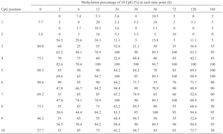
In this study the methylation status of 10 CpG sites in EGF promoter region was identified at 10 time points (Ta-ble 1). Among these positions, the methylation percentage of four CpGs was found to be significantly changed during liver regeneration in the PH group compared to the SO group (positions 3, 4, 7 and 8, p < 0.05); in particular, the difference in positions three and four was extremely strik-ing (p < 0.01). Positions one and two were unmethylated or low methylated; The methylation ratio of positions 5, 6, 9 and 10 did not show statistically significant differences. The relative changes in EGF mRNA levels at nine time points during liver regeneration were as follows: 1.05; 0.64; 0.29; 0.45; 0.36; 0.22; 1.17; 1.16 and 0; the normal control was set to 1.
Epigenetic events are involved in heritable gene ex-pression patterns. DNA methylation and demethylation in regulatory regions represents an epigenetic change that pro-foundly affects gene expression, and the transcriptional ac-tivity of a gene is inversely correlated with DNA methyl-ation of its promoter region (Cedar and Bergman, 2009). In normal mammalian somatic cells, most CpG sites are meth-ylated and associated with gene silencing, and methylation is also thought to prevent chromatin instability(Grunstein, 1997; Szyf, 2005). As previous studies had indicated that EGF plays a crucial role in the regulation of liver regenera-tion(Mead et al., 1990); we asked whether CpG methylation may play a role in the regulation of EGF ex-pression during liver regeneration.
which represses the expression of EGF. When using the on-line software TFSEARCH to predict TF binding sites we found a DNA-binding specificity for GATA family tran-scription factors. EGF expression was down-regulated in total during liver regeneration, and our data is consistent with the microarray analysis of Xu and Zhang (2009), but contrary to that of Mullhauptet al.(1994), which used mice as an experimental model. These contradictory results cer-tainly need further investigation.
The methylation mean percentage of positions 4, 7 and 8 was increased, especially at position 4 (Figure 2). The
methylation change of sites 7 and 8 was positively related with the changes of EGF mRNA levels(p < 0.01 r = 0.675 and r = 0.675).
DNA methylation can lead to changes in the 3D struc-ture of DNA, where by cytosine methylation can recruit methyl binding proteins (MBPs) and generate a repressed chromatin environment, so that the expression of genes be-comes directly regulated by the status of DNA methylation and consequent change in DNA structure (Delgado-Olguin and Recillas-Targa, 2011). We think that the increase in methylation that we observed at three sites probably helps to prevent the binding of an inhibiting factor.
Figure 1- Comparison of CpG3 methylation change (x axis) to EGF mRNA expression (y axis) in the PH samples. A positive correlation was revealed by a Spearman’s correlation analysis, r = 0.868, p < 0.01.
Figure 2- Methylation mean percentage of 10 CpG points in the EGF pro-moter region determined at nine time points during liver regeneration. Black column represent PH group; gray column represent SO group. Table 1- Methylation ratio of each CpG at different time points. The upper-row of each CpG position refers to the PH group, the lower one is for the SO group.
Methylation percentage of 10 CpG (%) at each time point (h)
CpG position 0 2 6 12 24 30 36 72 120 168
0 7.4 5.3 5.6 0 10.5 5 0 5
1 7.7 5 0 20 5.3 5.3 10 5 5.3 5
0 3.7 10.5 5.6 0 5.3 0 0 5
2 3.8 0 5 10 5.3 5.3 5 10 0 0
56.5 29.6 26.3 11.1 5 15.8 5 11.1 5
3 80.8 60 25 55 52.6 21.1 50 35 36.8 35
65.2 48.1 78.9 100 90 89.5 100 83.3 95
4 73.1 70 75 60 52.6 68.4 40 45 42.1 45
82.6 70.4 100 100 100 94.7 100 100 100
5 88.5 85 90 90 84.2 84.2 70 85 89.5 100
69.6 63 94.7 100 95 89.5 100 88.9 100
6 80.8 90 85 90 84.2 73.7 55 70 73.7 80
47.8 66.7 84.2 94.4 90 78.9 90 88.9 90
7 69.2 55 85 85 63.2 78.9 65 60 52.6 60
47.8 74.1 78.9 100 90 89.5 100 88.9 95
8 73.1 55 85 75 63.2 89.5 80 55 68.4 50
30.4 44.4 84.2 83.3 95 100 95 94.4 95
9 46.2 55 85 55 68.4 94.7 50 55 52.6 35
56.5 70.4 84.2 94.4 90 89.5 90 94.4 95
When we compared the SO group and the normal group (0 h) we found that the methylation ratios in sites 3, 4, 9 and 10 were also changed. In the SO group the changes inEGF mRNA expression was as follows: 0.56, 0.30, 0.33, 0.36, 037, 0.35, 0.5, 0.22 and 0.67, and there was no differ-ence when compared to the PH group (p > 0.05). The methylation change at the sites 4, 9 and 10 was inversely correlated with the changes in EGF expression(p < 0.01 r = 0.868, r = 0.612 and r = 0.696), this also being in agree-ment that methylation is a modification that represses gene expression. The methylation change at site 3 was positively associated with changes in EGF mRNA levels(p < 0.01 r = 0.868), and this was similar in the PH group. This leads us to conclude that also in the SO group EGF expression was probably affected by an alteration in DNA methyl-ation. When tissue was damaged there was an inflamma-tory response, an EGF is considered as a pro-inflammainflamma-tory cytokine (Kasza, 2013). In the PH and SO group, the ex-pression of EGF were both down-regulated, this perhaps contributing to relieve the inflammatory response.
Based on our data, it seems likely that DNA in the rat EGF promoter region is regulated by methylation variation during liver regeneration. In a next step we will investigate mechanism of methylation and demethylation and other epigenetic modification of EGF and their effect on expres-sion and translation of EGF, so as to gain further insight into their effect on liver regeneration.
Acknowledgments
This work was supported by the National Basic Re-search 973 Pre-research Program of China (No. 2012CB722304), the Basic and Frontier Technology Re-search Program of Henan province (No.092300410015) and Biology key discipline of Henan province. Editor helped us to revise style and English grammar of the manu-scripts.
References
Bock C, Reither S, Mikeska T, Paulsen M, Walter J and Lengauer T (2005) BiQ Analyzer: Visualization and quality control for DNA methylation data from bisulfite sequencing. Bio-informatics 21:4067-4068.
Calvanese V, Fernandez AF, Urdinguio RG, Suarez-Alvarez B, Mangas C, Perez-Garcia V, Bueno C, Montes R, Ramos-Mejia V, Martinez-Camblor P, et al. (2012) A promoter DNA demethylation landscape of human hematopoietic dif-ferentiation. Nucleic Acids Res 40:116-131.
Carpenter G (2000) The EGF receptor: A nexus for trafficking and signaling. Bioessays 22:697-707.
Cedar H and Bergman Y (2009) Linking DNA methylation and histone modification: Patterns and paradigms. Nat Rev Genet 10:295-304.
Cressman DE, Diamond RH and Taub R (1995) Rapid activation of the Stat3 transcription complex in liver regeneration. Hepatology 21:1443-1449.
Delgado-Olguin P and Recillas-Targa F (2011) Chromatin struc-ture of pluripotent stem cells and induced pluripotent stem cells. Brief Funct Genomics 10:37-49.
Fausto N (2000) Liver regeneration. J Hepatol 32:19-31. Fausto N, Campbell JS and Riehle KJ (2006) Mechanisms of liver
regeneration and their clinical implications. Hepatology 43:45-53.
Fausto N, Laird AD and Webber EM (1995) Role of growth fac-tors and cytokines in hepatic regeneration. FASEB J 9:1527-1536.
Grunstein M (1997) Histone acetylation in chromatin structure and transcription. Nature 389:349-352.
Guo JU, Su Y, Zhong C, Ming GL and Song H (2011a) Hydr-oxylation of 5-methylcytosine by TET1 promotes active DNA demethylation in the adult brain. Cell 145:423-434. Guo JU, Ma DK, Mo H, Ball MP, Jang MH, Bonaguidi MA,
Balazer JA, Eaves HL, Xie B, Ford E, et al. (2011b) Neuronal activity modifies the DNA methylation landscape in the adult brain. Nat Neurosci 14:1345-1351.
Haber BA, Mohn KL, Diamond RH and Taub R (1993) Induction patterns of 70 genes during 9 days after hepatectomy detine the temporal course of liver regeneration. J Clin Invest 91:1319-1326.
Higgins GM and Anderson RM (1931) Experimental pathology of the liver: Restoration of the liver of the white rat following partial surgical removal. Arch Pathol 12:186-202.
Kasza A (2013) IL-1 and EGF regulate expression of genes im-portant in inflammation and cancer. Cytokine 62:22-33. Li LC and Dahiya R (2002) MethPrimer: Designing primers for
methylation PCRs. Bioinformatics 18:1427-1431.
Ma DK, Jang MH, Guo JU, Kitabatake Y, Chang ML, Pow-Anpongkul N, Flavell RA, Lu B, Ming GL and Song H (2009) Neuronal activity-induced Gadd45b promotes epige-netic DNA demethylation and adult neurogenesis. Science 32:1074-1077.
Mead JE, Braun L, Martin DA and Fausto N (1990) Induction of replicative competence (“priming”) in normal liver. Cancer Res 50:7023-7030.
Michalopoulos GK (2011) Liver regeneration: Alternative epithe-lial pathways. Int J Biochem Cell B 43:173-179.
Miranda TB and Jones PA (2007) DNA methylation: The nuts and bolts of repression. J Cell Physiol 213:384-390.
Morrish DW, Dakour J, Li H, Xiao J, Miller R, Sherburne R, Berdan RC and Guilbert LJ (1997) In vitro cultured human term cytotrophoblast: A model for normal primary epithelial cells demonstrating a spontaneous differentiation program-me that requires EGF for extensive developprogram-ment of syncy-tium. Placenta 18:577-585.
Mullhaupt B, Feren A, Fodor E and Jones A (1994) Liver expres-sion of epidermal growth factor RNA. Rapid increases in immediate-early phase of liver regeneration. J Biol Chem 269:19667-19670.
Nadori F, Lardeux B, Rahmani M, Bringuier A, Durand-Schneider A-M and Bernuau D (1997) Presence of distinct AP-1 dimers in normal and transformed rat hepatocytes un-der basal conditions and after epiun-dermal growth factor stim-ulation. Hepatology 26:1477-1483.
Ramos JW (2008) The regulation of extracellular signal-regulated kinase (ERK) in mammalian cells. Int J Biochem Cell Biol 40:2707-2719.
Riehle KJ, Dan YY, Campbell JS and Fausto N(2011) New con-cepts in liver regeneration. J Gastroent Hepatol 26(Suppl 1):203-212.
Sambrook J and Russell DW (2001) Molecular Cloning: A Labo-ratory Manual. 3rd edition. Cold Spring Harbor Press, Cold Spring Harbor, NY, 463 pp.
Shearstone JR, Pop R, Bock C, Boyle P, Meissner A and Soco-lovsky M (2011) Global DNA demethylation during mouse erythropoiesisin vivo. Science 334:799-802.
Szyf M (2005) DNA methylation and demethylation as targets for anticancer therapy. Biochemistry (Mosc) 70:533-549. Weber M and Schübeler D (2007) Genomic patterns of DNA
methylation: Targets and function of an epigenetic mark. Curr Opin Cell Biol 19:273-280.
Xu CS and Zhang JB (2009) Research on the Functional Geno-mics of the Rat Regeneration Liver. Higher Education Press, BeiJing, 45 pp.
Zeng F, Lee H and Allen C(2006) Epidermal growth factor-conjugated poly(ethyle-neglycol)-block-poly(valerolac-tone) copolymer micelles for targeted delivery of chemo-therapeutics. Bioconjugate Chem 17:399-409.
Zwick E, Hackel PO, Prenzel N and Ullrich A(1999)The EGF re-ceptor as central transducer of heterologous signalling sys-tems. Trends Pharmacol Sci 20:408-412.
Internet Resources
http://asia.ensembl.org/Rattus_norvegicus/Gene/Se-quence?g=ENSRNOG00000032707;r =
2:88549746-88627376;t=ENSRNOT00000046113(March 4, 2013).
http://www.urogene.org/methprimer/ (March 4, 2013).
http://biq-analyzer.bioinf.mpi-sb.mpg.de/exam-ple.php(August10, 2013).
http://www.ncbi.nlm.nih.gov/nuccore/NM_012842.1 (October 10, 2013).
http://www.ncbi.nlm.nih.gov/nuccore/NM_017008.4 (October 10, 2013).
http://www.cbrc.jp/research/db/TFSEARCH.html(February 8, 2014).
Supplementary Material
The following online material is available for this article: Figure S1: The sequence of the sites with altered CpG
methylation.
This material is available as part of the online article from http://www.scielo.br/gmb
Associate Editor: Carlos R. Machado