Functional Neuroanatomy of Executive
Function after Neonatal Brain Injury in
Adults Who Were Born Very Preterm
Anastasia K. Kalpakidou1*, Matthew P. G. Allin1, Muriel Walshe1, Vincent
Giampietro2, Philip K. McGuire1, Larry Rifkin1, Robin M. Murray1, Chiara Nosarti1
1.Department of Psychosis Studies, Institute of Psychiatry, King’s Health Partners, King’s College London, London, United Kingdom,2.Department of Neuroimaging, Institute of Psychiatry, King’s Health Partners, King’s College London, London, United Kingdom
Abstract
Individuals who were born very preterm (VPT;,33 gestational weeks) are at risk of experiencing deficits in tasks involving executive function in childhood and beyond. In addition, the type and severity of neonatal brain injury associated with very preterm birth may exert differential effects on executive functioning by altering its neuroanatomical substrates. Here we addressed this question by investigating with functional magnetic resonance imaging (fMRI) the haemodynamic response during executive-type processing using a phonological verbal fluency and a working memory task in VPT-born young adults who had experienced differing degrees of neonatal brain injury. 12 VPT individuals with a history of
periventricular haemorrhage and ventricular dilatation (PVH+VD), 17 VPT individuals with a history of uncomplicated periventricular haemorrhage (UPVH), 13 VPT individuals with no history of neonatal brain injury and 17 controls received an MRI scan whilst completing a verbal fluency task with two cognitive loads (‘easy’ and ‘hard’ letters). Two groups of VPT individuals (PVH+VD; n510, UPVH; n58) performed an n-back task with three cognitive loads (1-, 2-, 3-back). Results demonstrated that VPT individuals displayed hyperactivation in frontal, temporal, and parietal cortices and in caudate nucleus, insula and thalamus compared to controls, as demands of the verbal fluency task increased, regardless of type of neonatal brain injury. On the other hand, during the n-back task and as working memory load increased, the PVH+VD group showed less engagement of the
frontal cortex than the UPVH group. In conclusion, this study suggests that the functional neuroanatomy of different executive-type processes is altered following VPT birth and that neural activation associated with specific aspects of executive OPEN ACCESS
Citation:Kalpakidou AK, Allin MPG, Walshe M, Giampietro V, McGuire PK, et al. (2014) Functional Neuroanatomy of Executive Function after Neonatal Brain Injury in Adults Who Were Born Very Preterm. PLoS ONE 9(12): e113975. doi:10. 1371/journal.pone.0113975
Editor:Yu-Feng Zang, Hangzhou Normal University, China
Received:August 21, 2014
Accepted:November 1, 2014
Published:December 1, 2014
Copyright:ß2014 Kalpakidou et al. This is an open-access article distributed under the terms of theCreative Commons Attribution License, which permits unrestricted use, distribution, and repro-duction in any medium, provided the original author and source are credited.
Data Availability:The authors confirm that all data underlying the findings are fully available without restriction. All relevant data are within the paper.
Funding:The study was funded by the March of Dimes Birth Defects Foundation, USA (12-FY03-41) (www.marchofdimes.com/) and the Cerebra Foundation For The Brain Injured Infant (www. cerebra.org.uk/). The funders had no role in study design, data collection and analysis, decision to publish, or preparation of the manuscript.
function (i.e., working memory) may be particularly sensitive to the extent of neonatal brain injury.
Introduction
‘Executive function’ in the neuropsychological literature refers to a variety of cognitive operations that permit the adaptive balance of maintenance and shifting of cognitive and behavioural responses to environmental demands, allowing the control of action and long-term goal-directed behavior [1]. These include inhibitory control, attention allocation, task initiation, working memory, mental flexibility, planning and problem-solving [2]. Individuals who were born very preterm (VPT; ,33 gestational weeks) are at increased risk of experiencing impairments in executive function [3], especially those with a history of neonatal brain injury [4,5].
Periventricular haemorrhage (PVH) is well-recognized on cranial neonatal ultrasounds and occurs in up to a quarter of VPT infants [6]. It originates in the subependymal germinal matrix, a transient metabolically-rich structure of the developing brain proliferating neuronal and glial precursor cells, that disappears by term. Anatomically, the germinal matrix lies predominantly adjacent to the head of the caudate nucleus during the last trimester of gestation [7]. PVH has been associated with impaired cortical growth in VPT infants [8], as well as with structural and functional alterations in caudate nucleus in VPT individuals in adolescence [9,10]. Therefore, PVH may lead to alterations in areas subserving the cognitive operations involved in executive function processing by disrupting the development of fronto-striatal circuits [11] and specifically the connections between the caudate nucleus, the hippocampus and the frontal and parietal association cortices [12,13].
PVH may occur either in isolation (i.e., uncomplicated periventricular haemorrhage-UPVH) or may be associated with ventricular dilatation (VD; PVH+VD) following extension of the haemorrhage in the lateral ventricles [14,15]. PVH+VD is likely to cause the greatest alterations in grey and white matter volumes in VPT individuals [16], with increased ventricular volume being associated with regional volume loss in brain areas in key nodes of the ‘executive’ network, i.e., hippocampus, caudate nucleus and frontal and parietal cortices [17].
In terms of long-term functional brain alterations following neonatal brain injury, we recently demonstrated that frontal and parietal blood-oxygen-level dependent (BOLD) signal linearly decreased with increasing neonatal ultrasound abnormalities in VPT young adults during completion of a verbal paired
In this paper we focus on two cognitive tasks, which tap different aspects of executive processing. The first is phonological verbal fluency, which involves processes such as attention allocation, response initiation, response monitoring and working memory [19,20] and is mainly subserved by frontal and striatal brain regions [21,22]. Individuals who were born VPT typically score approximately half a standard deviation below controls’ scores on this type of task [23]. We previously studied the functional neuroanatomy of phonological verbal fluency in a VPT-born adult heterogeneous sample (with no history of neonatal PVH+VD), using an fMRI task with differing cognitive loads (‘easy’, ‘hard’ letters) [24]. In our previous study, we reported differential patterns of brain activation in a fronto-striatal neural network between VPT individuals and controls. In the current study, we investigate the haemodynamic response to the same verbal fluency task in three groups of VPT young adults with the following neonatal ultrasound classifications: 1) PVH+VD 2) UPVH and 3) normal ultrasographic findings, and a group of term-born controls.
The second task is an n-back task, which assesses working memory. N-back paradigms typically engage neural networks that subserve processes of executive control of verbal encoding and retrieval and active maintenance processes i.e., frontal and parietal brain regions, respectively [25,26]. At the behavioural level, significant group differences of typically 0.4 standard deviation are observed in VPT samples in favour of term-born controls [23]. Only a few fMRI studies of working memory with VPT samples have been conducted to date and have reported significant neuroanatomical differences between VPT individuals and controls. A pioneering study of spatial working memory by Curtis and co-workers (2006) [27] showed decreased activation in the caudate nucleus of early
adolescents who were born very preterm compared to controls. Furthermore, a recent investigation by Griffiths and colleagues (2013) [28], employing a selective attention/working memory task, demonstrated decreased activation in a working memory network comprising fronto-parietal cortices, as well as in occipital areas, in children born extremely preterm (,28 gestational weeks) compared to controls. Here, we used a verbal n-back task with differential working memory loads (1-, 2-, 3-back) in two groups of VPT individuals: 1) PVH+VD and 2) UPVH.
We hypothesise that there would be differential activation in VPT individuals with differing degrees of neonatal brain injury [18] in selective components of the ‘executive’ network, specifically, in caudate, dorsolateral prefrontal cortex, superior frontal and medial parietal brain regions, especially as the cognitive load of the tasks increased [29–31].
Materials and Methods
Participants
Neonatal Unit at the University College London Hospital, where they received neonatal ultrasound scans daily for the first 4 days of life, at 1 week, and weekly until they were discharged from hospital [14]. These infants were all enrolled for participation in longitudinal follow-up studies [32–34]. At 14–15 years, 269 individuals of the original cohort agreed to be assessed. Results of the adolescent assessment have been previously published [35–37]. At 19–20 years, 94 individuals of those assessed in adolescence underwent further neuropsychological assessment [38]. Out of 94 individuals, 87 agreed to receive structural MRI scan as well [39]. A sub-sample of these individuals participated in a series of fMRI studies [24,40–
42]. The current study included 42 VPT individuals; 22 VPT individuals who had previously participated in fMRI studies (mean age at assessment: 20.28 years) [24,40–42] and 20 newly-recruited VPT individuals i.e., they had not been involved in the fMRI studies (mean age at assessment: 25.2 years), who had been part of the original cohort. The VPT individuals who participated in previous fMRI studies did not significantly differ from those VPT individuals who did not participate in these studies (but were assessed at 19–20 years) in full-scale intelligence quotient (IQ), as measured with Wechsler Abbreviated Scale of Intelligence (WASI) [43] (z521.09, p50.28). All participants were chosen on the basis of their neonatal ultrasonographic findings i.e., normal results (normal VPT, n513; 11 previously studied, 2 newly-recruited), UPVH (n517; 9 previously studied, 8 newly-recruited), and PVH+VD (n512; 2 previously studied, 10 newly-recruited) [18,36]. All VPT individuals performed a phonological verbal fluency task, while only newly-recruited VPT individuals (UPVH, n58; PVH+VD, n510) completed a working memory n-back task. Exclusion criteria were: severe head injury, stroke, epilepsy, multiple sclerosis, severe eyesight impairment, hearing and/or motor impairment, and pregnancy for female participants. Researchers were not blind to neonatal ultrasonographic findings at time of assessment. All data analyses were performed blind to group membership up until group level statistics.
Term-born control data (37–42 gestational weeks, n517, mean age at
assessment: 20.75 years) were available for the verbal fluency task [24]. Exclusion criteria, other than those common to the VPT participants, were: birth
complications (e.g., low birth weight defined as ,2500 grams, endotracheal mechanical ventilation) and history of psychiatric illness.
Ethical approval was granted by the local ethical committee i.e., the Institute of Psychiatry Research Ethics Committee (reference number: 149/02) and King’s College London Ethics Committee (reference number: 06/Q0703/97). Written informed consent was acquired by all participants, who were adults with capacity to provide informed consent. Consent documentation procedure was approved by the above ethical committees.
Neonatal, socio-demographic and neuropsychological data
Neonatal data i.e., birth weight (grams) and gestational age at birth (weeks) were collected for VPT study participants only. Information about sex, age at
assessment, and socio-economic status (SES) [44] was available for all study participants.
Four subtests from theWASI [43] (vocabulary, block design, similarities and matrix reasoning) were used to estimate verbal, performance and full-scale IQ. A measure of executive function, the Stockings of Cambridge test (SoC), from the Cambridge Neuropsychological Test Automated Battery (CANTAB)
(CANTABeclipse version, 2003), focusing on problem solving, was administered on VPT individuals who performed the n-back task.
fMRI Tasks
Verbal Fluency
All participants completed a phonological verbal fluency task, which we have previously used [24]. They were instructed to overtly generate a word beginning with the letter presented on the screen, avoiding proper names and repetitions and grammatical variations of a previous word [45]. On failure to generate a word, participants were asked to articulate the word ‘pass’.
The task was made up of two conditions (‘hard’, ‘easy’) and a baseline (‘rest’), which were presented in a total of 15, 35-second blocks. Each block consisted of 7 consecutive presentations of a given letter-stimulus (task conditions) or the word ‘rest’ (baseline) with a 5 s inter-stimulus interval (ISI). Each condition and the baseline were repeated five times. The ‘hard’ and the ‘easy’ conditions involved the following set of letters, respectively: I, F, O, N, E and C, P, S, T, L. Letter selection was made on the basis of sufficient power provided by this number of stimuli for detecting regional brain activation [46,47]. Stimuli were divided into ‘easy’ and ‘hard’ letters [24], according to the frequency of English words beginning with those letters [48]. High frequency letters (‘easy’) evoke a larger number of automated responses compared to low frequency letters (‘difficult’) and are therefore more discriminative in group comparisons [48].
The order of letter presentation was reversed for alternate participants. During baseline, participants were asked to read the word ‘rest’ aloud. Verbal responses were recorded using an MRI-compatible microphone using Cool Edit 2000 (Syntrillium Software Corporation).
N-back
baseline (0-back), participants were instructed to press a button whenever they saw the letter ‘X’ on the screen.
Task performance was recorded on-line and individual scores were calculated using a signal detection (d9 prime) measure, which takes into account correct responses and false alarms [50]. d9 prime is calculated as: Z(hit rate) - Z(false alarm rate). Reaction times were also recorded on-line.
Image acquisition
Scans were performed using a 1.5 Tesla GE MR Signa System at the Maudsley Hospital, London. T2*-weighted functional volumes were acquired (repetition time-TR52000 ms, echo-time-TE540 ms, flip angle590o, in-plane resolu-tion53.752) in 22 axial slices (slice thickness55 mm, gap50.5 mm) for the verbal fluency task (109 volumes) and, in 16 axial slices (slice thickness57 mm,
gap50.7 mm) for the n-back task (270 volumes). A 43-slice high-resolution gradient echo structural image was also collected (slice thickness53 mm, gap50.3 mm, TR53000 ms, TE540 ms, flip angle5900, in-plane resolu-tion51.882) and was used to normalize the individual functional data into standard space.
fMRI data analysis
The data were analysed using the XBAM (version 4.1) software, developed at King’s College London, Institute of Psychiatry, which uses a non-parametric approach based on permutation strategies (for a full description and references see
http://www.brainmap.co.uk). Following motion correction and smoothing with a Gaussian filter (FWHM 8.8 mm), single subject analyses in native space were performed.
The estimated BOLD effect was modelled using two Gamma variate functions and the sum of squares (SSQ) ratio, a goodness-of-fit statistic, was computed at each voxel. The data were then permuted and individual brain activation maps for each task condition were created [51]. To reduce the possible confounding effects of differential task performance on BOLD signal, only activations related to correct responses were considered for both verbal fluency and n-back tasks. Individual brain activation maps were then transformed into a standard Talairach space [52]. Group brain activation maps were then computed for each task condition using the median of the SSQ ratio over all individuals at each voxel and comparing them to those obtained from repeating the process with the permuted (null) data. The analysis was then extended from the voxel to the 3D cluster level.
For the verbal fluency task, we used a monotonic trend to compare the four groups (PVH+VD, UPVH, normal VPT, controls) across conditions (‘easy’, ‘hard’). This was a 4 (group) x 2 (task condition) factorial analysis of variance (ANOVA). Age at assessment was used as a covariate in the analyses as
participants’ age statistically differed between the groups (see Table 1).
For the n-back task, we explored the interaction of group (PVH+VD, UPVH) and task condition (working memory load) using 2 (group) x 3 (task condition) factorial ANOVA. Given that there were no significant BOLD signal differences between the 2- and 3-back conditions within each group, a 2 (group) x 2 (task condition; 1-, 3-back) factorial ANOVA was performed.
For both tasks, resulting maps were statistically thresholded in such a way as to yield less than 1 false positive 3D cluster per map. SSQ values were extracted from cluster mean where significant interaction effects were observed, in order to be used for graphical representation of the data in the results section.
Statistical analysis of non-imaging data
Statistical analyses were carried out with SPSS v20.0 (Chigago, USA). To explore possible between-group differences in sex and SES [44] distribution, a Chi-square test for independence (x2) was used. Between-group comparisons in terms of age
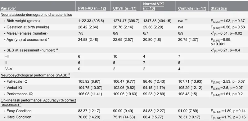
Table 1.Neonatal, socio-demographic, neuropsychological and on-line behavioural data of the study groups performing a verbal fluency task.
Variable+ PVH
+VD (n512) UPVH (n517)
Normal VPT
(n513) Controls (n517) Statistics
Neonatal/socio-demographic characteristics
NBirth-weight (grams) 1122.33 (395.6) 1274.47 (396.7) 1347.38 (404.15) n/a++ F(2,39)51.03, p50.37
NGestation at birth (weeks) 28.42 (2.64) 28.76 (2.14) 29.38 (2.29) n/a F(2,39)50.56, p50.58
NMales/Females (number) 7/5 8/9 6/7 8/9 x2(3)50.51, p50.92
NAge (yrs) at assessment * 24.58 (2.48) 22.65 (2.57) 20.80 (1.8) 20.75 (1.37) F(3,55)59.99,
p,0.001
NSES at assessment (number)a x2(6)56.21, p50.4
I–II 6 10 4 7
III 6 5 7 5
IV–V 0 2 2 4
Neuropsychological performance (WASI)b
NFull-scale IQ 105.92 (6.97) 106.47 (9.77) 96.46 (12.43) 107.71 (13.93) F(3,51)52.53, p50.07 NVerbal IQ 104.75 (10.07) 102.06 (9.62) 94.15 (11.79) 105.29 (12.12) F(3,51)52.5, p50.07
NPerformance IQ 106.08 (11.41) 109.06 (10.63) 99.23 (12.89) 108.43 (15) F(3,51)51.61, p50.2
On-line task performance: Accuracy (% correct responses)c
NEasy Condition 83.37 (12.17) 90.09 (9.49) 84.83 (12.27) 91.09 (7.89) F(3, 54)51.89, p50.14 NHard Condition 70.66 (14.29) 75.11 (14.63) 66.4 (15.77) 78.31 (10.17) F(3, 54)51.79, p50.16
+Mean and standard deviation (SD) are presented, unless otherwise stated++n/a5non-applicable; neonatal data were not available for controls *p,0.001,
Post-hoc comparisons with a Games-Howell test showed that the PVH+VD group was significantly older than the normal VPT [Mean difference; MD53.78, p,0.005] and the control groups [MD53.83, p,0.005]aFor controls n51 missing databFor controls n53 missing data; age at assessment was used as a covariatecFor PVH
+VD, n511; 1 participant was excluded from fMRI data analysis due to problems with scan acquisition.
at assessment, neonatal, neuropsychological [43] and on-line behavioural data were performed using one-way univariate ANOVA for comparison of 3 or more groups, or a student’s t test or a Mann-Whitney test, according to data
distribution, for comparisons of 2 groups.
To explore the link between task performance and fMRI data, correlation analysis was performed between SSQ values extracted from brain regions where significant interaction effects were observed and measures of task performance.
Results
Sample characteristics
Table 1 summarises neonatal, socio-demographic and neuropsychological data for groups of participants that performed the verbal fluency task. Groups significantly differed in age at assessment. There were no significant between-group differences in IQ.
Study groups that performed the n-back task did not significantly differ in neonatal, socio-demographic and neuropsychological data (Table 2).
On-line task performance
There were no significant between-group differences in the mean number of correct responses given during the ‘easy’ and the ‘hard’ condition of the verbal fluency task (Table 1).
There were significant differences in the d9 prime measure of on-line task performance between the PVH+VD and the UPVH groups during the 2- and 3-back conditions of the working memory task, but no significant differences in the mean reaction time during the n-back task (Table 2).
fMRI results
Table 3 shows the between-group fMRI results.
Verbal fluency task
There was a significant effect of group (i.e., VPT study groups.controls) as cognitive demands increased (‘hard’.‘easy’) in a cluster with local maxima in the right caudate nucleus. No significant effects of brain injury on BOLD signal were observed (Figure 1a).
N-back task
There were significant between-group differences (PVH+VD,UPVH) in the haemodynamic response to an increasing working memory load (3-back.1-back) in a cluster with local maxima in the left inferior frontal gyrus (Brodmann area-BA9) (Figure 1b).
condition, and SSQ values where significant interaction effects were noted, correlation analysis was carried out. Results showed that SSQ values in the left inferior frontal gyrus were not significantly associated with task performance (r50.43, p50.07).
Discussion
Verbal fluency
Our findings demonstrated that VPT-born young adults, regardless of extent of neonatal brain injury, exhibited a pattern of increased haemodynamic response to a phonological verbal fluency task as its cognitive demands increased (‘hard’.
Table 2.Neonatal, socio-demographic, neuropsychological and on-line behavioural data of the PVH+VD and UPVH groups performing an n-back task.
Variable+ PVH
+VD (n510) (n513) UPVH (n58) Statistics
Neonatal/socio-demographic characteristics
NBirth-weight (grams) 1150.6 (380.86) 1208 (322.75) F(16)50.35, p50.74
NGestation at birth (weeks) 28.7 (2.45) 28.75 (2.44) F(16)50.003, p50.97
NMales/Females (number) 6/4 2/6 x2(1)51.02, p50.31
NAge (yrs) at assessment 25.4 (1.71) 25.13 (1.25) F(16)51.2, p50.71
NSES at assessment (number) x2(2)52.61, p50.27
I–II 6 6
III 4 1
IV–V 0 1
Neuropsychological performance
NWASI
Full-scale IQ 106.7 (7.42) 104.25 (11.34) F(16)51.52, p50.59
Verbal IQ 105 (10.81) 100 (9.75) F(16)50.002, p50.32
Performance IQ 107.3 (12.07) 107.25 (11.7) F(16)50.06, p50.99
NCANTAB – Stockings of Cambridge
Planning time (ms)a 14084.8 (10912.79) 10135.16 (7703.62) F(16)52.01, p50.4
Execution time (ms)b 1366.25 (2169.7) 2074.28 (2134.27) F
(16)50.008, p50.5
Perfect solutions (number)c 9.2 (2.74) 9.13 (1.13) F
(16)54.81, p50.94 On-line task performance
Nd9prime
1-back 3.12 (1.07) 3.5 (0.47) Z533.5, p50.57
2-back 2.68 (1.28) 3.67 (0.17) F(16)52.18, p50.02
3-back 2.02 (0.83) 2.89 (0.83) Z514, p50.02
NReaction time (milliseconds)
1-back 682.13 (258.98) 550.03 (154.43) Z51.78, p50.83
2-back 625.99 (173.75) 570.26 (107.55) F(16)50.63, p50.44
3-back 707.53 (206.69) 659.11 (141.3) F(16)50.35, p50.58
+Mean and standard deviation (SD) are presented, unless otherwise stateda
The time taken to initiate the problemsbThe time taken to complete the problemscThe number of problems solved in minimum movesa,bThe planning and execution times were calculated at the highest level of difficulty (5 moves) to minimize possible ceiling effects.
Table 3.Between-group differences in regional brain activation with inreasing task difficulty during a verbal fluency and an n-back task.
Brain Region (Brodmann area)
Peak Talairach Coordinates
(x, y, z) Cluster size Cluster p value
Verbal fluency task (‘hard’>‘easy’): VPT groups>controls
R caudate nucleus extending to: 21,230, 17 203 0.00027
NLaterally - R insula (13) and superior temporal gyrus (BA 41) NInferiorly - L caudate body and bilaterally to thalamus
NSuperiorly - R posterior cingulate gyrus (31), R precuneus (31), R superior
temporal gyrus (39), R precentral gyrus (6) and R postcentral gyrus (2)
N-back task (3-back>1-back): PVH+VD#UPVH
L Inferior frontal gyrus (9) extends: 240, 4, 26 192 0.00039
NAnteriorly - L middle frontal gyrus (9) and L superior frontal gyrus (9) NInferiorly - L middle frontal gyrus (46)
NSuperiorly - L precentral gyrus (6) and L middle frontal gyrus (8/6)
R5right; L5left.
doi:10.1371/journal.pone.0113975.t003
Figure 1. Between-group differences in regional brain activation as cognitive load increased during fMRI tasks.The numbers at the top of each row of slices represents the z coordinate in Talairach space. The right side of the brain corresponds to the right side of each slice.
‘easy’) compared to term-born controls, in a large cluster that included the caudate nucleus, thalamus, insula, frontal, temporal and parietal cortices.
These results may partly be explained by structural alterations in the same brain regions that often accompany VPT birth [36,53,54], which are likely to exert some influence on activation patterns [41]. The lack of significant differences in caudate/thalamic activation between the three VPT samples grouped according to neonatal ultrasound classification is somewhat surprising, as alterations to these regions have been associated with cerebral haemorrhage [36,55,56]. Increasing cognitive load on a phonological verbal fluency task may have exceeded the capacities of the existing neural resources in the VPT group, resulting in the functional recruitment of additional neural resources [57]. Results from a previous fMRI investigation from our group using the same task, reported increased activation during ‘hard’ letters in the anterior cingulate gyrus and decreased activation during ‘easy’ letters in the same brain region in VPT young adults compared to controls [24]. Although different analyses methods were used in our previous study, these findings support the idea of increased recruitment of task-related brain regions with increased cognitive load. As task performance was similar in the four study groups, the between-group differences in BOLD signal that we observed here may not simply reflect differential task performance [58], although the strategies used by the four groups to complete the on-line task were not recorded and may indeed have differed.
Evidence from fMRI studies suggests that activation of the caudate nucleus, thalamus, insula and precentral and postcentral gyri is associated with articulatory demands [59–62] and that the superior temporal gyrus forms a part of the verbal fluency network sub-serving phonological aspects of the task [63–66]. Therefore, the greater engagement of these regions in the VPT groups seen here may have been necessary to achieve satisfactory on-line task performance. Increased activation of the caudate nucleus, precentral and postcental gyri has also been linked to increased cognitive demand, possibly relating to executive components of the verbal fluency task (15).
Furthermore, caudate nucleus has been involved in the suppression of irrelevant words, as well as of the activation of brain regions that may interfere with goal-oriented language production [67–69]. Therefore, the increased activation of the caudate nucleus in the VPT groups, as the cognitive demands of the task increased, may also reflect an increasing effort to maintain attention on task-related procedures and to suppress the generation of irrelevant words.
Taken together, the finding of altered activation in the VPT group during increasing cognitive load associated with phonological verbal fluency processing demonstrate long- term effects of preterm birth on the cortico-striatal-thalamo-cortical circuitry. These findings extend our previous research, which demon-strated altered activation in fronto-striatal pathways using the same task but considering ‘easy’ and ‘hard’ letter trials separately [24]. Alterations in the cortico-striatal-thalamo-cortical circuitry are likely to underlie the risk for specific motor, executive-type and emotional problems, which are part of the long-term sequelae of very preterm birth [71–73].
Working memory
Our results showed that VPT young adults who sustained severe neonatal brain injury displayed decreased brain activation compared to VPT young adults with less-severe neonatal brain injury (UPVH) in left frontal brain regions in response to increasing working memory load of an n-back task (3-back.1-back). The cluster in which this significant interaction was observed was centred in the inferior/middle/superior frontal gyri (BA9), extending to the dorsolateral
prefrontal cortex (DLPFC), which is a brain area thought to be centrally involved in working memory processing [74]. Hypoactivity in the DLPFC has been described in developmentally delayed populations, such as individuals with attention deficit hyperactivity disorder during executive-type tasks [75,76]. A load-dependent role of the DLPFC in working memory maintenance has also been reported (15).
Between-group differences (PVH+VD,UPVH) that were linked to increasing task difficulty were also evident in the left precentral gyrus (BA6), and left middle frontal gyrus (BA6/8) i.e., supplementary motor cortex. Results from neuroima-ging studies have shown that the precentral gyrus (BA6) and supplementary motor cortex may be involved in a sub-vocal rehearsal system of the phonological loop of working memory [29,62], whereby maintenance of verbal information is achieved.
At a behavioural level, the PVH+VD group had lowerd9scores than the UPVH group during the 3-back condition of the task, which is the most difficult, although there were no significant between-group differences in reaction time. The lack of significant association between d9 scores achieved during the 3-back condition and functional data suggests that our fMRI results may not be solely attributable to on-line performance differences, and may instead reflect the long-term effects of neonatal brain injury.
Limitations
The fMRI data reported here may not generalise to VPT populations at large, as VPT individuals with compromised cognitive function were not studied. A further limitation of this study relating to the interpretation of on-line behavioural data arises from the fact that the study groups are relatively small for behavioural data analysis [77]. Finally, the population who undertook n-back task was small and included neither healthy controls nor VPT individuals with normal neonatal ultrasound results, which may prompt a caveat in the interpretation of the results.
Conclusions
The results of the current study show that neonatal brain injury may exert differential effects on the functional neuroanatomy of various executive-type processes. Functional activation of cortico-striatal-thalamo-cortical circuitry associated with phonological verbal fluency appears to be increased following VPT birth, regardless of the extent of neonatal brain damage. This finding is in line with the results of other studies in VPT samples, which have documented the existence of functional neuroplastic adaptation in relation to language processing, reflected by the recruitment of additional task-related neural resources [24,78]. On the other hand, the presence of severe neonatal brain injury may be associated with decreased engagement of the frontal cortex during verbal working memory processes, possibly because of its limited resource capacities due to maturational delays [28,57,79]. These data increase our understanding of the long-term consequences of early brain injury in ex-preterm individuals and may aid the development of neuro-protective treatments designed to improve long-term sequelae.
Acknowledgments
The authors wish to thank Professor Mick Brammer and Mr Jeffrey Dalton for providing us with scientific advice, and all participants for their on-going cooperation.
Author Contributions
Conceived and designed the experiments: CN VG PM RMM LR MPA. Performed the experiments: AKK. Analyzed the data: AKK VG. Contributed reagents/ materials/analysis tools: VG AKK CN. Wrote the paper: AKK CN. Provided help with participants’ recruitment: MW.
References
2. Anderson PJ, Reidy N(2012) Assessing executive function in preschoolers 2. Neuropsychol Rev 22: 345–360.
3. Mulder H(2009) Development pf Executive Function and attention in Preterm Children: A Systematic Review. Developmental Neuropsychology 34: 393–421.
4. Luu TM, Ment L, Allan W, Schneider K, Vohr BR(2011) Executive and memory function in adolescents born very preterm. Pediatrics 127: e639–e646.
5. Nosarti C, Giouroukou E, Micali N, Rifkin L, Morris RG, et al.(2007) Impaired executive functioning in young adults born very preterm. J Int Neuropsychol Soc 13: 571–581.
6. Larroque B, Marret S, Ancel PY, Arnaud C, Marpeau L, et al. (2003) White matter damage and intraventricular hemorrhage in very preterm infants: the EPIPAGE study 1. J Pediatr 143: 477–483.
7. Ballabh P(2010) Intraventricular hemorrhage in premature infants: mechanism of disease 1. Pediatr Res 67: 1–8.
8. Vasileiadis GT, Gelman N, Han VK, Williams LA, Mann R, et al.(2004) Uncomplicated intraventricular hemorrhage is followed by reduced cortical volume at near-term age. Pediatrics 114: e367–e372.
9. Nagy Z, Ashburner J, Andersson J, Jbabdi S, Draganski B, et al. (2009) Structural correlates of preterm birth in the adolescent brain 1. Pediatrics 124: e964–e972.
10. Salvan P, Froudist WS, Allin MP, Walshe M, Murray RM, et al.(2013) Road work on memory lane-Functional and structural alterations to the learning and memory circuit in adults born very preterm 1. Neuroimage.
11. Chow TW, Cummings JL(1999) Frontal-subcortical circuits. In: Miller BL, Cummings JL, editors. The human frontal lobes: Functions and Disorders. New York: Guilford Press. pp. 25–43.
12. Evrard P, Gressens P, Volpe JJ(1992) New concepts to understand the neurological consequences of subcortical lesions in the premature brain. Biol Neonate 61: 1–3.
13. Alexander GE, DeLong MR, Strick PL(1986) Parallel organization of functionally segregated circuits linking basal ganglia and cortex. Annu Rev Neurosci 9: 357–381.
14. Stewart AL, Thorburn RJ, Hope PL, Goldsmith M, Lipscomb AP, et al. (1983) Ultrasound appearance of the brain in very preterm infants and neurodevelopmental outcome at 18 months of age. Arch Dis Child 58: 598–604.
15. Paneth N, Rudelli R, Kazam E, Monte W(1994) Brain Damage in the Preterm Infant. London: Mac Keith Press.
16. Nosarti C, Nam KW, Walshe M, Murray RM, Cuddy M, et al.(2014) Preterm birth and structural brain alterations in early adulthood. NeuroImage: Clinical 6: 180–191.
17. Allin M, Henderson M, Suckling J, Nosarti C, Rushe T, et al.(2004) Effects of very low birthweight on brain structure in adulthood. Dev Med Child Neurol 46: 46–53.
18. Kalpakidou AK, Allin MP, Walshe M, Giampietro V, Nam KW, et al.(2012) Neonatal brain injury and neuroanatomy of memory processing following very preterm birth in adulthood: an fMRI study. PLoS One 7: e34858.
19. Indefrey P, Levert WJ(2000) The neural correlates of language production. In: Gazzaniga MS, editors. The New Cognitive Neurosciences. Cambridge: MIT Press. pp. 845–865.
20. Ruff RM, Light RH, Parker SB, Levin HS(1997) The psychological construct of word fluency. Brain Lang 57: 394–405.
21. Rubia K, Smith AB, Woolley J, Nosarti C, Heyman I, et al. (2006) Progressive increase of frontostriatal brain activation from childhood to adulthood during event-related tasks of cognitive control. Hum Brain Mapp 27: 973–993.
22. Woodward LJ, Edgin JO, Thompson D, Inder TE(2005) Object working memory deficits predicted by early brain injury and development in the preterm infant. Brain 128: 2578–2587.
24. Nosarti C, Shergill SS, Allin MP, Walshe M, Rifkin L, et al.(2009) Neural substrates of letter fluency processing in young adults who were born very preterm: Alterations in frontal and striatal regions. Neuroimage.
25. Cohen JD, Perlstein WM, Braver TS, Nystrom LE, Noll DC, et al.(1997) Temporal dynamics of brain activation during a working memory task. Nature 386: 604–608.
26. Baddeley AD(1986) Working memory. New York: Oxford University Press.
27. Curtis WJ, Zhuang J, Townsend EL, Hu X, Nelson CA (2006) Memory in early adolescents born prematurely: a functional magnetic resonance imaging investigation. Dev Neuropsychol 29: 341–377.
28. Griffiths ST, Gundersen H, Neto E, Elgen I, Markestad T, et al.(2013) fMRI: blood oxygen level-dependent activation during a working memory-selective attention task in children born extremely preterm 1. Pediatr Res 74: 196–205.
29. Rypma B, Prabhakaran V, Desmond JE, Glover GH, Gabrieli JD(1999) Load-dependent roles of frontal brain regions in the maintenance of working memory. Neuroimage 9: 216–226.
30. Kirschen MP, Chen SH, Schraedley-Desmond P, Desmond JE(2005) Load- and practice-dependent increases in cerebro-cerebellar activation in verbal working memory: an fMRI study 1. Neuroimage 24: 462–472.
31. Low KA, Leaver EE, Kramer AF, Fabiani M, Gratton G(2009) Share or compete? Load-dependent recruitment of prefrontal cortex during dual-task performance 1. Psychophysiology 46: 1069–1079.
32. Costello AM, Hamilton PA, Baudin J, Townsend J, Bradford BC, et al. (1988) Prediction of neurodevelopmental impairment at four years from brain ultrasound appearance of very preterm infants. Dev Med Child Neurol 30: 711–722.
33. Roth SC, Baudin J, McCormick DC, Edwards AD, Townsend J, et al. (1993) Relation between ultrasound appearance of the brain of very preterm infants and neurodevelopmental impairment at eight years. Dev Med Child Neurol 35: 755–768.
34. Stewart AL, Costello AM, Hamilton PA, Baudin J, Townsend J, et al.(1989) Relationship between neurodevelopmental status of very preterm infants at one and four years. Dev Med Child Neurol 31: 756– 765.
35. Nosarti C, Al-Asady MH, Frangou S, Stewart AL, Rifkin L, et al.(2002) Adolescents who were born very preterm have decreased brain volumes. Brain 125: 1616–1623.
36. Nosarti C, Giouroukou E, Healy E, Rifkin L, Walshe M, et al.(2008) Grey and white matter distribution in very preterm adolescents mediates neurodevelopmental outcome. Brain.
37. Stewart AL, Rifkin L, Amess PN, Kirkbride V, Townsend JP, et al. (1999) Brain structure and neurocognitive and behavioural function in adolescents who were born very preterm. Lancet 353: 1653– 1657.
38. Allin M, Walshe M, Fern A, Nosarti C, Cuddy M, et al.(2008) Cognitive maturation in preterm and term born adolescents. J Neurol Neurosurg Psychiatry 79: 381–386.
39. Allin MP, Kontis D, Walshe M, Wyatt J, Barker GJ, et al.(2011) White matter and cognition in adults who were born preterm 1. PLoS One 6: e24525.
40. Lawrence EJ, Rubia K, Murray RM, McGuire PK, Walshe M, et al. (2009) The neural basis of response inhibition and attention allocation as mediated by gestational age. Hum Brain Mapp 30: 1038– 1050.
41. Lawrence EJ, McGuire PK, Allin M, Walshe M, Giampietro V, et al.(2010) The Very Preterm Brain in Young Adulthood: The Neural Correlates of Verbal Paired Associate Learning. The Journal of Pediatrics.
42. Narberhaus A, Lawrence E, Allin MP, Walshe M, McGuire P, et al.(2009) Neural substrates of visual paired associates in young adults with a history of very preterm birth: Alterations in fronto-parieto-occipital networks and caudate nucleus. Neuroimage.
43. Wechsler D (1999) Wechsler Abbreviated Scale of Intelligence. New York: The Psychological Corporation.
44. HMSO(1991) Her Majesty’s Stationary Office (HMSO), Standard Occupational Classification. London: HMSO.
46. Bullmore E, Brammer M, Williams SC, Rabe-Hesketh S, Janot N, et al.(1996) Statistical methods of estimation and inference for functional MR image analysis. Magn Reson Med 35: 261–277.
47. Curtis VA, Bullmore ET, Brammer MJ, Wright IC, Williams SC, et al. (1998) Attenuated frontal activation during a verbal fluency task in patients with schizophrenia. Am J Psychiatry 155: 1056–1063.
48. Borkowski J, Benton A, Spreen O(1967) Word fluency and brain damage. Neuropsychologia 5: 135– 140.
49. Broome MR, Matthiasson P, Fusar-Poli P, Woolley JB, Johns LC, et al.(2009) Neural correlates of executive function and working memory in the ‘at-risk mental state’. Br J Psychiatry 194: 25–33.
50. Sala-Llonch R, Pena-Gomez C, renaza-Urquijo EM, Vidal-Pineiro D, Bargallo N, et al.(2012) Brain connectivity during resting state and subsequent working memory task predicts behavioural performance 11. Cortex 48: 1187–1196.
51. Bullmore E, Long C, Suckling J, Fadili J, Calvert G, et al.(2001) Colored noise and computational inference in neurophysiological (fMRI) time series analysis: resampling methods in time and wavelet domains. Hum Brain Mapp 12: 61–78.
52. Talairach J, Tournoux P(1988) A Co-planar Stereotactic Atlas of the Human Brain. New York: Thieme Medical Publishers.
53. Kesler SR, Ment LR, Vohr B, Pajot SK, Schneider KC, et al.(2004) Volumetric analysis of regional cerebral development in preterm children. Pediatr Neurol 31: 318–325.
54. Taylor HG, Filipek PA, Juranek J, Bangert B, Minich N, et al.(2011) Brain volumes in adolescents with very low birth weight: effects on brain structure and associations with neuropsychological outcomes. Dev Neuropsychol 36: 96–117.
55. Srinivasan L, Dutta R, Counsell SJ, Allsop JM, Boardman JP, et al.(2007) Quantification of deep gray matter in preterm infants at term-equivalent age using manual volumetry of 3-tesla magnetic resonance images 1. Pediatrics 119: 759–765.
56. Volpe JJ (1995) Neurological Evaluation Hypoxic-ischemic Encephalopathy and Intracranial Hemorrhage. In: Neurology of the newborn. Philadelphia: Saunders. pp. 95–463.
57. Just MA, Varma S(2007) The organization of thinking: what functional brain imaging reveals about the neuroarchitecture of complex cognition. Cogn Affect Behav Neurosci 7: 153–191.
58. Fu CH, McGuire PK(1999) Functional neuroimaging in psychiatry 1. Philos Trans R Soc Lond B Biol Sci 354: 1359–1370.
59. Fu CH, Morgan K, Suckling J, Williams SC, Andrew C, et al.(2002) A functional magnetic resonance imaging study of overt letter verbal fluency using a clustered acquisition sequence: greater anterior cingulate activation with increased task demand. Neuroimage 17: 871–879.
60. Murphy K, Corfield DR, Guz A, Fink GR, Wise RJ, et al.(1997) Cerebral areas associated with motor control of speech in humans. J Appl Physiol 83: 1438–1447.
61. Klein D, Zatorre RJ, Milner B, Meyer E, Evans AC(1994) Left putaminal activation when speaking a second language: evidence from PET. Neuroreport 5: 2295–2297.
62. Paulesu E, Frith CD, Frackowiak RS(1993) The neural correlates of the verbal component of working memory. Nature 362: 342–345.
63. Fu CH, Suckling J, Williams SC, Andrew CM, Vythelingum GN, et al.(2005) Effects of psychotic state and task demand on prefrontal function in schizophrenia: an fMRI study of overt verbal fluency. Am J Psychiatry 162: 485–494.
64. Yetkin FZ, Hammeke TA, Swanson SJ, Morris GL, Mueller WM, et al. (1995) A comparison of functional MR activation patterns during silent and audible language tasks. AJNR Am J Neuroradiol 16: 1087–1092.
65. Lurito JT, Kareken DA, Lowe MJ, Chen SH, Mathews VP(2000) Comparison of rhyming and word generation with FMRI. Hum Brain Mapp 10: 99–106.
66. Burton MW, Locasto PC, Krebs-Noble D, Gullapalli RP (2005) A systematic investigation of the functional neuroanatomy of auditory and visual phonological processing. Neuroimage 26: 647–661.
68. Crosson B, Benefield H, Cato MA, Sadek JR, Moore AB, et al.(2003) Left and right basal ganglia and frontal activity during language generation: contributions to lexical, semantic, and phonological processes 1. J Int Neuropsychol Soc 9: 1061–1077.
69. Ali N, Green DW, Kherif F, Devlin JT, Price CJ (2010) The role of the left head of caudate in suppressing irrelevant words 3. J Cogn Neurosci 22: 2369–2386.
70. Menon V, Adleman NE, White CD, Glover GH, Reiss AL(2001) Error-related brain activation during a Go/NoGo response inhibition task 2. Hum Brain Mapp 12: 131–143.
71. Anderson PJ, Doyle LW (2004) Executive functioning in school-aged children who were born very preterm or with extremely low birth weight in the 1990s. Pediatrics 114: 50–57.
72. Bolisetty S, Dhawan A, bdel-Latif M, Bajuk B, Stack J, et al.(2014) Intraventricular hemorrhage and neurodevelopmental outcomes in extreme preterm infants 1. Pediatrics 133: 55–62.
73. Johnson S, Marlow N(2011) Preterm birth and childhood psychiatric disorders 1. Pediatr Res 69: 11R– 18R.
74. Owen AM, McMillan KM, Laird AR, Bullmore E(2005) N-back working memory paradigm: a meta-analysis of normative functional neuroimaging studies. Hum Brain Mapp 25: 46–59.
75. Dickstein SG, Bannon K, Castellanos FX, Milham MP(2006) The neural correlates of attention deficit hyperactivity disorder: an ALE meta-analysis. J Child Psychol Psychiatry 47: 1051–1062.
76. Valera EM, Brown A, Biederman J, Faraone SV, Makris N, et al. (2010) Sex differences in the functional neuroanatomy of working memory in adults with ADHD. Am J Psychiatry 167: 86–94.
77. Rubia K, Smith AB, Brammer MJ, Taylor E(2007) Temporal lobe dysfunction in medication-naive boys with attention-deficit/hyperactivity disorder during attention allocation and its relation to response variability. Biol Psychiatry 62: 999–1006.
78. Constable RT, Vohr BR, Scheinost D, Benjamin JR, Fulbright RK, et al. (2013) A left cerebellar pathway mediates language in prematurely-born young adults 1. Neuroimage 64: 371–378.