Molecular cloning, characterization and expression analysis of a novel
PDRG1
gene from black tiger shrimp (
Penaeus monodon)
Chao Zhao
1,3, Wenting Dai
1,2,3and Lihua Qiu
1,3,41
South China Sea Fisheries Research Institute, Chinese Academy of Fishery Sciences, Guangzhou, China.
2
College of Aqua-life Science and Technology, Shanghai Ocean University, Shanghai, China.
3
Key Laboratory of South China Sea Fishery Resources Exploitation & Utilization, Ministry of Agriculture,
Guangzhou, China.
4
Tropical Aquaculture Research and Development Center of South China Sea Fisheries Research Institute,
Sanya, China.
Abstract
P53 And DNA Damage-Regulated Gene 1 (PDRG1) is a novel gene which plays an important role in chaperone-mediated protein folding. In the present study, the full-length complementary DNA (cDNA) sequence of thePDRG1 gene fromPenaeus monodon (PmPDRG1) was cloned by the rapid amplification of cDNA ends (RACE) method. The cDNA ofPmPDRG1 spans 1,613 bp, interrupted by only one short intron, and encodes a protein of 136 amino acids with calculated molecular weight of 15.49 kDa. The temporal expression profile ofPmPDRG1 in different tis-sues and in different developmental stages of the ovary was investigated by real-time quantitative PCR (RT-qPCR). An RNA interference (RNAi) experiment was performed to study the relationship betweenP. monodon p53 (Pmp53) and PmPDRG1, and the results showed that the relative expression level of PmPDRG1 mRNA was notably up-regulated from 12 h to 96 h afterPmp53 was silenced both in ovary and hepatopancreas. To further explore the role of PmPDRG1 in ovarian development, dopamine (DA) and 5-hydroxytryptamine (5-HT)-injected shrimps were analyzed by RT-qPCR, indicating that PmPDRG1 may be involved in the regulation of ovarian development ofP. monodon.
Keywords:PDRG1, gene cloning, RT-qPCR, ovarian development, black tiger shrimp.
Received: May 23, 2016; Accepted: September 28, 2016.
Introduction
P53 And DNA Damage-Regulated Gene 1(PDRG1, namely was first identified in 2003 by Luoet al. (2003). The humanPDRG1resides at the long arm of chromosome 20 and encodes a protein of 133 amino acids that is present within a distinct subcellular compartment of the cytoplasm (Deloukaset al., 2001; Luoet al., 2003).PDRG1is usually strongly over-expressed in multiple human malignancies (Jianget al., 2011; Wanget al., 2015a). PDRG1 protein was identified as a subunit of the R2TP/prefoldin-like com-plex, which is involved in the assembly of the RNA poly-merase II complex (Pol II) in the cytoplasm of eukaryotic cells (Sardiuet al., 2008; Boulonet al., 2012; Mitaet al., 2013). The tumor suppressor protein p53 can down-regu-late the expression of PDRG1 mRNA, while ultraviolet (UV) radiation has the opposite effect (Luoet al., 2003). As certain interactions between PDRG1 and p53 exist, some
scholars believe that PDRG1 has the potential to be a novel valuable tumor biomarker that could play a role in cancer development and/or progression or to be a DNA dam-age-associated maker (Jianget al., 2011; Saigusaet al., 2012; Wanget al., 2015a). Furthermore, PDRG1 is proven to be involved in apoptosis and cell cycle regulation (Jiang
et al., 2011; Wang et al., 2014). Although the roles of PDRG1 in DNA damage and tumor cell growth in verte-brates have been widely studied, the functions of PDRG1 in invertebrates, especially in crustaceans, are poorly under-stood.
The black tiger shrimp (P. monodon) is one of the most important aquatic commercial animals in Asia, espe-cially in southern China. Because the eyestalk of P. monodoncan secrete ovarian suppression hormones, uni-lateral eyestalk ablation is usually adopted to induce ovar-ian maturation ofP. monodon, but this technique leads to the death of parent shrimps and lowers spawning quality (Benzie, 1998; Phinyoet al., 2013). Therefore, it is impera-tive to explore alternaimpera-tive technologies to eyestalk ablation and to understand the molecular mechanisms that control
DOI: http://dx.doi.org/10.1590/1678-4685-GMB-2016-0144
Send correspondence to Lihua Qiu. The South China Sea Fisheries Research Institute, Chinese Academy of Fishery Sciences, 231 Xingangxi Road, Guangzhou 510300, P. R. China. E-mail: [email protected]
the development and maturation of ovaries/oocytes (Hiransuchalertet al., 2013; Phinyo et al., 2014). In our previous study, we found that Pmp53 plays an important role in the development and maturation of the ovaries inP. monodon(Daiet al., 2016). In the present study, we si-lenced Pmp53 to investigate its relationship with
PmPDRG1and the role that PmPDRG1 may play in the ovarian development ofP. monodon. Biogenic amines such as dopamine (DA) and serotonin (5-hydroxytryptamine, 5-HT) are able to affect numerous physiological processes in crustaceans through their actions as neuroregulators. Both DA and 5-HT have been shown to be involved in the synthesis and release of neurohormones, such as crustacean hyperglycemic hormone (CHH), vitellogenesis-inhibiting hormone (VIH) and molt-inhibiting hormone (MIH) (Chen
et al., 2003). It has been demonstrated that injected 5-HT can induce ovarian maturation in shrimp (Vaca and Alfaro, 2000), while dopamine depressed vitellogenin synthesis (Chenet al., 2003). The relationship between PDRG1 and ovarian maturation and the effects of DA and 5-HT on the expression levels of PmPDRG1 in ovaries and hepato-pancreas ofP. monodonare presented in this paper.
In this study, we cloned and characterized the full-length cDNA ofP. monodon PmPDRG1and assessed the distribution ofPmPDRG1 transcripts in different tissues and ovary developmental stages. In addition, we investi-gated the expression profiles ofPmPDRG1 mRNA in se-lected tissues after injection of Pmp53-dsRNA and exposure to 5-HT and DA. Results from this study will con-tribute to a better understanding ofPDRG1and its function in ovarian development ofP. monodon.
Materials and Methods
Experimental animals and sample collection
Healthy black tiger shrimp,P. monodon(100±18 g), cultivated in aerated seawater (salinity of 30 PSU) for three days at 25±1oC in the Shenzhen Base of South China Sea Fisheries Research Institute (Shenzhen, Guangdong prov-ince, China) were used as the material in the experiment. Various tissues (ovary, heart, intestine, brain, muscles, stomach and gills) from male and female individuals were dissected, snap frozen in liquid nitrogen, and stored at -80oC. Five shrimp in each ovarian maturation stages were selected. The different ovarian stages used in this study were classified according to the morphology reported by Huanget al.(2006), as ovogonium stage (I), chromatin nu-cleolus stage (II), perinunu-cleolus stage (III), yolky stage (IV), and cortical rod stage (V).
Total RNA extraction, first strand cDNA synthesis and DNA extraction
Total RNA was isolated from the examined tissue (about 100 mg) of the shrimp using TRIzol (Invitrogen, Shanghai, China) reagent following the manufacturer’
pro-tocol, resuspended in DEPC-treated water and stored at –80oC (Jianget al., 2009). The concentration of RNA was determined using a NanoDrop 2000 spectrometer (Thermo, USA), and RNA integrity was assessed by 1% agarose gel electrophoresis. The cDNA was synthesized from 1mg of mRNA using a PrimeScript Reverse Transcriptase kit (TaKaRa, Dalian, China) following the manufacturer’s protocol, as previously described (Wanget al., 2015b). The cDNA was used as the template for PCR reactions in gene cloning. The phenol-chloroform-isoamyl alcohol method was used to isolate total genomic DNA, which was then used as template to amplify introns.
Gene cloning and sequencing
A partial sequence ofPDRG1(970 bp) was isolated from the transcriptome database. Initially, PCR was carried out using the cDNA described above as template, using the primers PaF and PaR (Table 1) designed according to the partial sequence ofPDRG1, for verification. Then, the 3’ end cDNA sequence of thePDRG1gene was isolated using a SMARTTM RACE cDNA amplification kit (Clontech, Takara) (Jiang et al., 2009; Wang et al., 2015b). In the 3’RACE PCR, the touchdown PCR step was performed with the gene-specific primer pdrg-sp1 and a universal primer UPM (a mix of UPX-long and UPX-short, Table 1). The PCR cycling parameters were as follows: an initial de-naturation at 94oC for 3 min, followed by 30 cycles at 94oC for 30 s, 60oC for 30 s, and 72oC for 3 min, and the last cy-cle was followed by 10 min extension at 72 oC. Addi-tionally, a nested PCR with pdrg-sp2 and NUP was carried out (PCR profile was as follows: 94oC for 3 min; 94oC for 30 s, 55oC for 30 s, 72oC for 3 min in 35 cycles; 72oC for 10 min). The PCR products were purified using a PCR puri-fication kit (Sangon, Shanghai, China) and cloned into the pMD18-T vector (TaKaRa). After transformtion into com-petent cells (E.coli DH5a), the positive clones were se-quenced in both directions (Invitrogen, Guangzhou, China), and the resulting sequences were verified and sub-jected to cluster analysis.
Sequence analysis, multiple sequence alignment, and phylogenetic analysis
per-formed using the ClustalX 2.0.11 software. The signal peptide was predicted using the Signal P 4.1 program (http://www.cbs.dtu.dk/services/SignalP/) and the SMART 4.0 program (http://smart.embl-heidelberg.de/) was used to predict functional sites or domains in the amino acid se-quence. A phylogenetic tree was constructed by the neigh-bor-joining (NJ) method and support of a bootstrap analysis with 1,000 replications implemented in the MEGA 5.0 package (Chenet al., 2013).
RNA interference
To investigate the relationship between PmPDRG1 and Pmp53 in ovarian development, an RNAi experiment was carried out using dsRNA specific forPmp53. Anin vi-troexpression system was adopted to obtain dsRNA. In brief, the recombined pD7 vector containing a bidirectional T7 RNA polymerase promoter was constructed using the pUC18 vector as described in previous studies (Robalinoet al., 2007; Wanget al., 2010).
Subsequently, a recombinant plasmid (pD7-p53) containing a reverse complement ofp53 was established and used as PCR template. Two separate PCR assays were set up with the primers pF/i53-R and pR/i53-F (Table 1). The PCR products of 763 bp and 590 bp were excised, gel-purified and used for in vitro transcription. Subse-quently, dsRNA-p53 was synthesized using the in vitro
Transcription T7 kit (TaKaRa) according to the manufac-turer’s instructions. The quality of dsRNAs was verified by 1.5% agarose gel electrophoresis and quantified using UV
spectrophotometry. The dsRNA was stored at -80oC until the experiment.
P. monodonshrimps (100±2 g, 14 months old) were acclimatized for 2 days before dsRNA-p53 injection. The shrimps were injected with 300 mg of dsRNA-p53 dis-solved in 40mL of sterilized saline solution (10 mM Tris-HCl pH 7.5, 400 mM NaCl). Shrimps injected with steril-ized saline solution were used as vehicle control (VC). Ovaries and hepatopancreas of the shrimp were randomly collected at 0, 12, 24, 48, 72, 96 h after injection, and frozen in liquid nitrogen for future RT-qPCR analysis.
5-HT and dopamine challenge
To examine the effects of 5-HT and DA on the ex-pression levels of PmPDRG1, seven groups of female shrimp (100±18 g, 14 months old) were injected intramus-cularly into the first abdominal segment with 5-HT and DA (50mg/g body weight). Shrimp injected with sterilized sa-line solution at 0 h were included as control. The ovaries and hepatopancreas were collected at 0, 6, 12, 24, 48, 72, 96 h post injection, and preserved in liquid nitrogen for future RT-qPCR analysis.
RT-qPCR for gene expression profile analysis
RT-qPCR was used to detect the temporal expression of the genes. cDNA was synthesized using the PrimeScriptTMRT reagent Kit with gDNA Eraser (Perfect Real Time) (TaKaRa) and used as templates for RT-qPCR assays. RT-qPCR was performed using SYBR®Premix Ex Taq TM II(TaKaRa) as described in our previous study
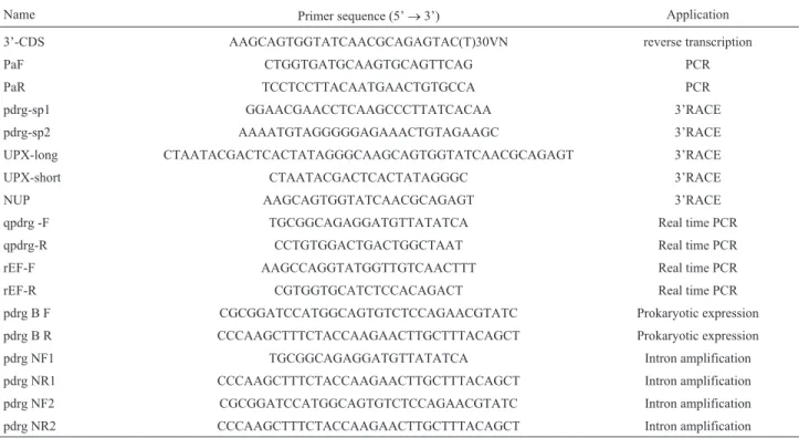
Table 1- Primers used for gene cloning and expression analysis.
Name Primer sequence (5’®3’) Application
3’-CDS AAGCAGTGGTATCAACGCAGAGTAC(T)30VN reverse transcription
PaF CTGGTGATGCAAGTGCAGTTCAG PCR
PaR TCCTCCTTACAATGAACTGTGCCA PCR
pdrg-sp1 GGAACGAACCTCAAGCCCTTATCACAA 3’RACE
pdrg-sp2 AAAATGTAGGGGGAGAAACTGTAGAAGC 3’RACE
UPX-long CTAATACGACTCACTATAGGGCAAGCAGTGGTATCAACGCAGAGT 3’RACE
UPX-short CTAATACGACTCACTATAGGGC 3’RACE
NUP AAGCAGTGGTATCAACGCAGAGT 3’RACE
qpdrg -F TGCGGCAGAGGATGTTATATCA Real time PCR
qpdrg-R CCTGTGGACTGACTGGCTAAT Real time PCR
rEF-F AAGCCAGGTATGGTTGTCAACTTT Real time PCR
rEF-R CGTGGTGCATCTCCACAGACT Real time PCR
pdrg B F CGCGGATCCATGGCAGTGTCTCCAGAACGTATC Prokaryotic expression
pdrg B R CCCAAGCTTTCTACCAAGAACTTGCTTTACAGCT Prokaryotic expression
pdrg NF1 TGCGGCAGAGGATGTTATATCA Intron amplification
pdrg NR1 CCCAAGCTTTCTACCAAGAACTTGCTTTACAGCT Intron amplification
pdrg NF2 CGCGGATCCATGGCAGTGTCTCCAGAACGTATC Intron amplification
(Jiang et al., 2009). The reference gene elongation fac-tor-1alpha(EF-1a) (GenBank: DQ021452.1) was used as internal control for normalizing the cDNA template by menas of the primers rEF-F and rEF-R (Table 1). Each 25
mL reaction solution contained: 12.5mL of 2SYBR® Pre-mix Ex TaqII, 0.5mL of forward primer (10mM), 0.5mL of reverse primer (10mM), 2mL of cDNA template equivalent to 70 ng total RNA, and 9.5mL sterile distilled water. Each reaction was carried out simultaneously in three separate tubes and the test was repeated three times (Liet al., 2013). Thermal cycling conditions were 95oC for 30 s, followed by 42 cycles of 95oC for 5 s, 60oC for 30 s. A melting curve analysis was added (95oC for 1 s, 65oC for 15 s, 95oC for continuous acquisition) to demonstrate the specificity of the PCR products, as revealed by a single peak. The 2-DDCT method was used to calculate relative gene expression lev-els (Livak and Schmittgen, 2001; Qiuet al., 2010).
Statistical analysis
Statistical analyses were carried out using SPSS soft-ware (SPSS Inc, USA). Data are reported as mean± stan-dard error (SE). Results obtained from qRT-PCR analysis were subjected to one-way analysis of variance (one-way ANOVA) followed by an unpaired, two-tailedt-test. Dif-ferences were considered significant at P < 0.05.
Results
Characterization of thePmPDRG1full-length cDNA
The full-length cDNA sequence ofPmPDRG1 was obtained by RACE-PCR, and the complete nucleotide se-quence and the deduced amino acid sese-quence are shown in Figure 1a (GenBank: KX156929). The cDNA sequence of
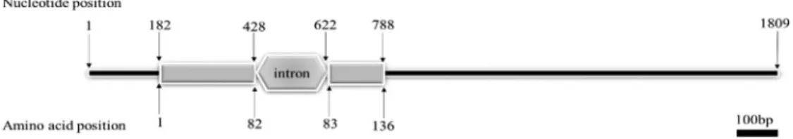
PmPDRG1is 1,613 bp in length, including an open reading frame of 411 bp (position 182-592 bp), a 5’UTR of 181 bp and a 3’UTR of 1,021 bp. Two eukaryotic polyadenylation signals AATAAA were located between nucleotides 926-931 bp and 956-961 bp and a poly (A) tail 28 bp down-stream. The ORF sequence was predicted to encode a pro-tein of 136 amino acids with a calculated molecular mass of about 15.49 kDa, and a theoretical isoelectric point of 8.62.
The structure prediction results showed that PmPDRG1 contains 66.17% ofa-helix, 5.15% ofb-pleated sheet, and 28.68% of random coil. These conserved regions include seven phosphorylation sites and one N-glyco-sylation site (Figure 1a). Signal P 4.1 analysis revealed that PmPDRG1 does not contain a typical signal peptide se-quence.PmPDRG1is encoded in the nuclear genome, and has only one intron in the ORF (Figure 1b).
Phylogenetic analysis of PmPDRG1
The predicted amino acid sequence shared homology with previously published PDRGR1 sequences of other species in the GenBank database, detected using BLAST
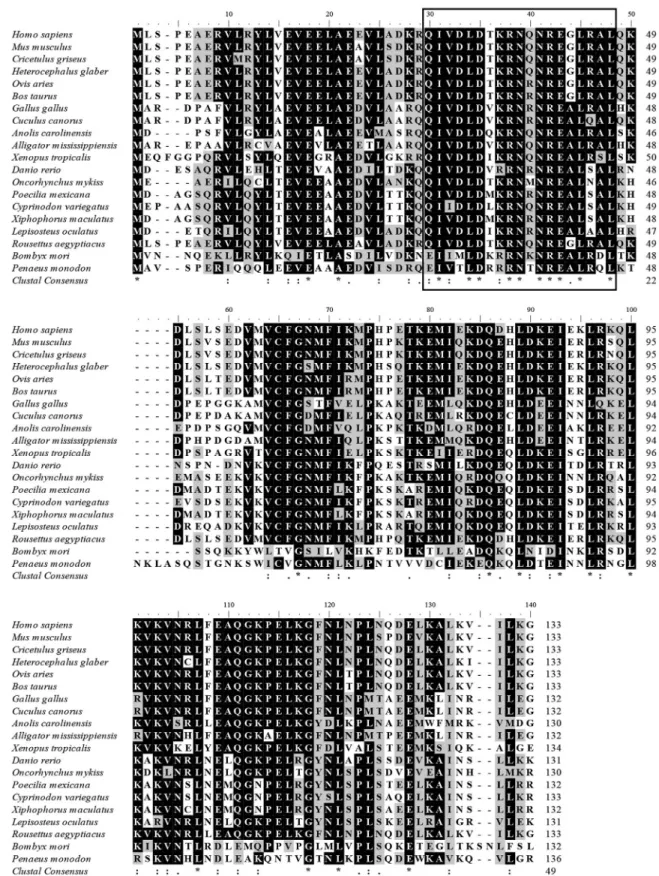
program. These include a 49% identity withAnoplopoma fimbria(ACQ58385.1), 48% identity withOncorhynchus mykiss(NP_001154150.1), and 47% identity with Danio rerio (NP_001017757.1). The putative amino acid se-quence of PmPDRG1 was aligned with other species in conserved regions. The deduced amino acid sequence QIVDLDTKRNQNREALRAL (30-48aa) of PmPDRG1 shared high homology with other species (Figure 2).
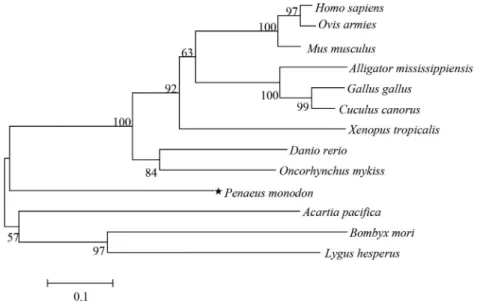
As shown in Figure 3, the PDRG1 phylogenetic tree comprised two main clusters: the upper cluster contained vertebrate PDRG1 sequences, while the second cluster con-tained invertebrate PDRG1 sequences. Vertebrate PDRG1 proteins appeared closely related to each other and con-verged into one subgroup, which include PmPDRG1. Al-though the shrimp PmPDRG1 was more similar to the vertebrate subgroup than the other main branch that in-cludes the majority of the invertebrates, it still was an out-lier in the main branch of the vertebrate subgroup.
Tissue expression analysis ofPmPDRG1
The tissue distribution pattern ofPmPDRG1mRNA is shown in Figure 4. The RT-qPCR results proved that the
PmPDRG1gene was expressed in all the examined tissues, with relatively high levels in the ovary, gill and intestine, moderate levels in the heart and brain, and low levels in th muscle and stomach (Figure 4).
Expression profiles ofPmPDRG1mRNA during ovarian maturation stages
The relative expression levels ofPmPDRG1mRNA in different ovarian stages ofP. monodonwere investigated by RT-qPCR. The expression level in stage III ovaries was about 14.5-fold higher than in other stages (P < 0.05) as shown in Figure 5. The expression among stages I, II, IV, and V were not significantly different.
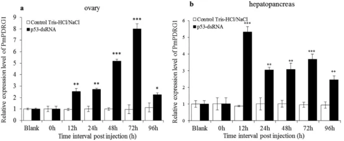
PmPDRG1mRNA expression profiles afterPmp53 gene silencing by Pmp53-dsRNA
To investigate the relationship between Pmp53 and PmPDRG1, the Pmp53 gene was silenced by Pmp53-dsRNA. In ovary, the silencing efficiency ofPmp53at 12, 24, 48 and 72 h of dsRNA-p53 post-injection were 65.86, 85.35, 64.06 and 25.45%, respectively, and in hepatopan-creas, the silencing efficiency from 12, 24, 48, 72 and 96 h post injection were 37.70, 39.29, 64.24, 88.67 and 20.37%, respectively. Detailed data onPmp53gene silencing have been published in our previous study (Daiet al., 2016). Af-ter Pmp53 was successfully silenced, the PmPDRG1
Figure 2- Multiple alignments of the deduced amino acid sequence ofPmPDRG1with other knownPDRG1aligned by Clustal X 2. 0. 11. Identical and similar sites are indicated with asterisks (*) and dots (. or :) , respectively. Black rectangles represent the phylogenetically conserved domain in different species. The species names and GenBank accession numbers are as follows:Homo sapiens(NP_110442.1);Mus musculus(NP_849270.1);Cricetulus griseus (ERE71423.1); Heterocephalus glaber (EHB11715.1); Ovis aries (XP_004014522.1); Bos taurus (NP_001071583.1); Gallus gallus
(XP_015151961.1);Cuculus canorus(XP_009560934.1);Anolis carolinensis(XP_008119059.1);Alligator mississippiensis(KYO24169.1);Xenopus tropicalis(NP_001015688.1);Danio rerio(NP_001017757.1);Oncorhynchus mykiss(NP_001154150.1);Poecilia mexicana (XP_014829745.1);
topancreas ofP. monodon, the relative expression levels of
PmPDRG1mRNA were notably up-regulated from 12 to 96 h post-injection of Pmp53-dsRNA compared to the con-trol group (Figure 6b). The relative expression levels of
PmPDRG1 mRNA were 5.3, 3.0, 3.1, 3.7 and 2.5-fold higher compared to the control group at 12, 24, 48, 72 and 96 h respectively.
PmPDRG1mRNA expression profiles after stimulation by 5-HT and DA
The expression levels ofPmPDRG1at 12–96 h post injection of Tris-HCl/NaCl were not significantly different
from the untreated group. After the shrimp were injected with DA, the expression levels ofPmPDRG1were signifi-cantly reduced from 12–96 h in the ovary ofP. monodon
(Figure 7a) and at 48, 72 and 96 h in hepatopancreas ofP. monodon(Figure 7c). In ovary, the expression levels of
PmPDRG1were significantly increased after injection of 5-HT at 12, 24, 48 and 72 h compared to the control group (Figure 7b), and in hepatopancreas, at 12–96h post injec-tion. Specifically, after injection of 5-HT the expression level ofPmPDRG1 was 6.98-fold higher compared with the control group at 72h (Figure 7d).
Figure 3- NJ phylogenetic tree based on amino acid sequence encoded by PDRG1 as revealed by MEGA 5. 0 software. The numbers above the branches represent bootstrap values (1000 replicates). The following PDRG1 proteins family members were used in the phylogenetic analysis:Homo sapiens
(NP_110442.1); Ovis aries (XP_004014522.1); Mus musculus (NP_849270.1); Alligator mississippiensis (KYO24169.1); Gallus gallus
(XP_015151961.1);Cuculus canorus (XP_009560934.1);Xenopus tropicalis (NP_001015688.1);Danio rerio(NP_001017757.1);Oncorhynchus mykiss(NP_001154150.1);Penaeus monodon (KX156929);Acartia pacifica (ALS04393.1);Bombyx mori (XP_004928502.1);Lygus Hesperus
(JAG33482.1).
Figure 4- Relative expression levels ofPmPDRG1mRNA in tissues. Tis-sue distribution ofPDRG1transcripts in the shrimp by RT-qPCR analysis usingEF-1aas an internal reference. Vertical bars represented mean±SD (n = 5). Significant differences between the experimental and the control group are indicated by asterisks * (P< 0.05); ** (P< 0.01).
Figure 6-PmPDRG1mRNA expression profiles after silencing by Pmp53-dsRNA. (a)PmPDRG1relative expression levels in ovary tissue post treat-ment with Pmp53-dsRNA; (b)PmPDRG1relative expression levels in hepatopancreas tissue post treatment with Pmp53-dsRNA. Vertical bars represent the mean±SD (n =3). Significant differences between the experimental and the control group are indicated by asterisks. *P< 0.05; **P< 0.01; ***P< 0.001).
Discussion
In the present study, the full-length cDNA sequence of the P. monodon PmPDRG1 gene was identified and characterized (Figure 1). Two potential polyadenylation signal sequences (AATAAA) were found in the 3’UTR of
PmPDRG1, however, only one is found in PDRG1 of
Apostichopus japonicus(Wang and Yang, 2013), and hu-man (Luoet al., 2003). Although the deduced amino acid sequence QIVDLDTKRNQNREALRAL (30-48aa) of PmPDRG1 shares high homology with other species (Fig-ure 2) (Wang and Yang, 2013), the total protein sequence of PmPDRG1 consists of 136 amino acids, compared to 133 amino acids deduced for PDRG1 of both human and mouse (Luoet al., 2003; Wang and Yang, 2013). Luoet al.(2003) reported that a helix-turn-helix motif (LNQDELKALKVILKG) exists at the C-terminal end of both human and mouse PDRG proteins, which is involved in protein-protein and protein-DNA interactions. However, we could not find such a motif in PmPDRG1, and further research is needed to study this difference. So far, the func-tion of PDRG1 is still unclear because research about ani-malPDRG1genes is relative rare and lacks thoroughness.
To study the evolutionary relationships of PmPDRG1 with other invertebrate and vertebrate PDRG1 family members, a phylogenetic analysis of the PDRG1 was per-formed. Vertebrate PDRG1 proteins are closely related to each other and converge into one subgroup, and even though PmPDRG1 was included in this vertebrate sub-group, the relationship was not very obvious.The results of the Blast and phylogenetic analysis suggested that PmPDRG1 iss a new member of the PDRG1 family. But the reason why PmPDRG1 was included in the vertebrate subgroup still needs further study.
The expression pattern in different tissues can indi-cate to some extent the main function(s) of the respective target gene. The results showed thatPmPDRG1is widely expressed in all the examined tissues, but especially high relative expression levels were detected in the ovary, gill and intestine (Figure 4). The results further indicate that the
PmPDRG1gene may play diverse roles inP. monodon,and that its main function sites may be the ovary, gill and intes-tine. The results forPmPDRG1expression patterns during different maturation stages of the ovaries showed that
PmPDRG1mRNA increases sharply in stage III, and this is similar to previous reports in which the peak expression levels ofPmCyclin AandPmCDK2, involved in ovarian development, were found at stage III (Visudtipholeet al., 2009; Daiet al., 2015). As stage III of ovary development is marked by massive cell proliferation and the presence of oocytes that have accumulated yolk substances in the cyto-plasm (Huang et al., 2006), the results indicate that PmPDRG1 may be related to the oogenesis stage of ovarian development. In our previous study, we found that Pmp53 plays an important role in the development and maturation of the ovaries inP. monodon(Daiet al., 2016). To now
study the relationship betweenPmp53andPmPDRG1we successfully silenced thePmp53 gene (Dai et al., 2016) causing an up-regulation of the relative expression of
PmPDRG1both in the ovary and hepatopancreas, indicat-ing that Pmp53 could down-regulatePmPDRG1transcript levels. The molecular regulatory mechanisms, however, still need to be further studied.
The study of molecular regulatory mechanisms re-lated to promotion of reproductive development and matu-ration have begun to receive more attention,, especially in shrimp reproduction. Oocyte development includes a series of complex cellular events, in which differential genes ex-press in a temporal and spatial fashion to guarantee the proper development of the oocytes or to store transcripts and proteins as maternal factors for early embryogenesis (Qiuet al., 2005). Vitellogenin (Vg) is synthesized in both the ovary and the hepatopancreas ofP. monodon(Urtgam
et al., 2015), and is a nutritive resource, playing an impor-tant role in embryonic growth and gonadal development (Baiet al., 2015). That is the reason why we selected ovary and hepatopancreas to perform the DA and 5-HT challenge assay. Molecular effects of DA and 5-HT on the relative ex-pression levels of thePmpPDRG1 in ovaries and hepa-topancreas are first reported in this study. The expression levels ofPmPDRG1mRNA were reduced after injection of DA, and increased after injection of 5-HT both in ovaries and hepatopancreas. Previous studies have shown that DA depresses vitellogenin synthesis and inhibits ovarian matu-ration. The expression level change ofPmPDRG1mRNA after DA or 5-HT injection may imply thatPmPDRG1is implicated in the regulation of ovarian maturation of P. monodon. However, knowledge on the detailed functional mechanisms of PmPDRG1 in ovarian maturation are still limited and require further research.
In summary, the complete cDNA sequence of
PmPDRG1was isolated and characterized inP. monodon. Subsequently, the mRNA distribution pattern of
PmPDRG1in different tissues and ovarian stages was stud-ied to explore its role in the development and maturation of the ovaries. In addition, the expression pattern of
PmPDRG1post Pmp53-dsRNA was studied to explore the possible relationship betweenPmp53andPmPDRG1. Mo-lecular effects of 5-HT and DA on the expression regula-tion ofPmPDRG1in ovaries and hepatopancreas are first reported in this study, which should help to improve our un-derstanding of the molecular mechanisms of ovarian devel-opment in shrimp.
Acknowledgments
the special projects of the marine fisheries science and tech-nology and the industrial development in Guangdong Prov-ince (A201501A11).
References
Altschul SF, Madden TL, Schäffer AA, Zhang J, Zhang Z, Miller W and Lipman DJ (1997) Gapped BLAST and PSI-BLAST: A new generation of protein database search programs. Nu-cleic Acids Res 25:3389-3402.
Bai H, Qiao H, Li F, Fu H, Sun S, Zhang W, Jin S, Gong Y, Jiang S and Xiong Y (2015) Molecular characterization and devel-opmental expression of vitellogenin in the oriental river prawnMacrobrachium nipponenseand the effects of RNA interference and eyestalk ablation on ovarian maturation. Gene 562:22-31.
Benzie JA (1998) Penaeid genetics and biotechnology. Aqua-culture 164:23-47.
Boulon S, Bertrand E and Pradet-Balade B (2012) HSP90 and the R2TP co-chaperone complex: Building multi-protein ma-chineries essential for cell growth and gene expression. RNA Biol 9:148-154.
Chen J, Liu P, Li Z, Chen Y and Qiu GF (2013) The cloning of the cdk2 transcript and the localization of its expression during
gametogenesis in the freshwater giant prawn,
Macro-brachium rosenbergii. Mol Biol Rep 40:4781-4790. Chen Y, Fan H, Hsieh S and Kuo C (2003) Physiological
involve-ment of DA in ovarian developinvolve-ment of the freshwater giant
prawn, Macrobrachium rosenbergii. Aquaculture
228:383-395.
Dai W, Qiu L, Zhao C, Fu M, Ma Z, Zhou F and Yang Q (2016) Characterization, expression and silencing by RNAi of p53 from Penaeus monodon. Mol Biol Rep 43:549-561. Dai WT, Fu MJ, Zhao C, Zhou FL, Yang QB, Wang Y, Shi JX and
Qiu LH (2015) Molecular cloning and expression analysis
of CDK2 gene from black tiger shrimps (Penaeus
monodon). South China Fish Sci 11:1-11.
Deloukas P, Matthews L, Ashurst J, Burton J, Gilbert J, Jones M, Stavrides G, Almeida J, Babbage A and Bagguley C (2001) The DNA sequence and comparative analysis of human chromosome 20. Nature 414 865-871.
Hiransuchalert R, Thamniemdee N, Khamnamtong B, Yamano K and Klinbunga S (2013) Expression profiles and localization of vitellogenin mRNA and protein during ovarian develop-ment of the giant tiger shrimp Penaeus monodon. Aqua-culture 412:193-201.
Huang JH, Zhou FL, Ma ZM, Ye L and Jiang SG (2006) Morpho-logical and histoMorpho-logical observation on ovary development ofPenaeus monodonfrom northern South China Sea. J Trop Oceanogr 25:47-52.
Jiang L, Luo X, Shi J, Sun H, Sun Q, Sheikh MS and Huang Y (2011) PDRG1, a novel tumor marker for multiple malig-nancies that is selectively regulated by genotoxic stress. Cancer Biol Ther 11:567-573.
Jiang S, Qiu L, Zhou F, Huang J, Guo Y and Yang K (2009) Mo-lecular cloning and expression analysis of a heat shock
pro-tein (Hsp90) gene from black tiger shrimp (Penaeus
monodon). Mol Biol Rep 36:127-134.
Li WX, Huang HY, Huang JR, Yu JJ, Ma J and Ye HH (2013) Molecular cloning, expression profiles and subcellular
lo-calization of cyclin B in ovary of the mud crab, Scylla paramamosain. Genes Genomics 35:185-195.
Livak KJ and Schmittgen TD (2001) Analysis of relative gene ex-pression data using real-time quantitative PCR and the 2-DDCTmethod. Methods 25:402-408.
Luo X, Huang Y and Sheikh MS (2003) Cloning and characteriza-tion of a novel gene PDRG that is differentially regulated by p53 and ultraviolet radiation. Oncogene 22:7247-7257. Mita P, Savas JN, Ha S, Djouder N, Yates III JR and Logan SK
(2013) Analysis of URI nuclear interaction with RPB5 and components of the R2TP/prefoldin-like complex. PloS One 8:e63879.
Phinyo M, Nounurai P, Hiransuchalert R, Jarayabhand P and Klinbunga S (2014) Characterization and expression analy-sis of Cyclin-dependent kinase 7 gene and protein in ovaries of the giant tiger shrimpPenaeus monodon. Aquaculture 432:286-294.
Phinyo M, Visudtiphole V, Roytrakul S, Phaonakrop N, Jarayabhand P and Klinbunga S (2013) Characterization and expression of cell division cycle 2 (Cdc2) mRNA and pro-tein during ovarian development of the giant tiger shrimp
Penaeus monodon. Gen Comp Endocrinol 193:103-111. Qiu G, Yamano K and Unuma T (2005) Cathepsin C transcripts
are differentially expressed in the final stages of oocyte mat-uration in kuruma prawnMarsupenaeus japonicus. Comp Biochem Physiol B Biochem Mol Biol 140:171-181. Qiu L, Ma Z, Jiang S, Wang W, Zhou F, Huang J, Li J and Yang Q
(2010) Molecular cloning and mRNA expression of
peroxiredoxin gene in black tiger shrimp (Penaeus
monodon). Mol Biol Rep 37:2821-2827.
Robalino J, Bartlett TC, Chapman RW, Gross PS, Browdy CL and Warr GW (2007) Double-stranded RNA and antiviral im-munity in marine shrimp: Inducible host mechanisms and evidence for the evolution of viral counter-responses. Dev Comp Immunol 31:539-547.
Saigusa S, Tanaka K, Toiyama Y, Matsushita K, Kawamura M, Okugawa Y, Hiro J, Inoue Y, Uchida K and Mohri Y (2012) Gene expression profiles of tumor regression grade in lo-cally advanced rectal cancer after neoadjuvant chemoradio-therapy. Oncol Rep 28:855-861.
Sardiu ME, Cai Y, Jin J, Swanson SK, Conaway RC, Conaway JW, Florens L and Washburn MP (2008) Probabilistic as-sembly of human protein interaction networks from label-free quantitative proteomics. Proc Natl Acad Sci U S A 105:1454-1459.
Urtgam S, Treerattrakool S, Roytrakul S, Wongtripop S, Prommoon J, Panyim S and Udomkit A (2015) Correlation between gonad-inhibiting hormone and vitellogenin during ovarian maturation in the domesticatedPenaeus monodon. Aquaculture 437:1-9.
Vaca AA and Alfaro J (2000) Ovarian maturation and spawning the white shrimp,Penaeus vannamei, by serotonin injection. Aquaculture 182:373-385.
Visudtiphole V, Klinbunga S and Kirtikara K (2009) Molecular characterization and expression profiles of cyclin A and cyclin B during ovarian development of the giant tiger
shrimpPenaeus monodon. Comp Biochem Physiol A Mol
Integr Physiol 152:535-543.
Wang J, Zhang X, Wang L, Yang Y, Dong Z, Wang H, Du L and
Wang C (2015a) MicroRNA-214 suppresses oncogenesis and exerts impact on prognosis by targeting PDRG1 in
blad-der cancer. PloS One 10:e0118086.
Wang KH-C, Tseng C-W, Lin H-Y, Chen I-T, Chen Y-H, Chen
Y-M, Chen T-Y and Yang H-L (2010) RNAi knock-down of
theLitopenaeus vannamei Tollgene (LvToll) significantly
increases mortality and reduces bacterial clearance after
challenge with Vibrio harveyi. Dev Comp Immunol
34:49-58.
Wang T and Yang H (2013) Cloning and characterization of PDRG gene from sea cucumberApostichopus japonicusand the expression in intestine during aestivation. Marine Sci 37:1-9.
Wang Y, Fu MJ, Zhao C, Zhou FL, Yang QB, Jiang SG and Qiu LH (2015b) Molecular cloning and expression analysis of
MAT1gene in black tiger shrimp (Penaeus monodon). Genet Mol Res5:1-13.
Associate Editor: Luiz F. Z. Batista