Effect of Citrinin and in Association with Aflatoxin B
1on the Infectivity and
Proliferation of
Toxoplasma gondii
in vitro
Joanna D’Arc A Herzog-Soares and Ronald Bastos Freire Parasitologic Section, Institute of Tropical Pathology and Public Health, Federal University of Goiás, Goiás, GO, Brazil
Macrophages exposed to 10 µµµµµg/mL citrinin (CTR) or 0.01 µµµµµg CTR mixed with 0.04 µµµµµg aflatoxin B1 (AFB1) for a period of 2 h at 37oC, were infected with 106Toxoplasma gondii tachyzoites/µµµµµL.
The parasites were treated with mycotoxins (2 h at 37oC) before being added to the macrophage
culture. The number of tachyzoites was quantified 2, 24, 48, 72 and 96 h after infection. During the first 2 hours, 59% infectivity was observed in the control. After exposure to CTR or the mixture of toxins (CTR-AFB1), macrophages were infected with 77.5% and 75% of the inoculated tachyzoites, respectively. Similarly, 72.3% of the cells were infected when cultured together with previously treated parasites. The treatment with CTR-AFB1 gave rise to 2.9 times more tachyzoites than the control at 72 h. An increased number of parasites was recovered from macrophages exposed to CTR after 96 h, and to CTR-AFB1 after 72 h of culture; The number of tachyzoites recovered from the supernatant was 1.94 and 2.06 times higher, respectively, than in the control (5 x 10⁵ ±±±±± 0.054 /mL).
Key Words: Aflatoxins, imunossuspression, macrophages, citrinin, Toxoplasma gondii.
Received on 13 August 2003; revised 25 January 2004. Address for correspondence: Dr.Joanna D´Arc Soares. Setor de Parasitologia, Instituto de Patologia Tropical e Saúde Pública, Universidade Federal de Goiás, Rua Delenda Resende de Melo s/n0, Setor Universitário,74605-050 Goiânia , GO, Brasil
Fax: (55 62) 261-64 14. E-mail: [email protected]
The Brazilian Journal of Infectious Diseases 2004;8(1):101-108 © 2004 by The Brazilian Journal of Infectious Diseases and Contexto Publishing. All rights reserved.
Inadequately stored products and agricultural by-products exposed to high humidity and high temperatures facilitate the development of fungi. The presence of these microorganisms, in addition to spoiling the products, reduces their quality and favors the development of mycotoxins, which are fungal secondary metabolites. These substances are important, since some are responsible for serious health problems for animals and man. It is known that citrinin, produced by
various species of Penicilium and Aspergillus, when
ingested in low concentrations can cause nephropathy in both animals [1,2] and man [3]. The aflatoxins,
produced by Aspergillus flavus and Aspergillus
parasiticus, are the most powerful hepatocarcinogens
found as natural contaminants of food and animal rations [2,4-8]. When ingested in very low concentrations they
cause an immunosuppressive effect, leading to a
reduction in the natural and acquired resistance to illnesses [9-12]. Mycotoxins are reported to be one of the main causes of outbreaks of coccidioses in domestic
animals [13-15].Since immunosuppressor drugs are
of great public health importance, studies on natural Brazilian immunotoxins that are common in the environment are of extreme importance and relevance.
Toxoplasma gondii is an opportunist parasite that
affects not only man, but also various species of domestic and wild animals. In immunocompetent individuals, toxoplasmosis usually assumes a benign character, and infection induces a humoral and cellular response that efficiently restricts the pathogenic action, controlling the diffusion of the parasite. In individuals with chronic infection and a compromised immune
system, the Toxoplasma is freed of the immunological
where it reproduces, causing the serious forms of
toxoplasmosis [16,17]. Since T. gondii is an
intracellular parasite that attacks macrophages, alterations in this host system can can cause antigenic variations, or even alterations in the course of natural infections, which can cause a reactivation of infections in individuals carrying chronic infections [18, 19]. We studied the imunomodulating activity of citrinin (CTR) and its association with aflatoxin
B1 (AFB1) on macrophages and T. gondii
tachyzoites, in vitro.
Material and Methods
Mycotoxins
Purified and crystallized citrinin (CTR), supplied by the Center of Mycology and Mycotoxicology of the Rural Federal University of Rio de Janeiro, and aflatoxin
B1 (AFB1) (SIGMA, St. Louis, ME, USA), were
solubilized at a ratio of 10 mg/mL of 1M carbonate-bicarbonate buffer solution, pH 9, and were sterilized
by filtering through a Millipore Membrane (0.22 µ) into
a sterile flask. Solutions containing 10 mg/mL AFB1
and CTR were diluted before use to a concentration
of 100 µg/mL in phosphate buffered saline (PBS).
Successive dilutions of these solutions were made to
provide final concentrations of 10 µg/mL and 0.01 µg/
mL CTR and 0.04 µg/mL AFB1 per 106 cells/mL of
cell culture medium.
Animals
Female mice, fed with commercial ration, free of mycotoxins, and with access to drinking water, were supplied by the animal rearing facility of the Rural Federal University of Rio of Janeiro (UFRRJ).
Isolation and culture of macrophages
Six swiss albino mice weighing approximately 20 g
were injected intraperitoneallywith 0.1 mL/10 g live
weight of a 3% Sephadex G-50 suspension in 0.85%
saline solution. After 40 h, the mice were sacrificed and their peritoneal cavity washed with 3 mL of solution of 0.3% sodium citrate and this material was then poured into previously cooled tubes. Exudates were centrifuged at 1500 rpm, 10°C for 15 min. The sediment was resuspended in 1 mL of Mit-Glutamin Ohne-NaHCO
3 (RPMI 1640) supplemented with 5% fetal
calf serum, penicillin (100 U/mL), and streptomycin
(50 µg/mL). The viability of the cells was determined
by Trypan Blue exclusion [20] in a Newbauer chamber [21]. The macrophages were quantified and kept in
suspension at a concentration of 106 viable cells/mL in
RPMI-1640. Aliquots of 1 mL were placed on cover glasses (5.5 x 22 mm) in sterile Leighton tubes. The tubes were incubated for 48 h at 37°C, 90% humidity
and 5% CO2. After this period, the cell cultures were
washed with sterile PBS (pH 7.2) to remove non-adhered cells, and cover glasses with non-adhered
macrophages were used for in vitro experiments.
Preparation of inocule for in vitro infection
Tachyzoites were obtained by washing the
peritoneal cavities of mice infected with the T. gondii
(C strain), kindly donated by the Fundação Oswaldo Cruz of Rio de Janeiro (FIOCRUZ), with sterile PBS. Peritoneal washings were centrifuged at 500 rpm, 37°C for 5 min to separate the tachyzoites from the
cells. The supernatant was recovered and quantified.
Parasite viability was measured by Trypan Blue
exclusion. Suspensions containing 1.2x10⁶ tachyzoites/
mL were kept under refrigeration until use.
Exposure to mycotoxins
Macrophage cultures were exposed to 10 µg/ml
CTR and 0.01 µg/ml CTR associated with 0.04 µg/
ml AFB1 for 2 h at 37°C. Assays in which the
tachyzoites (106 tachyzoites of T. gondii) were
previously treated with mycotoxins (association of
CTRand AFB1, 2 h at 37°C) before being added to
Evaluation of infectivity and proliferation potential
of tachyzoitesin vitro
The cell cultures were washed with PBS (pH 7.2) before incubation with 1 mL of the tachyzoites
suspension (containing 1.2x10⁶) for 2 h at 37°C. The
supernatants were then removed and the number of tachyzoites quantified. The macrophage cultures were washed again with PBS (pH 7.2), 1 mL RPMI-1640 added and incubated again at 37°C. This procedure was repeated at intervals of 24, 48, 72 and 96 h after infection, and the number of tachyzoites was scored to determine the relative quantity of parasites delivered to the milieu as a result of its proliferation. The number of intracellular forms of the parasite (infectivity) was estimated by the difference between the median values in the inocule and the tachyzoites delivered per each time point during the course of the experiment.
Statistical analysis of results
Analysis of variance and the Tukey test(estimate of the degrees of freedom as a function of p, Vieira & Hoffman 1989) werer used.
Results
CTR and CTR-AFB
1 repeatedly interfered with the
infectivity of the tachyzoites (Table 1). The lowest activity was seen 2 h after infection in the control, in which it was estimated that 59% of the tachyzoites had penetrated the cells. After exposure of macrophages to CTR and to CTR-AFB
1, the tachyzoites percentiles
of infection were 77.5% and 75%, respectively. The
treatment of infective forms of T. gondii with
CTR-AFB
1, followed by amendment to macrophage cultures
gave rise to the internalization of 72.3% tachyzoites after 2 h of infection. When the parasitic recovery was evaluated in the macrophages exposed to CTR, a significant increase was observed only after 96 h, when
1.94 (9.7 x 10⁵ ± 0.07 tachyzoites/mL) times more
tachyzoites were recovered than in the control (5 x
105 ± 0.054 tachyzoites/mL). Macrophages exposed
to CTR-AFB1, started to give increased tachyzoite
recovery at 72 h after infection, when 12 x 105 ± 0.58
tachyzoites/mL were recovered, which was 2.06 times the number of tachyzoites observed in the control
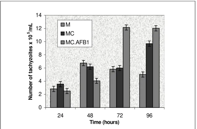
system (5.8 x 10⁵ ± 0.18 tachyzoites/mL) (Figure 1).
In this treatment 12 x 105 ± 0.18 tachyzoites/mL were
recovered, or 2.4 times more parasites than the control
(5 x 10⁵ ± 0.18 tachyzoites/mL) at 96 h. Tachyzoites
treated with CTR-AFB
1 lead to a greater recovery of
parasites during the entire period of the time course experiment. In this case, the most significant results were registered after 72 h of infection, when 2.9 times more tachyzoites were recovered in the cell cultures (17 x
105 ± 0.42 tachyzoites/mL) than was detected in the
control (5.8 x 10⁵± 0.75 tachyzoites/mL) (Figure 2).
The results were closely related, showing a huge reproducibility with a standard deviation never above 0.8 and a significance as large as 99.99%. It demonstrated that a single dose of mycotoxins at concentrations as low as 1 DL50 of CTR or 0.01 DL50
of both CTR and AFB1 in the mixture CTR- AFB1,
might act on the macrophages, favoring the infectivity
and consequent proliferation of T. gondii.
Discussion
Macrophages play a crucial role in both non-specific and acquired immune responses. They have a role in the direct destruction of microorganisms [23] and tumoral cells [24,25]. Cell-mediated immunity is the main line of defense against infection by coccidians [26],
however the infecting forms of T.gondii modify cell
functions and the immuneresponse when they penetrate
the macrophages, inhibiting the fusion of the lysosomes with vacuoles and, in turn, hindering the action of the
degradative enzymes [27]. T. gondii grows without
Table 1. Table 1 - Evaluation of the infectivity potential of tachyzoites of Toxoplasma gondii for peritoneal macrophages, in the different treatments with mycotoxins
Results are the mean of 4 repetitions (p<0.01). M- macrophages; M.C.- macrophages exposed to citrinin; M.C.AFB1- macrophages edposed to the association of citrinin and aflatoxin; T.C.AFB1 -tachyzoites exposed to the association of citrinin and aflatoxin.
Figure 1. Effect of citrinin and its association with aflatoxin B1 on the proliferation of Toxoplasma gondii in a
culture of macrophages at different time intervals, where the macrophages were previously exposed to 10 µg/mL
CTR and 0.01 µg/mL CTR associated with 0.04 µg/mL AFB
1 for a period of 2 h.
0 2 4 6 8 10 12 14
24 48 72 96
Time (hours)
N
u
m
b
er
o
f t
ac
h
yz
o
it
es
x
10
5 /m
L M
MC
MC.AFB1
Experimental Inocule x 106 Tachyzoites x105/ Infectivity %
systems 106 macrophages
M 1.2 ± 0.33 7.0 ± 0.38 59
M.C 1.2 ± 0.33 9.3± 0.35 77.5
M.C.AFB1 1.2± 0.33 9.0 ± 0.1 75
T.C. AFB
0 2 4 6 8 10 12 14 16 18 20
24 48 72 96
Time (hours)
Nu
mb
e
r o
f
ta
c
h
y
z
o
it
e
s
x
10
5 /m
L
T
T.C.AFB1
Figure 2. Effect of the association of citrinin and aflatoxin B1 on the proliferation of the Toxoplasma gondii in a
culture of macrophages at different time intervals, where the tachyzoites were previously exposed to 0.01 µg/mL
of CTR associated with 0.04 µg/mL AFB1 for a period of 2 h.
peroxide and peroxide ions, promoting powerful inactivation effects against these microrganisms [28].
Some fungal toxins are known to be
immunossuppressors; amongst these, AFB1 is
particularly known for its hepatoxic,
hepatocarcinogenic and mutageniceffects in man and
several other animal species [21,29-31], and CTR is known for its nephrotoxic effects [2,3]. Although the toxic effects of these mycotoxins are known, there is a great lack of data regarding the effects of small concentrations of these toxins. There is a common notion that mycotoxin effects on the immune response might favor the appearance of serious infectious outbreaks [32], or even the reactivation of infections
by intracellular parasites, such as T. gondii in chronic
individuals [16]. Although numerous studies have shown
diseases of high prevalence and morbidity, such as toxoplasmosis.
We evaluated the effect of mycotoxins on the
intracellular parasitism of T. gondii. In the first series
of experiments, the effect of CTR and its association
with AFB1 upon tachyzoite infectivity in cells in culture
was evaluated. A significant increase in tachyzoite assimilation by the cells that had been priorly treated with mycotoxins was observed. Such increased assimilation of parasites seems to be directly related to active penetration by a larger number of parasites (Table 1). Previous studies have demonstrated that the cytotoxic action of CTR on macrophages limits the phagocytic processes [1] and that AFB
1 causes
significant cytotoxicity in these cells, provoking morphological alterations and causing a reduction in important functions, such as adhesion and phagocytic activity [35, 6], increasing the susceptibility to infectious diseases [10,33,37]. Although the mechanisms by which these mycotoxins exert these effects on the macrophages are not entirely clear, preliminary adhesion
of T. gondii to the cell’s apical complex is known to
involve interactions between the parasite and the surface receptors of the target cell [38,39]. Cellular invasion also requires parasite motility, which is dependent upon the extra-cellular pH gradient, which is determined by ions; the internal pH is greater than the external pH [39,40]. The fact that these mycotoxins favor tachyzoite infectivity indicates that they act upon the cellular receptors, increasing the ligation points between the parasites and the cell, facilitating their adhesion, or they may decrease intracellular pH, stimulating the motility of the tachyzoites. When we evaluate the proliferation of the tachyzoites in the cultured macrophages, an increase in tachyzoites in the experimental systems exposed to the mycotoxins was observed. In the cells treated with CTR, there was a significant increase in the proliferation of the tachyzoites,
which started after 96 h. In vitro studies have
demonstrated that CTR has various effects on mitocondrial function and macro-molecule biosynthesis [41], acting on oxidative metabolism [42] and increasing the production of reactive oxygen, in turn, stimulating the production of the superoxide anion in
the respiratory chain [43]. The increase in parasitic proliferation in cultured cells exposed to CTR might result from oxidative stress of host cells, which would not display any mechanism of parasite destruction. In
cells treated with CTR-AFB1, a similar increase of
parasitic proliferation was observed after 72 h. Such an earlier effect seemed to be related to an addictive toxicity, leading to an increase in host cell mortality. Previous studies have demonstrated that the effect of the combination of mycotoxins affects immunocompetent cells [44], being able to significantly increase toxicity to myelocytes [34]. The recovery of integrally viable parasites after direct treatment with mycotoxins, leading to an increased recovery of parasites delivered by the host cells during the time course experiment indicated that the toxic chemicals interact with tachyzoites trough distinct mechanisms. The similarity in the parasite recovery rate in the two systems of cells-toxins and parasites-toxins might not be related to any ordinary mechanism. One possibility would be the formation of surface complexes that facilitate the host parasite interaction, since a tendency to form cellular agglutinates was observed when in the presence of these mycotoxins (data not shown). This possibility is reinforced by the fact that the entire cellular system exposed to mycotoxins was washed
three times by centrifugation (600 x g/10 min/4 ± 1°C)
with mycotoxin-free RPMI prior to the infection experiments and that all inoculations were carried out with previously quantified live infective forms. The combined results might also be related to the decrease in the primary functions of the macrophages, as
previously demonstrated for AFB1 [35,36].
Macrophages, when activated, increase their phagocytic activity and liberate products, such as cytokines and intermediate reagents for non-specific primary defense against infectious agents [17, 19].
AFB1 modifies the functions of the macrophage,
decreasing the secretion of IL-1 and IL-6 and the
production of TNF-α, nitric oxide, superoxide anion
and hydrogen peroxide [36]. Nevertheless, a significant increase in tachyzoites in the experimental
systems exposed to the association of CTR and AFB1
any kind of memory inhibition of the parasites, but rather a direct host cell cytotoxic effect. Otherwise, a reduction of IL-6, TNF and nitric oxide, which are important in the control of tachyzoite replication in the acute phase of the infection, should also be expected in long-term infections in individuals exposed to low doses of common, naturally occurring mycotoxins. In our experimental model a plateau of recovery was observed following emission of viable parasites. This could be related to limitations of the experimental system itself. The diminution of viable target host cells increased with time, and there was a significant diminution in the capabilities of capture and metabolism for the proliferation of the parasites. These data indicate that there is selection by apoptosis; further investigations are required to clarify the possible mechanisms of the actions of these
mycotoxins on T. gondii. Unfortunately specific
anti-metabolites, which could be used to isolate the pathways involved in parasitism, are still unknown. Due to the easy reproducibility of our experimental model, it may provide comparative results, which should be useful to better understand this modality of environmental interaction. Cultures of macrophages from chronically infected individuals, as well as from acute and sub clinical infections would be of great interest. The possibilities of studying the genetic expressions of chemokines, as well as the mechanisms of immunity and genetic sensitivity to different environmental toxicants are also of great importance. Our results reinforce the suggestion that mycotoxins, even at low concentrations, act on tachyzoites and macrophages to favor the infectivity and proliferation
of T. gondii, and that association with these mycotoxins
enhances pathologies to the immune cells. It also appears clear that increased monitoring and control, as well as revision of the legal acceptable levels for these toxins, is necessary, since in Brazil, despite the current legislation, there aflatoxins are abundant, and there is a high level of incidence in foods used for human and animal consumption, such as maize, peanuts and their derivatives, putting immune compromised individuals, such as children and those who have AIDS, at risk, especially in the rural areas of the country.
Acknowledgements
We sincerely thank Prof. Dr. José Clecildo Barreto Bezerra who gave inconditional help with this research project. CNPq (PROC 200559/00-1) provided financial support.]
References
1. FrankH.K. Citrinin. Z Ernahrungswiss 1992;31:164-77. 2. Pitt J.L. Toxigenic fungi and mycotoxins. Br Med Bull
2000;56:184-92.
3. Fink-Gremmels. Mycotoxins: their implications for human and animal health. Vet Q J 1999;21:115-20.
4. Robens J.F., Richard J.L. Aflatoxins in animal and human health. Rev Environ Contam Toxicol 1992;127:69-94. 5. D’Mello J.P.F., Macdonald A.M.C. Mycotoxins. Animal
Feed Science and Technol 1997;69:155-66.
6. Wang J.S., Huang T., Su J., et al. Hepatocellular carcinoma and aflatoxin exposure in Zhuqing Village, Fusui County, People’s Republic of China. Cancer Epidemiol Biomarkers and Prevent 2001;10:143-6.
7. Midio A.F., Campos R.R., Sabino M. Occurrence of aflatoxins B1, B2, G1 and G2 in cooked food components of whole meals marketed in fast food outlets of the city of São Paulo, Brazil. Food addit Contam 2001;18:445-8. 8. Maia P.P., Pereira Bastos de Siqueira M. Occurrence of aflatoxins B1,B2, G2 in some Brazilian pet foods. Food addit Contam 2002;19(12):1180-3.
9. Sharma R.P. Immunotoxicity of mycotoxins. J Dairy Sci
1993;76:892-7.
10. Bondy G.S., Pestka J.J. Immunomodulation by fungal toxins. J Toxicol Environ Heallth B Crit Rev
2000;3:109-43.
11. Shivachandra S.B., Sah R.L, Singh S.D, et al. Immunosuppression in broiler chicks fed aflatoxin and inoculated with fowl adenovirus sorotype-4 (FAV-4) associated with hydropericardium syndrome. Vet Res Commun 2003;27(1):39-51.
12. Raisuddin S., Singh K.P., Zaidi S.I., et al. Immunosuppressive effects of aflatoxin in growing rats. Mycopathol 1993,124:189-94.
13. Smith I.E., Moss M.O. Micotoxins: formation, analysis and significance. Bohn Wiley & Sons Ltd., U.K., 1985. 14. Corrier E. Micotoxicosis mecanisms of immunossupression. Vet Immunol and Immunopathol
1991;30:73-87.
16. Luft B.J., Remington J.S. Toxoplasmic encephalitis in AIDS. Clin Infect Dis 1992;15:211-22.
17. Alexander J., Scharton-Kerten T.M., Yap G., et al. Mechanisms of innate resistence to Toxoplasma gondii. Phils Trans R Soc Lond B Biol Sci 1997;352: 1355-9. 18. Venturini M.C., Quiroga M.A., Risso M.A., et al.
Mycotoxin t-2 and aflatoxin b1 as immunosuppressors in mice chronically infected with Toxoplasma gondii. J Comp Pathol 1996;115:229-37.
19. Dlugonska H. Immunity in Toxoplasma gondii infections. Postepy Hig Med Dosw 2000;54:53-65.
20. Phillips H.J. Dye exclusion test cell viability. In; Kruse P.F., Patterson M,K. Tissue Culture Methods and applications,. Academic Press, New York, NY, 1973. 21. Qureshi M.A., Hagler W.M. Effects of fumonisin-B1
exposure on chicken macrophages functions in vitro. Poultry Sci 1992;71:104-12.
22. Vieira S., Hoffman R. Estatística Experimental. Ed. Atlas, São Paulo, 1989.
23. Macmicking J.D., Nathan C., Xie Q.W. Nitric oxide and macrophage function. An Rev Immunol
1997;15:323-50.
24. Qureshi M.A., Miller L. Signal requirements for the acquisition of tumoricidal competence by chicken peritoneal macrophages. Poultry Sci 1991;70:530-8. 25. Chang C.I., Liao J.C., Kuo L. Macrophage arginase
promotes tumor cell growth and suppresses nitric oxide-mediated tumor cytotoxicity. Cancer Res
2001;61:1100-6.
26. Lillehoj H.S., Trout J.M. CD8+ T-cell-coccidia interations.
Parasitol Today 1994;10:10-3.
27. Sibley L.D., Boothdoyds J.D. Calcium regulated secretion and modification of host-cell endocytic compartments by Toxoplasma. J Cell Biol 1991;115(5a).
28. Krahenbuhl J.L., Remington J.S. Cytotoxic and microbicidal properties of macrophages, In: R van Furth, Molecular Phagocytes Functional Aspects, Martinus Nijhaff Publishers The Hague, Germany. 1980. 29. Sahoo P.K., Mukherjee S.C., Nayak S.K., et al. Acute and subchronic toxicity of aflatoxin B1 to rohu, Labeo rohita (Hamilton). Indian J Exp Biol. 2001;39:453-8.
30. Dimitri R.A., Gabal M.A., Saleh, N. Effect of aflatoxin ingestion in feed on body weight gain and tissue residues in rabbits. Mycoses 1998;41:87-91.
31. Quist C.F., Bounous D.I., Kilburn J.V., et al. The effect of dietary aflatoxin on wild turkey poults. J Wildl Dis.
2000;36(3):436-44.
32. Schuch M. The significance of mycotoxin assimilation for the productivity and health of animal. Otch Tierarzth wuchenschr 1989;96:353-5.
33. Pier A.C., Richard J.L., Cyzewski S.J. Implication of micotoxins in animal disease. J Am Vet Med Assoc
1980;176:719-24.
34. Terse P.S., Madryastra M.S., Zurovac O.,et al. Comparison of in vitro and in vivo biological activity of micotoxins. Toxicon 1993;31:913-9.
35. Neldon-Ortiz D.L., Qureshi M.A. Effect of AFB1 embryonic exposure on mononuclear phagocytic cell functions. Dev Comp Immunol 1992;16:187-96.
36. Moon E.Y., Rhee D.K., Pyo S. In vitro suppressive effect of aflatoxin B1 on murine peritoneal macrophage functions. Toxicology 1999;133:171-9.
37. Pestka J.J., Bondy G.S. Alteration of immune function following dietary mycotoxins in different animal species. Fed Cosmet Toxicol 1990;68:1009-16.
38. Minco J.R., Kasper L.H. Attachment of Toxoplasma gondii to host cells involves major surface protein, SAG-1(P30). Exp Parasitol 1994;79:11-20.
39. Bonhomme A., Bouchot, A., Pezzella N., et al. Signaling during the invasion of host cells by Toxoplasma gondii. Microb Rev 1999;23:551-61.
40. Endo T., Yagita K. Effect of extracellular ions on motility and cell entry in Toxoplasma gondii. J Protozool
1990;37:133-8.
41. Braumberg R.C., Gantt O., Barton C., et al. In vitro effects of the nephrotoxins ochratoxin A and citrinin upon biochemical functions of porcine kidney. Arch Environ Contam Toxicol 1992;22:464-70.
42. Chagas G.M., Oliveira M.B.M., Campello A.P., et al. Mechanism of citrinin-induced dysfunction of mitochondria. III. Effects on renal cortical and liver mitochondrial swelling. J Appl Toxicol 1995;15:91-5. 43. Ribeiro S.M., Chagas G.M., Campello A.P., et al. Mechanism
of citrinin-induced dysfunction of mitochondria. V. Effects on the homeostasis of the reactive oxygen species. Cell Biochem Funct 1997;15: 203-9.