Actin-interacting and flagellar proteins in
Leishmania spp
.:
Bioinformatics predictions to functional assignments in phagosome formation
Michely C. Diniz
1, Marcília P. Costa
1, Ana C.L. Pacheco
1, Michel T. Kamimura
1, Samara C. Silva
1,
Laura D.G. Carneiro
1, Ana P.L. Sousa
1, Carlos E.A. Soares
1, Celeste S.F. Souza
2and Diana Magalhães de Oliveira
11
Núcleo Tarcísio Pimenta de Pesquisa Genômica e Bioinformática, Universidade Estadual do Ceará,
Fortaleza, CE, Brazil.
2Laboratório de Imunomodulação e Protozoologia, Fundação Oswaldo Cruz, Manguinhos, RJ, Brazil.
Abstract
Several motile processes are responsible for the movement of proteins into and within the flagellar membrane, but lit-tle is known about the process by which specific proteins (either actin-associated or not) are targeted to protozoan flagellar membranes. Actin is a major cytoskeleton protein, while polymerization and depolymerization of parasite actin and actin-interacting proteins (AIPs) during both processes of motility and host cell entry might be key events for successful infection. For a better understanding the eukaryotic flagellar dynamics, we have surveyed genomes, transcriptomes and proteomes of pathogenicLeishmania spp. to identify pertinent genes/proteins and to build in silico models to properly address their putative roles in trypanosomatid virulence. In a search for AIPs involved in flagellar activities, we applied computational biology and proteomic tools to infer from the biological meaning of coronins and Arp2/3, two important elements in phagosome formation after parasite phagocytosis by macrophages. Results presented here provide the first report ofLeishmania coronin and Arp2/3 as flagellar proteins that also might be involved in phagosome formation through actin polymerization within the flagellar environment. This is an issue worthy of furtherin vitro examination that remains now as a direct, positive bioinformatics-derived inference to be presented.
Key words:actin-interacting proteins (AIPs), flagellar proteins,Leishmania, coronin and Arp2/3, phagosome.
Received: January 14, 2009; Accepted: May 25, 2009.
Introduction
Leishmania spp.is a trypanosomatid protozoan re-sponsible for a complex group of diverse clinical forms of classically neglected diseases collectively known as leishmaniases (Peters and Killick-Kendrick, 1987). As an insinuating and persistent pathogen,Leishmaniahas a spe-cialized organelle for motility, the flagellum, which is es-sential for parasite migration, invasion and persistence on host tissues. Several motile processes have been reported in the literature that could be responsible for the movement of proteins into and within the flagellar membrane (Koz-minskiet al., 1993). However, little is known about the pro-cess by which specific proteins (either actin-associated or not) are targeted to protozoan flagellar membranes, with few exceptions such as the work by Snapp and Landfear (1999) which targets a motif for a flagellar integral
mem-brane protein in Leishmania enriettii. Actin is a major cytoskeleton protein, while polymerization and depoly-merization of parasite actin and actin motor-associated pro-teins during both processes of motility and host cell entry might be key events for successful infection.
To gain a better understanding of flagellar dynamics inLeishmania, we have surveyed genomes, transcriptomes and proteomes of these pathogens to identify pertinent genes/proteins and to buildin silicomodels to properly ad-dress their putative roles in trypanosomatid virulence. We have applied computational tools (hidden Markov models, Viterbi algorithm and comparative modeling) to infer bio-logical meaning through detailed sequence-structural-functional analyses on actinteracting proteins (AIPs) in-volved in flagellar activities ofLeishmania spp. The actin system evolved to be fail-safe with multiple proteins shar-ing overlappshar-ing but novel roles, whereas these multifaceted roles are likely to provide the versatile scenario involving actin-interacting activities.
Proteins such as coronin and Arp2/3 complex have a common feature of actbinding activity and might be in-www.sbg.org.br
Send correspondence to Diana Magalhães de Oliveira. Núcleo Tarcísio Pimenta de Pesquisa Genômica e Bioinformática, Univer-sidade Estadual do Ceará, Av. Paranjana 1700, Bloco UHV/NUGEN, Campus do Itaperi, 60740-000 Fortaleza, CE, Brazil. E-mail: [email protected].
volved inLeishmaniaintraflagellar pathways (Broadhead et al., 2006; Costaet al., 2007). The coronin family com-prises two groups of evolutionary conserved WD-repeat proteins known to interact with Arp2/3 complex that help regulate the nucleation dynamics of actin filaments (Ryba-kin and Clemen, 2005). Invasion and differentiation pro-cesses inLeishmaniadirectly involve phagocytosis and the formation of an intracellular membrane-bounded organelle called a phagosome, sinceLeishmania promastigotes must first evade complement-mediated lysis until they are en-gulfed by a mammalian host macrophage (Puenteset al., 1989; Olivieret al., 2005).
By phagocytosis we mean the process of ingestion per se. A phagosome is the endocytic compartment that contains a non-interfering particle, as opposed to a vacu-ole which describes a compartment containing a particle, such as a pathogen likeLeishmania that diverts normal phagosome maturation. Phagosome maturation refers to the process of intracellular phagosome development after closure of the phagocytic cup (reviewed by Haas, 2007). Phagocytosis has evolved into a highly complex and regu-lated process in multicellular eukaryotes by which mi-crobes and other particles are taken up into the phagosome (Griffiths and Mayorga, 2007). The precise mechanisms by which coronin and Arp2/3 contribute to phagocytosis are not known (Yanet al., 2005), but it is crucial to inves-tigate elements that will shed more light on these mecha-nisms.
Although not clearly understood, it has been shownin vitrothat, after the phagocytosis ofL. donovaniby macro-phage-like cells J774, parasites are transiently located in phagosomes with poor fusogenic properties towards late endocytic compartments (Desjardins and Descoteaux, 1997). In contrast, after internalization, they are found in compartments that rapidly fuse with late endocytic orga-nelles (Dermineet al., 2000). A possibility is that these dis-tinctive features of the early phagosomes could be linked to different proteins expressed on the parasite surface, which may modify the fusion capacity of the phagosomal mem-brane (Desjardins and Descoteaux, 1997; Dermineet al., 2000). Furthermore, most of the Leishmania-containing phagosomes have been shown to accumulate F-actin, which is noted around these phagosomes (Holm et al., 2001).
We should recall, then, the early events following phagocytosis ofLeishmania, pre-adapted to the encounter within intracellular conditions of mammals. As reported (Courret et al., 2002), young phagosomes containing Leishmaniarapidly acquire a competence to fuse with late endosomes/lysosomes. If we take this version, AIPs have a greater chance of being key elements on the phagosome maturation than any of the many surface membrane pro-teins that have been widely investigated. For that reason, we have focused our studies on AIPs because they are rich in motif-binding activities, besides being excellent models
for sequence and structural comparisons, predictions and examinations to be carried out with all kinds of restraints on spatial structure of the amino acid sequence(s) and ligands. Our coronin and Arp2/3 model restraints were derived from known related protein structures (comparative modeling) and from rules of secondary structure packing (combina-torial modeling). Ourin silicoexaminations provide infor-mation about AIPs in the specific domain of phagosome function in Leishmania spp. infection. The role of the phagosome is to deliver particulate material to a hydrolytic environment that will lead to its degradation. The matura-tion process of the phagocytic compartment is linked inti-mately to both digestive housekeeping processes and innate sensing of molecules associated with infection (Vieiraet al., 2002). Therefore, mechanisms and regulation behind this maturation process are strategic for both hosts and pathogens, whereas here we present and discuss results concerningLeishmaniaAIPs that are also flagellar proteins that might be targeted in phagosome formation.
Methods
Biological databases and bioinformatics tools
As previously (Oliveiraet al., 2005; Gouveiaet al., 2007; Vasconceloset al., 2007, 2008), we have used pub-licly available datasets of individual/clusters of gene/pro-tein data onLeishmania spp. (from GeneDB, a core part of Sanger Institute Pathogen Genomics) and from other avail-able eukaryotic organisms (at NCBI and UniProt/Swiss-Prot/trEMBL knowledge DB), including sequences from the genome projects ofC. reinhardtiiin the U.S. Depart-ment of Energy (JGI) and all available data at The ChlamydomonasFlagellar Proteome Project. For databases (DB) searches, accession numbers/identifiers are those used in four main sources: NCBI, GeneDB, PDB and UniProt. As previously described (Oliveira et al., 2005; Gouveia et al., 2007; Vasconcelos et al., 2007, 2008), BLAST and its variants (Altschul et al., 1997) and MUSCLE (Edgar, 2004) were used for sequence similarity searches and comparisons/analysis through pairwise and multiple alignments. As previously detailed (Costaet al., 2007), for pattern recognition tasks of gene/protein predic-tions, motif finding and refinements for defining core do-mains of interest, HMM implementations such as HMMER (Eddy, 1998) and a structural prediction adaptation (Yoshizawaet al., 2006) were employed, with the addi-tional improvement of the Viterbi algorithm (VA) (Forney, 1973; Black, 2004) for best possible alignments after re-cursion (Alexandrov and Gerstein, 2004). As previously (Costaet al., 2007), we assume that the HMMs generated the input sequence and are looking for the highest probabil-ity path. For sequence positioni= 0, 1 ,…,L+1; for state l= 0, 1, …,n:
In-silicosurvey
As described in previous studies (Oliveira et al., 2005; Gouveiaet al., 2007; Vasconceloset al.2008), we took alignments created with FASTA/BLAST as input and computed alignment tables, providing hierarchical and suc-cessive correlations between each of the two sets of se-quences. FASTA files for amino-acid (aa) sequences of coding regions were downloaded from sources detailed above. All flagellar proteins and AIPs inLeishmaniawere collected from experimental papers and from the compre-hensive GeneDB database. An illustration of the tools used is seen in Figure 1. Briefly, these sequences were used as queries against genomic datasets with PSI-BLAST (BLASTP2.2.10) (Altschulet al., 1997) to determine se-quence similarity among all possible sese-quences, followed by multiple sequence alignments (MSA) performed with MUSCLE (Edgar, 2004) on target entries of main source DBs searched against various collections of protein motifs and families. Results of MSA were used as training datasets for HMM profiles (Eddy, 1998; Yoshizawaet al., 2006) and the SMART (Letunic et al., 2006) motif patterns of cluster motifs. The quality of trained HMM predictions and SMART pattern matching was examined. The HMM pro-files for cluster motifs were trained, calibrated, and used for DB searches using HMMER package ver.2.3.1 with default parameters. Gene ontology (GO) terms were assigned, based on top matches to proteins with GO annotations from Swiss-Prot/trEMBL, AMIGO after GeneDB and TargetP access. Functional assignment of these genes/gene prod-ucts was inferred using an RPS-BLAST search against con-served domain DBs (CDD) (Marchler-Baueret al., 2005); information was taken into account about subcellular local-ization (Emanuelssonet al., 2000), sequence and structural features, domains/motifs conservation (von Meringet al., 2005; Letunic et al, 2006), andin vitro characterization (Avidor-Reisset al., 2004; Tull et al., 2004). For three-dimensional (3D) modeling of AIPs we employed
Model-ler (Sali and Blundell, 1993; Marti-Renomet al., 2000) and ESyPred3D (Lambertet al., 2002). For visualization of 3D modeled structures, we employed the browser plug-in Chi-me. For addressing particular sites of structural-functional relationships of AIPs we used Cn3D (NCBI) and STING (Higaet al., 2004). Best models were evaluated by using PROCHECK (Laskowskiet al., 1993) and a thorough anal-ysis of consensus AIPs structures was carried out for model dissection, superposition and rms deviation (rmsd) calcula-tions.
Proteomic analyses
Samples. Purified fractions of flagella from Leishmania amazonenesis, strain H21 (MHOM/BR/76/ MA-76), were kindly provided by Fiocruz (Fundação Oswaldo Cruz, Manguinhos, Rio de Janeiro). An amount of 250mg of flagellar protein extract was normalized for
use.
Two-dimensional (2D) gel electrophoresis. We per-formed proteomic analyses through 2D polyacrylamide gel electrophoresis (2D-PAGE), as previously described (Drummelsmithet al., 2003, Brobeyet al., 2006). Briefly, proteins were treated with a lysis buffer with 7 M urea, 2 M thiourea, 2% NP-40, 2% DTT and 2% ampholytes (pH 3 to 10). In the first dimension, a protein separation by iso-electric point was performed applying IPG strips, soaked in the sample solution, at 300V for 5 min, 3,500 V for 5 h through EttanIPGphor IIIsoelectric Focusing System (GE Healthcare). To promote efficient protein transfer from the first to the second dimension, IPG strips were incubated in reducing buffer (1% DTT, 6M urea, 30% glycerol, 2% SDS, 50 mM Tris, pH 8.8) for 15 min, followed by 15 min incubation in alkylation buffer (2.5% iodoacetamide, 6 M urea, 30% glycerol, 2% SDS, 50 mM Tris, pH 8.8). The strips were immediately applied to the second dimension 12.5% polyacrylamide gel. Strips were, then, overlaid on freshly poured 10% Tricine-SDS gels (18 x 16 cm) and sealed with agarose solution (0.5% agarose, plus a few grains of bromophenol blue in a Tris-tricine cathode buf-fer). Protein standards of molecular mass range 14400 Da to 97000 Da were used (GE Healthcare). Gel electrophore-sis was carried out at 250 V for 1 h, and then at 500 V until the dye front reached the bottom of the gel (around 8 h). Gels were silver-stained as previously described (Gromova and Celis, 2006).
After running and scanning the gels, the resulting protein gel was analyzed using ImageMaster 2-D Plati-num 6.0® (Amersham Biosciences, Uppsala, Sweden). The spots detected automatically by the software were vi-sually inspected. Spot filtering and editing were per-formed manually to remove artifacts and to correct for spots that did not split correctly or were not detected by the software’s automatic spot detection process. Molecu-lar mass (MW) and isoeletric point (pI) were predicted for the best spots.
Results
To identify putative flagellar proteins ofLeishmania participating in the actin system, we have taken into ac-count all actin-binding, interacting or related/regulated pro-teins as source data to investigate their possible involve-ment in assembly of F-actin (the principal driving force behind many forms of parasite locomotion, including those regarding or underlying internalization into the host cell). Our underlying hypothesis is that polymerization and depo-lymerization of parasite actin and actin motor-associated proteins, during both processes of motility and host cell en-try, might be key events for successful infection, including the parasite survival within phagosomes. Recently (Vas-concelos et al., 2007, 2008) we reported an actin-poly-merization Leishmania protein, profilin, and its partner, formin, as putative flagellar proteins due to their likely in-volvement in axonemal assembly/disassembly, therefore, flagellar dynamics/remodeling. In addition, we predicted otherLeishmaniaAIPs, coronins and Arp2/3 complex as being flagellar proteins after detailed in silico structural alignments (Costa et al., 2007). Now, we focus on Leishmania coronin and Arp2/3 complex proteins as flagellar elements to phagosome formation in an attempt to directly associate the flagellum to the phagosomes, which are pivotal organelles in the ability of macrophages to per-form several of their key functions, such as the handling of apoptotic cells, tissue remodeling, and restriction of the es-tablishment and spread of intracellular pathogens (Méresse et al., 1999), such asLeishmania. Comparative bioinfor-matics analyses (performed afterin silicogenomic searches andin vitroproteomic screenings) have led to the identifi-cation of flagellar proteins that are involved not only in
cytoskeleton activity, signaling, endocytosis, and lytic ac-tivity, but also putatively in the phagosome formation. With this study, we are able to confirm previous data on proteins already shown to be involved in actin systems, but not previously reported as Leishmania- or as flagellar-associated.
Flagellar coronin identification inLeishmania
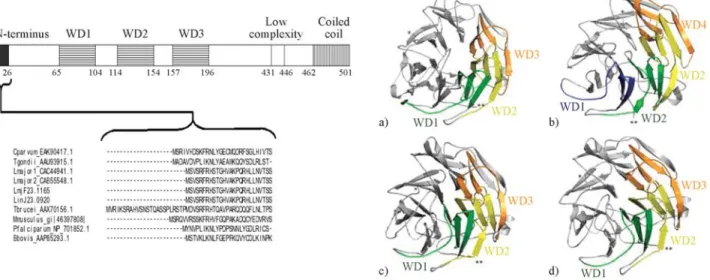
Coronins constitute an evolutionary conserved family of WD40-repeat (WDR) actin-binding proteins (reviewed by de Hostos, 1999; Rybakin and Clemen, 2005), originally described in the social amoebaDictyostelium discoideum (de Hostoset al., 1991). They can be divided, according to structure and function, into two subfamilies: one of shorter proteins (45-50kDa) implicated in, among other functions, nucleation of F-actin; and the other of longer, around 900 aa length, highly homologous coronins (POD-1 and Crn7) found in C. elegans, Drosophila and D. discoideum (Appletonet al., 2006; Rybakinet al., 2006). Until now, biochemical activities of coronins have been largely un-known, although one report (Humphrieset al., 2002) iden-tified how coronin and Arp2/3 complex interactin vitro. The comparative alignment of primary aa sequences of Leishmania coronins showed that they are highly con-served and have similar domain structures (Figure 2a), sharing ~45% aa identity withD. discoideum. Our models for the predicted 3D structure ofLeishmaniacoronins, seen in Figure 2b, also reinforce their close proximity to PDB solved structures for coronin.
The proteomic screening performed on the flagellar fraction of L. amazonensis enabled us to anticipate evi-dence of coronin, among other proteins, as at least six puta-tive spots, as shown in Figure 3 which provides a fairly
good insight onLeishmaniaflagellar proteins (manuscript in preparation) that are also classical AIPs, such as coronin. Since coronin can be required for an early step in phago-some formation (Yanet al., 2005), which is consistent with its role in actin polymerization and accumulation around phagosomes formed during the ingestion of mycobacteria (Ferrari et al., 1999) and also in preventing phagosome maturation and mycobacterial killing (Ferrariet al., 1999), here we have considered this information as an important element for assigning a possible phagosome role for Leishmania coronin as well. We must recall that Leishmaniahas differential levels of survival inside phago-somes, as has been elegantly reported by Gueirardet al. (2007), whose work with L. donovani shows degraded parasites in spacious phagosomes, in contrast to morpho-logically intact parasites in tight compartments within neu-trophils. These results suggest that the survival of parasites is linked to their ability to be targeted to tight/non-cidal and non-degradative compartments.
It is of paramount interest to mention that a phago-some, when it normally matures into a phagolysophago-some, un-dergoes a transition that correlates with functional changes
from an organelle with early endosomal characteristics to a compartment with lysosomal, degradative properties. If we consider that persistent accumulation of coronin around phagosomes containing pathogens is believed to prevent phagosome maturation and mycobacterial killing (Ferrari et al., 1999), it would be conceivable to determine how coronin, actively recruited by these pathogens, may be in-volved in the modulation of the actin cytoskeleton, thereby influencing intracellular trafficking and survival (Jaya-chandranet al., 2008). While broad investigations into this are ongoing, the few pieces of information available stem largely from studies on phagosomes containing pathogens such asLeishmaniathat force the maturation machinery to slow down but will not permanently inhibit phagolysosome formation, with growth dependent upon eventual phago-lysosome formation (Haas, 2007).
To what extent these different killing pathways (layed or not) are used in a given phagosome certainly de-pends on various cellular and environmental conditions (usually a plethora of macrophage receptors at variable pro-portions that may influence phagosome fate to variable de-grees), including interactions with the flagellum apparatus.
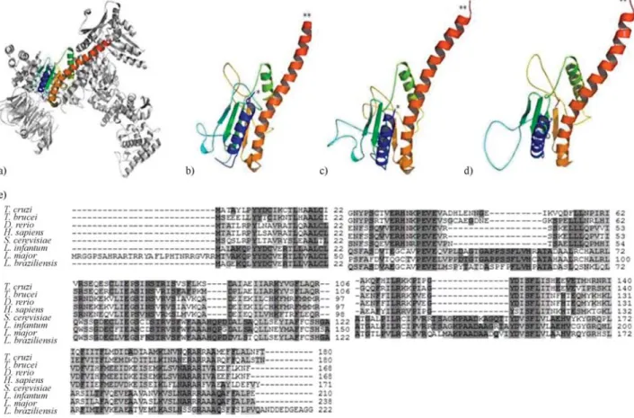
Figure 3- 3D visualization of Arp2/3 complex modeling, including possible binding site to a coronin WDR domain at central (blue) region and multiple alignment of the amino acid sequences of Arp2/3 complex proteins from different organisms. A) PDB solved structure ofBos taurusArp2/3 complex (1K8K) used as a template for building comparative models ofLeishmaniaArps with sequence from B)L. braziliensis(LbrM02.0360); C)L. infantum
(LinJ02.0520) and D)L. major(XP_822258.1). *N-terminus; **C-terminus. E) Multiple alignment of the amino acid sequences of Arp2/3 complex pro-teins. Accession numbers are:T. cruzi(XP_810627.1),T. brucei(XP_951567.1),D. rerio(NP_991100.1),H. sapiens(NP_005709.1),S. cerevisiae
We must bear in mind that flagella are dynamic structures that exchange up to 20% of their polypeptides within 3 h without any changes in their overall length (Song and Dentler, 2001; Wieseet al., 2003). Because flagellar micro-tubules are constantly turning over, the phagosomal mem-brane remains tightly associated with the parasites but the compartments become shorter (Marshal and Rosembaum, 2001). This reduction in size correlates with the progressive loss of the flagellum, suggesting that the parasite remodel-ing is accompanied by the removal of the phagosomal membrane.
Proteins involved in actin cytoskeleton dynamics that were recently implicated in phagocytosis in another proto-zoan,Entamoeba histolytica, are actobindin, coactosin and formin-like proteins (Marionet al., 2005), members of the so-called “core machinery” involved inde novo polymer-ization of actin filaments, which includes several subunits of the Arp2/3 complex, CAP protein, profilin and ADF/cofilin protein family (Loiselet al., 1999; Marionet al., 2005). ADF/cofilin proteins are essential to activate the turnover of actin filaments in dynamic regions of the cell and have already been shown to promote the remodeling of the actin network beneath the phagocytic cup (Bamburg, 1999; Bierneet al., 2001) and they have also been the sub-ject of our own bioinformatics analyses (Pachecoet al., 2007) and of a recentin vitroidentification (Tammanaet al., 2008) inLeishmaniaflagellum.
Here we have chosen to follow the hypothesis that the actin filaments beneath the phagocytic cup could be orga-nized as a bundle-like structure, rather than a dendritic net-work, which is necessary to form a stiff actin network around the phagocytic cup thereby facilitating the contrac-tile activity that closes the vacuole (Marionet al., 2005). It is tempting to recall the model in which Toxoplasma gondii-containing vacuoles possess deep invaginations supported by host microtubules that deliver host endo-somes and lysoendo-somes into the parasitophorous vacuole (Coppenset al., 2006), where the parasites feed on these endocytic organelles and not, as expected, on the Toxoplasmavacuole arising completely separate from the endocytic pathway (Sibley, 2003).
LeishmaniaArp2/3 complex genes
Arp2/3 complex has emerged as a central effector of actin assembly (Higgs and Pollard, 2001; Humphrieset al., 2002). It is composed of seven evolutionarily conserved subunits: two actin-related proteins (Arp2 and Arp3) and five other subunits (yeast Arc40, Arc35, Arc18, Arc19, and Arc15). Since infection of mammalian hosts with Leishmaniaprotozoa does depend on the ability of these parasites to replicate within macrophage phagolysosomes, it is noteworthy to link the function of Arps, such as the multiprotein Arp2/3 complex, and the phagosome forma-tion, as a key step in parasite persistence within the host. Previously we have catalogued a list ofLeishmania Arp2/3
complex genes comprising six putative sequences in L. braziliensisandL. infantum, and five inL. major(Costaet al., 2007), which are highly homologous with well-charac-terized Arp2/3 proteins in different organisms (as seen in Figure 4a). In Figure 4b we show a panel of four modeled Arp2/3 proteins inLeishmaniawhere all of them possess the WDR, but no other known binding sites. WDR domains dominantly interfere with coronin function, acting as multi-molecular scaffolding domains by bringing together inter-acting proteins on a single surface (Okuet al., 2003; Yanet al., 2005). They are likely to interfere with normal cell function by scavenging interacting proteins away from en-dogenous, full-length coronin.
If Arp2/3 complex is, in fact, one of those proteins that interact with coronin (Humphrieset al., 2002; Yanet al., 2005), other portions (except WDR) of the protein are responsible for coronin recruitment to sites of actin remod-eling, which would, then, link coronin and Arp2/3 to the sites of dynamic actin remodeling, with the contradiction that the WDR domain of coronin would act in a domi-nant-negative manner by preventing Arp2/3 accumulation. Conflicting interpretations (Humphrieset al., 2002; Yanet al., 2005) pose a likely, unforeseen alternative: that coronin might play both positive (stimulating) and negative (inhib-iting) roles in actin polymerization, depending on whether Arp2/3 functions prevent branching of F-actin or not.
Since our flagellar proteomic analyses on L. amazonensisdid demonstrate clear support for the presence of at least four Arp2/3 proteins, as shown in Figure 3, we put forward an interesting point of view regarding the pos-sibility that different sources of Arp2/3 proteins, whether flagellar or from the cytoskeleton, might be responsible in part for such paradox on activities of the coroninArp2/3 in-teraction.
SomeLeishmaniaflagellar AIPs are homologous to phagosome proteins
To find out which AIPs inLeishmaniawere homolo-gous to known phagosome proteins, as characterized in other studies (Desjardinset al., 1994; Garin et al., 2001; Marionet al., 2005; Stuart et al., 2007; Haas, 2007), we have searched lists of reportedin vitrophagosome assays that gave us a panel ranging from 140-617 proteins (Garin
et al., 2001; Gotthardtet al., 2006; Stuartet al., 2007) that are studied due to their direct involvement in phagosome formation. Therefore, we have compared their data to that found inLeishmaniagenomes, proteomes and particularly in our L. amazonensis flagellar proteome. We then col-lected a secol-lected list of Leishmania proteins, including coronin and Arp2/3, ascribed as highly homologous to mammalian phagosome proteins (Table 1). We built a chart to emphasize these comparisons (Figure 5), which is a mod-ified view of the full virtual phagosome (Garinet al., 2001) to cope with our predictive analysis of parasite flagellar proteins as homologous to mammalian phagosome coun-terparts. Therefore, in this study, we attempt to make a small contribution to further comprehensive analysis of Leishmania-containing phagosomes from which most of proteins have yet to be identified.
Table 1- List of selectedLeishmaniaactin-interacting proteins (AIPs) that are seemingly homologous to the phagosome proteins, as described by Garin
et al.(2001). The proteins are presented with respective sequence ID (as on GeneDB), length (in number of amino acids), predicted molecular weight and isoelectric point (pI).
Protein Organism Accession number MW (kDa) pI Amino acids
Prohibitin L.braziliensis LbrM34.0070/LbrM16.1230 32.2/30.2 10.0/8.3 292/268
L. infantum LinJ16.1700/ LinJ35.0170 30.2/32.3 8.3/9.9 268/292
L. major LmjF35.0070/ LmjF16.1610 32.3/30.2 9.9/8.3 292/268 RAB2 L.braziliensis LbrM30.1540 25.6 8.3 235
L. infantum LinJ30.2050 25.6 7.3 235
L. major LmjF30.1710 25.6 7.3 235
RAB7 L.braziliensis LbrM18.0760/ LbrM10.0880 24.0/24.6 5.3/5.0 223/221
L. infantum LinJ10.1520 24.9 5.4 222
L. major LmjF10.1170/ LmjF18.0890 24.7/24.1 5.7/5.1 221/223
RAB11B L.braziliensis LbrM32.1770 24.8 6.6 219
L. infantum LinJ32.2250 25.2 7.1 224
L. major LmjF32.1840 25.2 7.1 224
Stomatin L.braziliensis LbrM05.0940 40.2 8.6 358
L. infantum LinJ05.1050 39.6 7.8 357
L. major LmjF05.1040 39.7 8.3 357
Ubiquitin C L.braziliensis LM24.42/ LbrM25.0230 34.5/25.1 4.7/5.3 307/233
LbrM32.0660 26.2 4.5 233
L. infantum LM24.42/ LinJ32.0850 34.3/26.3 4.9/4.3 307/234
LinJ25.0190 25.1 4.7 233
L. major LM24.42 / LmjF32.0700 34.4/26.4 4.8/4.4 307/234
Ubiquitin C L.major LmjF25.0190 25.1 4.9 233
14-3-3 L.braziliensis LbrM35_V2.3430/LbrM11_V2.0040 29.6/29.0 4.6/4.9 258/253
L.infantum LinJ36_V3.3360/ LinJ11_V3.0350 29.7/29.2 4.6/4.9 258/253
L.major LmjF36.3210/ LmjF11.0350 29.6/29.1 4.6/5.0 258/253 ARP3 L.braziliensis LbrM15_V2.1360 38.1 5.4 348
L.infantum LinJ15_V3.1410 43.7 6.1 405
L.major LmjF15.1360 43.7 5.9 405
Ash L.infantum LinJ30_V3.3620 18.6 5.7 162
L.major LmjF30.3560 18.5 6.1 162
Table 1 (cont.)
Protein Organism Accession number MW (kDa) pI Amino acids
Calreticulin L.infantum LinJ31_V3.2670 45.0 4.4 400
L.major LmjF31.2600 45.0 4.4 400
L.braziliensis LbrM31_V2.2940 48.4 4.1 422
Cathepsin L L.major LmjF08.1030/ LmjF08.1040 48.0/37.7 7.6/7.1 443/348
LmjF08.1010/ LmjF08.1020 37.9/37.7 7.3/6.7 348/348
LmjF08.1050/ LmjF08.1060 37.8/48.0 6.7/7.6 348/443
LmjF08.1070/ LmjF08.1080 37.8/48.0 6.7/7.6 348/443
L.infantum LinJ08_V3.0960/ LinJ08_V3.0950 47.9/41.3 7.2/7.5 443/381
L.braziliensis LbrM08_V2.0810/ LbrM08_V2.0820 47.9/47.9 6.0/7.1 441/441
LbrM08_V2.0830 47.9 6.7 441
Coronin L.infantum LinJ23_V3.1400 56.2 7.2 510
L.major LmjF23.1165 56.6 6.6 510
L.braziliensis LbrM23_V2.1260 57.0 6.9 510
Cytochrome P450 L.braziliensis LbrM20_V2.2920/LbrM34_V2.2490 61.7/91.3 7.8/8.0 546/832
LbrM20_V2.2230/ LbrM27_V2.0100 68.4/67.2 6.2/8.9 624/592
LbrM30_V2.3580 58.0 7.3 509
L.infantum LinJ35_V3.2600/ LinJ34_V3.3110 91.1/61.3 8.0/6.7 832/546
LinJ34_V3.3610/ LinJ34_V3.2500 22.8/67.9 10.1/5.6 197/624
LinJ27_V3.0090 66.7 8.6 592
L.major LmjF27.0090/ LmjF35.2560 68.5/90.9 8.1/7.9 606/831
LmjF34.3330/ LmjF30.3550 59.9/58.0 7.6/7.1 533/508
LmjF34.2670 68.0 6.1 624
HSP-60 L.braziliensis LbrM32_V2.2030/ LbrM30_V2.2790 64.3/58.2 6.0/5.4 594/539
LbrM35_V2.2240/ LbrM35_V2.2250 60.2/59.5 5.3/5.1 564/562
L. infantum LinJ32_V3.1940/ LinJ36_V3.2130 64.3/60.5 6.4/5.1 594/566
LinJ36_V3.2140/ LinJ30_V3.2830 59.3/58.1 5.1/5.2 562/538
L. major LmjF32.1850/ LmjF36.2020 64.3/60.1 6.3/5.2 594/565
LmjF36.2030/ LmjF30.2820 59.3/58.0 5.1/5.3 562/538
Prohibitin L.braziliensis LbrM34.0070/LbrM16.1230 32.2/30.2 10.0/8.3 292/268
L. infantum LinJ16.1700/ LinJ35.0170 30.2/32.3 8.3/9.9 268/292
L. major LmjF35.0070/ LmjF16.1610 32.3/30.2 9.9/8.3 292/268 RAB2 L.braziliensis LbrM30.1540 25.6 8.3 235
L. infantum LinJ30.2050 25.6 7.3 235
L. major LmjF30.1710 25.6 7.3 235
RAB7 L.braziliensis LbrM18.0760/ LbrM10.0880 24.0/24.6 5.3/5.0 223/221
L. infantum LinJ10.1520 24.9 5.4 222
L. major LmjF10.1170/ LmjF18.0890 24.7/24.1 5.7/5.1 221/223
RAB11B L.braziliensis LbrM32.1770 24.8 6.6 219
L. infantum LinJ32.2250 25.2 7.1 224
L. major LmjF32.1840 25.2 7.1 224
Stomatin L.braziliensis LbrM05.0940 40.2 8.6 358
L. infantum LinJ05.1050 39.6 7.8 357
L. major LmjF05.1040 39.7 8.3 357
Ubiquitin C L.braziliensis LM24.42/ LbrM25.0230 34.5/25.1 4.7/5.3 307/233
LbrM32.0660 26.2 4.5 233
L. infantum LM24.42/ LinJ32.0850 34.3/26.3 4.9/4.3 307/234
LinJ25.0190 25.1 4.7 233
Many groups are currently looking into these various aspects of phagocyte biology and progress is steady, al-though no comprehensive phagosome proteome or lipi-dome has been published for any pathogen-containing
vacuole (Haas, 2007). There is an amazing diversity of compartments that intracellular pathogens inhabit, and they appear to use a stunning number of different factors to es-tablish these compartments. The real challenge remains in Table 1 (cont.)
Protein Organism Accession number MW (kDa) pI Amino acids
Ubiquitin C L.major LmjF25.0190 25.1 4.9 233
14-3-3 L.braziliensis LbrM35_V2.3430/LbrM11_V2.0040 29.6/29.0 4.6/4.9 258/253
L.infantum LinJ36_V3.3360/ LinJ11_V3.0350 29.7/29.2 4.6/4.9 258/253
L.major LmjF36.3210/ LmjF11.0350 29.6/29.1 4.6/5.0 258/253 ARP3 L.braziliensis LbrM15_V2.1360 38.1 5.4 348
L.infantum LinJ15_V3.1410 43.7 6.1 405
L.major LmjF15.1360 43.7 5.9 405
Ash L.infantum LinJ30_V3.3620 18.6 5.7 162
L.major LmjF30.3560 18.5 6.1 162
L.braziliensis LbrM30_V2.3590 18.5 6.9 162
Calreticulin L.infantum LinJ31_V3.2670 45.0 4.4 400
L.major LmjF31.2600 45.0 4.4 400
L.braziliensis LbrM31_V2.2940 48.4 4.1 422
Cathepsin L L.major LmjF08.1030/ LmjF08.1040 48.0/37.7 7.6/7.1 443/348
LmjF08.1010/ LmjF08.1020 37.9/37.7 7.3/6.7 348/348
LmjF08.1050/ LmjF08.1060 37.8/48.0 6.7/7.6 348/443
LmjF08.1070/ LmjF08.1080 37.8/48.0 6.7/7.6 348/443
L.infantum LinJ08_V3.0960/ LinJ08_V3.0950 47.9/41.3 7.2/7.5 443/381
L.braziliensis LbrM08_V2.0810/ LbrM08_V2.0820 47.9/47.9 6.0/7.1 441/441
LbrM08_V2.0830 47.9 6.7 441
Coronin L.infantum LinJ23_V3.1400 56.2 7.2 510
L.major LmjF23.1165 56.6 6.6 510
L.braziliensis LbrM23_V2.1260 57.0 6.9 510
Cytochrome P450 L.braziliensis LbrM20_V2.2920/LbrM34_V2.2490 61.7/91.3 7.8/8.0 546/832
LbrM20_V2.2230/ LbrM27_V2.0100 68.4/67.2 6.2/8.9 624/592
LbrM30_V2.3580 58.0 7.3 509
L.infantum LinJ35_V3.2600/ LinJ34_V3.3110 91.1/61.3 8.0/6.7 832/546
LinJ34_V3.3610/ LinJ34_V3.2500 22.8/67.9 10.1/5.6 197/624
LinJ27_V3.0090 66.7 8.6 592
L.major LmjF27.0090/ LmjF35.2560 68.5/90.9 8.1/7.9 606/831
LmjF34.3330/ LmjF30.3550 59.9/58.0 7.6/7.1 533/508
LmjF34.2670 68.0 6.1 624
HSP-60 L.braziliensis LbrM32_V2.2030/ LbrM30_V2.2790 64.3/58.2 6.0/5.4 594/539
LbrM35_V2.2240/ LbrM35_V2.2250 60.2/59.5 5.3/5.1 564/562
L. infantum LinJ32_V3.1940/ LinJ36_V3.2130 64.3/60.5 6.4/5.1 594/566
LinJ36_V3.2140/ LinJ30_V3.2830 59.3/58.1 5.1/5.2 562/538
L. major LmjF32.1850/ LmjF36.2020 64.3/60.1 6.3/5.2 594/565
LmjF36.2030/ LmjF30.2820 59.3/58.0 5.1/5.3 562/538
*Predicted Molecular Weight and Isoeletric Point (pI).
14-3-3 proteins are a family of conserved regulatory molecules expressed in all eukaryotic cells. 14-3-3 proteins have the ability to bind a multitude of functionally diverse signaling proteins, including kinases, phosphatases, and transmembrane receptors, being considered evolved members of the Tetratricopeptide Repeat (TPR) superfamily.
understanding how phagocytes and their phagosomes co-operate in pathological situations to phagosome biogenesis, as well as how to turn a phagosome from a hospitable into a killing environment forLeishmania.
Discussion
Phagocytosis is initiated by recognition of a pathogen by a host cell (usually macrophages and neutrophils) recep-tors that trigger its engulfment into the phagosome. Follow-ing their attachment to the macrophage, Leishmania promastigotes are internalized to the relatively benign envi-ronment of the endosome, where they begin to differentiate into amastigotes. Unlike amastigotes, promastigotes are vulnerable to degradation by the acidic and hydrolytic envi-ronment of the phagolysosome (Olivieret al., 2005). They must therefore retard endosome maturation and phago-some-endosome fusion, a process that has been observed by the absence or delayed arrival of late endosomal markers such as rab7 and LAMP-1 (Scianimanicoet al., 1999) and is related to the accumulation of F-actin (Holmet al., 2001). The mechanism is not completely understood (Olivier et al., 2005), but it has been shown that the delay in phago-lysosome maturation provides a window during which pro-mastigotes can differentiate into the more resistant amastigotes.
Nascent phagosomes have a composition that resem-bles that of the plasmalemma and are unable to digest the
vacuolar contents. This capability is acquired by remodel-ing of the phagosomal membrane and contents through a complex series of fusion events with compartments of the endocytic and secretory pathways (Desjardins and Des-coteaux, 1997). The components delivered to maturing phagosomes include acid hydrolases, as well as vacuolar-type ATPases that are responsible for acidification of the phagosomal lumen. This maturation process culminates in the formation of the phagolysosome, a highly acidic orga-nelle (pH < 5.5) where degradation occurs.
Here we report the association of two classical AIPs (coronins and Arp2/3) to the flagellar compartment in Leishmania, certainly a reasonable indication to link fla-gellar proteins to a possible F-actin binding in this proto-zoan.In vitro, purified coronin is said to bind specifically to F-actin, to bundle actin filaments, and to weakly promote actin assembly (David et al., 1998; Fukui et al., 1999), whereas, inAcanthamoeba, coronin was localized in the cell’s periphery (on the leading edge) consistent with that of actin located around the phagocytic cups (Baldoet al., 2005). Understanding the molecular mechanisms of phago-some maturation is critical because a number of pathogens, particularlyLeishmania, survive inside host cells through subversion of this process (Rosenberger and Finlay, 2003). Keeping these facts in mind, together with our own results, we, then, believe that flagellar AIPs, such as coronin and Arp2/3/, might play a role in these subversion mechanisms that maintain the parasite viable within the phagosome.
Some reports have shown thatLeismaniacan persist in mammalian neutrophils for up to 48 h, a period that largely exceeds the normal life span of neutrophils, indicat-ing that the phagocytosis/infectious process might alter the ability of neutrophils to initiate their programmed cell death, increasing the lifetime of neutrophils by delaying apoptosis (Agaet al., 2002; Gueirardet al., 2007). It seems thatLeishmaniamay have co-evolved with their mamma-lian hosts (Table 1 and Figure 5) to take advantage of phagosomes to establish a privileged niche for the transient parasitism of leukocytes and their subsequent invasion of macrophages, since it has been shown that the delay in phagolysosome maturation provides a window during which promastigotes can differentiate into the more resis-tant amastigotes (Olivieret al., 2005).
One could expect that phagosomes containing a given type of particle that entered cells simultaneously via the same receptor would behave the same, at least in a single cell. Surprisingly, however, phagosomes formed via the same receptors are found in different chemical states even within the same macrophage (Griffiths, 2004), a notion that is very useful to help our speculations onLeishmaniaAIPs rich content and at the same time homologous to a few phagosome proteins. Each phagosome is an individual en-tity whose behavior depends on a finite number of stable equilibrium states in its membrane signaling networks (Griffiths, 2004), which would, thus, underline a diversity Figure 5- Schematic illustration ofLeishmania spp. proteins that are
of available elements to disturb effective phagosome for-mation.
As stated by Rasmusson and Descoteaux (2004),L. donovanipromastigotes inhibit phagolysosome biogenesis in a lipophosphoglycan (LPG)-dependent manner, which correlates with an accumulation of periphagosomal F-actin, forming a physical barrier that preventsL. donovani pro-mastigote-containing phagosomes from interacting with endocytic vacuoles. Such inhibition of phagosome matura-tion may constitute a strategy to provide an environment propitious to promastigote-to-amastigote differentiation. Interestingly, some molecules, known to be involved in apoptotic signaling, are thought to accumulate in phago-some membrane microdomains, suggesting that this loca-tion might be important to their specific funcloca-tion, such as LPG in a disruptive process in the phagosome (Gueirardet al., 2007), which could imply an involvement in the ability ofLeishmaniato inhibit neutrophil apoptosis and, thus, the posterior macrophage activity that also depends on actin.
Assuming that the driving force for host cell entry in-volves polymerization of parasite actin and its AIPs, plus the recognized subcellular localization of functional coronins and Arps shown in the phagosome (Bricheuxet al., 2000; Asanoet al., 2001; Baldoet al., 2005), then we can propose a possible viable role for flagellar coronin and Arp2/3 within phagosome. As Love et al. (1998) have shown, periphagosomal actin is rapidly lost when parasites are internalized. These authors have mentioned a higher percentage ofL. majorpromastigotes surrounded by actin after experimentally adding parasites to macrophages. These findings suggest a clear binding to F-actin and im-plicit evidence of intense actin-interacting activity during phagosome formation after host cell entry (promastigote phagocytosis).
It seems quite obvious that intracellular parasites liv-ing within the harsh environment of phagocytes have de-veloped strategies that allow them to adapt quickly, escape from first-line defense systems, and inhibit several func-tions of their host cells (Olivieret al., 2005). One of these strategies might be explained through the complex interac-tions of the actin system (Sturgill-Koszyckiet al., 1996; Tardieux et al., 1998; Ullrich et al., 1999; Vieira et al., 2002; Yanet al., 2005). Coronins were first identified as AIPs, but, although they are crucial to the dynamics of actin filaments and cell movement, their actin-binding sites have been difficult to pin down, which reinforces the signifi-cance of computational biology and proteomic predictions that focus on coronin direct ligands, such as Arp2/3. More-over, establishing relationships of subcellular/organellar localizations, such as those we now report for coronin and Arp2/3 on the flagellum ofL. amazonensis, will help to clarify unforeseen roles that parasite AIPs might play on phagocytosis and phagosome formation through direct actin polymerization within the flagellar dynamic environ-ment. This is an issue worthy of furtherin vitro
examina-tion that remains now as a direct, positive bioinformatics-derived inference to be presented. Research into phagosome biogenesis has flourished in recent years (Haas, 2007) and it will surely lead to a better understanding of Leishmaniapathogenesis and mechanisms of immune in-vasion that involve the flagellar apparatus.
Acknowledgments
We wish to thank Fundação Oswaldo Cruz (FIOCRUZ, Brazil) for providing biological samples and GeneDB (The Wellcome Trust Sanger Institute, Pathogen Sequencing Unit) for full access to bioinformatics tools and databases onLeishmaniagenomes. This work is supported in part by CNPq and FUNCAP through individual grants to DMO and graduate fellowships to MCD, MPC, ACLP and SCS. MCD is a PhD student from RENORBIO (Rede Nordeste de Biotenologia) and this research is part of her thesis work.
References
Aga E, Katschinski DM, van Zandbergen G, Laufs H, Hansen B, Muller K, Solbach W and Laskay T (2002) Inhibition of the spontaneous apoptosis of neutrophil granulocytes by the intracellular parasite Leishmania major. J Immunol 169:898-905.
Alexandrov V and Gerstein M (2004) Using 3D Hidden Markov Models that explicitly represent spatial coordinates to model and compare protein structures. BMC Bioinform 5:2. Altschul SF, Madden TL, Schaffer AA, Zhang J, Zhang Z, Miller
W and Lipman DJ (1997) Gapped BLAST and PSI-BLAST: A new generation of protein database search programs. Nu-cleic Acids Res 25:3389-402.
Appleton BA, Wu P and Wiesmann C (2006) The crystal structure of murine coronin-1: A regulator of actin cytoskeletal dy-namics in lymphocytes. Structure 14:87-96.
Asano S, Mishima M and Nishida E (2001) Coronin forms a stable dimer through its C-terminal coiled coil region: An impli-cated role in its localization to cell periphery. Genes Cells 6:225-235.
Avidor-Reiss T, Maer AM, Koundakjian E, Polyanovsky A, Keil T, Subramaniam S and Zuker CS (2004) Decoding cilia function: Defining specialized genes required for compart-mentalized cilia biogenesis. Cell 117:527-539.
Baldo ET, Moon EK, Kong HH and Chung DI (2005)
Acanthamoeba healyi: Molecular cloning and characteriza-tion of a coronin homologue an actin-related protein. Exp Parasitol 110:114-22.
Bamburg JR (1999) Proteins of the ADF/cofilin family: Essential regulators of actin dynamics. Annu Rev Cell Dev Biol 15:185-230.
Bierne H, Gouin E, Roux P, Caroni P, Yin HL and Cossart P (2001) A role for cifinin and LIM kinase in Listeria-induced phagocytosis. J Cell Biol 155:101-112.
Broadhead R, Dawe HR, Farr H, Griffiths S, Hart SR, Portman N, Shaw MK, Ginger ML, Gaskell SJ, McKean PG, et al.
(2006) Flagellar motility is required for the viability of the bloodstream trypanosome. Nature 440:224-227.
Brobey RKB, Mei FC, Cheng X and Soong L (2006) Comparative two-dimensional gel electrophoresis maps for promastigotes ofLeishmania amazonensisandLeishmania major. Braz J Infect Dis 10:1-6.
Coppens I, Dunn JD, Romano JD, Pypaert M, Zhang H, Boothroyd JC and Joiner KA (2006)Toxoplasma gondii se-questers lysosomes from mammalian hosts in the vacuolar space. Cell 125:261-274.
Costa MP, Lucas HR, Maia ARS, Pacheco ACL, Pinheiro DP, Kamimura MT, Araújo-Filho R and Oliveira DM (2007) Flagellar proteins prediction after sequence-structure align-ments of Coronin and Arp2/3 complex inLeishmania spp. In: Proceedings of the IEEE International Conference on Bioinformatics and Biomedicine - Workshop on Computa-tional Structural Bioinformatics. IEEE Press, Piscitaway, pp 80-88.
Courret N, Fréhel C, Gouhier N, Pouchelet M, Prina E, Roux P and Antoine JC (2002) Biogenesis ofLeishmania -harbour-ing parasitophorous vacuoles follow-harbour-ing phagocytosis of the metacyclic promastigote or amastigote stages of the para-sites. J Cell Sci 115:2303-2316.
David V, Gouin E, Troys MV, Grogan A, Segal AW, Ampe C and Cossart P (1998) Identification of cofilin, coronin, Rac and capZ in actin tails using aListeriaaffinity approach. J Cell Sci 111:2877-2884.
de Hostos EL (1999) The coronin family of actin-associated pro-teins. Trends Cell Biol 9:345-350.
de Hostos EL, Bradtke B, Lottspeich F, Guggenheim R and Gerisch G (1991) Coronin, an actin binding protein of
Dictyostelium discoideumlocalized to cell surface projec-tions has sequence similarities to G protein beta subunits. EMBO J 10:4097-104.
Dermine JF, Scianimanico S, Privé C, Descoteaux A and Desjardins M (2000) Leishmania promastigotes require lipophosphoglycan to actively modulate the fusion proper-ties of phagosomes at an early step of phagocytosis. Cell Microbiol 2:115-126.
Desjardins M and Descoteaux AJ (1997) Inhibition of phago-lysosomal biogenesis by the Leishmania lipophospho-glycan. Exp Med 185:2061-2068.
Desjardins M, Huber LA, Parton RG and Griffiths G (1994) Biogenesis of phagosomes proceeds through a sequential se-ries of interactions with the endocytic apparatus. J Cell Biol 124:677-688.
Drummelsmith J, Brochu V, Girard I, Messier N and Ouellette M (2003) Proteome mapping of the protozoan parasite
Leishmaniaand application to the study of drug targets and resistance mechanisms. Mol Cell Proteomics 2:146-155. Eddy SR (1998) Profile hidden Markov models. Bioinformatics
14:755-763.
Edgar RC (2004) MUSCLE: A multiple sequence alignment me-thod with reduced time and space complexity. Nucleic Acids Res 32:1792-1797.
Emanuelsson OH, Nielsen S, Brunak and Heijne GV (2000) Pre-dicting subcellular localization of proteins based on their N-terminal amino acid sequence. J Mol Biol 300:1005-1016.
Ferrari G, Langen H, Naito M and Pieters J (1999) A coat protein on phagosomes involved in the intracellular survival of my-cobacteria. Cell97:435-447.
Forney Jr DG (1973) The Viterbi algorithm. Proceedings of the IEEE 61 3:268-278.
Fukui Y, Engler S, Inoue S and de Hostos EL (1999) Architectural dynamics and gene replacement of coronin suggest its role in cytokinesis. Cell Motil Cytoskel 42:204-217.
Garin J, Diez R, Kieffer S, Dermine J-F, Duclos S, Gagnon E, Sadoul R, Rondeau C and Desjardins M (2001) The phago-some proteome: Insight into phagophago-some functions. J Cell Biol 152:165-180.
Gotthardt D, Blancheteau V, Bosserhoff A, Ruppert T, Delorenzi M and Soldati T (2006) Proteomic fingerprinting of phago-some maturation and evidence for the role of a G alpha dur-ing uptake. Mol Cell Proteomics 5:2228-2243.
Gouveia JJ, Vasconcelos EJR, Pacheco ACL, Araújo-Filho R, Maia AR, Kamimura MT, Costa MP, Viana DA, Costa RB, Maggioni R,et al.(2007) Intraflagellar transport (IFT) com-plex inLeishmania spp:In silicogenome-wide screening and annotation of gene function. Genet Mol Res 6:675-689. Gueirard P, Laplante A, Rondeau C, Milon G and Desjardins M
(2007) Trafficking ofLeishmania donovanipromastigotes in non-lytic compartments in neutrophils enables the subse-quent transfer of parasites to macrophages. Cell Microbiol 10:100-111.
Griffiths G (2004) On phagosome individuality and membrane signalling networks. Trends Cell Biol 14:343-51.
Griffiths G and Mayorga L (2007) Phagosome proteomes open the way to a better understanding of phagosome function. Genome Biology 8:207.
Gromova I and Celis JE (2006) Protein detection in gels by silver staining: A procedure compatible with mass-spectrometry. In: Celis JE, Carter N, Hunter T, Simons K, Small JV and Shotton D (eds) Cell Biology: A Laboratory Handbook. 3rd edition. v. 4. Elsevier. Academic Press, San Diego, pp 421-429.
Haas A (2007) The Phagosome: Compartment with a license to kill. Traffic 8:311-330.
Higa RH, Togawa RC, Montagner AJ, Palandrani JC, Okimoto IK, Kuser PR, Yamagishi ME, Mancini AL and Neshich G (2004) STING Millennium Suite: Integrated software for extensive analyses of 3d structures of proteins and their complexes. BMC Bioinform 5:107-111.
Higgs HN and Pollard TD (2001) Regulation of actin filament net-work formation through ARP2/3 complex: Activation by a diverse array of proteins. Annu Rev Biochem 70:649-676. Holm A, Tejle K, Magnusson KE, Descoteaux A and Rasmusson
B (2001)Leishmania donovanilipophosphoglycan causes periphagosomal actin accumulation: Correlation with im-paired translocation of PKCalpha and defective phagosome maturation. Cell Microbiol 3:439-447.
Humphries CL, Balcer HI, DAgostino JL, Winsor B, Drubin DG, Barnes G, Andrews BJ and Goode BL (2002) Direct regula-tion of Arp2/3 complex activity and funcregula-tion by the actin binding protein coronin. J Cell Biol 159:993-1004. Jayachandran R, Gatfield J, Massner J, Albrecht I, Zanolari B and
Kozminski KG, Johnson KA, Forscher P and Rosenbaum JL (1993) A motility in the eukaryotic flagellum unrelated to flagellar beating. Proc Natl Acad Sci USA 90:5519-5523. Lambert C, Leonard N, De Bolle X and Depiereux E (2002)
ESyPred3D: Prediction of proteins 3D structures. Bioin-formatics 18:1250-1256.
Laskowski RA, MacArthur MW, Moss DS and Thornton JM (1993) PROCHECK: A program to check the stereoche-mical quality of protein structures. J Appl Cryst 26:283-291. Letunic L, Copley RC, Pils B, Pinkert S, Schultz J and Bork P
(2006) SMART 5: Domains in the context of genomes and networks. Nucleic Acids Res 34:257-260.
Loisel TP, Boujemaa R, Pantaloni D and Carlier M-F (1999) Re-constitution of actin-based motility ofListeriaandShigella
using pure proteins. Nature 401:613-616.
Love DC, Kane MM and Mosser DM (1998) Leishmania amazonensis: The phagocytosis of amastigotes by macro-phages. Exp Parasitol 88:161-171.
Marchler-Bauer A, Anderson JB, Cherukuri PF, DeWeese-Scott C, Geer LY, Gwadz M, He S, Hurwitz DI, Jackson JD, Ke Z,
et al.(2005) CDD: A conserved domain database for protein classification. Nucleic Acids Res 33:D192-196.
Marion S, Laurent C and Guillén N (2005) Signalization and cytoskeleton activity through myosin IB during the early steps of phagocytosis inEntamoeba histolytica: A proteo-mic approach. Cell Microbiol 7:1504-1518.
Marshall WF and Rosenbaum JL (2001) Intraflagellar transport balances continuous turnover of outer doublet microtubules: Implications for flagellar length control. J Cell Biol 155:405-414.
Marti-Renom MA, Stuart A, Fiser A, Sánchez R, Melo F and Sali A (2000) Comparative protein structure modeling of genes and genomes. Annu Rev Biophys Biomol Struct 29:291-325.
Méresse S, Steele-Mortimer O, Moreno E, Desjardins M, Finlay BB and Gorvel JP (1999) Controlling the maturation of pathogen-containing vacuoles: A matter of life or death. Nat Cell Biol 1:E183-E188.
Oku T, Itoh SM, Okano M, Suzuki A, Suzuki K, Nakajin S, Tsuji T, Nauseef WM and Toyoshima S (2003) Two regions re-sponsible for the actin binding of p57, a mammalian coronin family actin-binding protein. Biol Pharm Bull 26:409-416. Oliveira DM, Gouveia JJS, Diniz NB, Pacheco AC, Vasconcelos
EJ, Diniz MC, Viana DA, Ferreira TD, Albuquerque MC, Fortier DC, et al. (2005) Pathogenomics analysis of
Leishmania spp: Flagellar gene families of putative viru-lence factors. OMICS 9:171-191.
Olivier M, Gregory DJ and Forget G (2005) Subversion mecha-nisms by whichLeishmaniaparasites can escape the host immune response: A signaling point of view. Clin Microbiol Rev 18:293-305.
Pacheco ACL, Araujo FF, Kamimura MT, Medeiros SR, Viana DA, Oliveira FCE, Araújo-Filho R, Costa MP and Oliveira DM (2007) Following the Viterbi path to deduce flagellar actin-interacting proteins of Leishmania spp.: Report on Cofilins and Twinfilins. In: Proceedings of Computational Models For Life Sciences - International Symposium on Computational Models of Life Sciences - AIP Proceedings of International Symposium on Computational Models of Life Sciences. American Institute of Physics, Melville, v. 952, pp 315-324.
Peters W and Killick-Kendrick R (1987) The Leishmaniases in Biology and Medicine v. II: Clinical Aspects and Control. Peters W and Killick-Kendrick R (eds) Academic Press, London, pp 551-941.
Puentes SM, Dwyer DM, Bates PA and Joiner KA (1989) Binding and release of C3 fromLeishmania donovanipromastigotes during incubation in normal human serum. J Immunol 143:3743-3749.
Rasmusson B and Descoteaux A (2004) Contribution of electron and confocal microscopy in the study of Leishmania -ma-crophage interactions. Microsc Microanal 10:656-61. Rosenberger CM and Finlay BB (2003) Phagocyte sabotage:
Dis-ruption of macrophage signalling by bacterial pathogens. Nat Rev Mol Cell Biol 4:385-396.
Rybakin V and Clemen CS (2005) Coronin proteins as multi-functional regulators of the cytoskeleton and membrane traf-ficking. BioEssays 27:625-632.
Rybakin V, Gounko NV, Späte K, Höning S, Majoul IV, Duden R and Noegel AA (2006) Crn7 interacts with AP-1 and is re-quired for the maintenance of Golgi morphology and protein export from the Golgi. Biol Chem 281:31070-31078. Sali A and Blundell TLJ (1993) Comparative protein modelling
by satisfaction of spatial restraints. Mol Biol 234:779-815. Scianimanico S, Desrosiers M, Dermine JF, Meresse S,
Des-coteaux A and Desjardins M (1999) Impaired recruitment of the small GTPase rab7 correlates with the inhibition of phagosome maturation by Leishmania donovani promas-tigotes. Cell Microbiol 1:19-32.
Sibley LD (2003)Toxoplasma gondii: Perfecting an intracellular life style. Traffic 4:581-586.
Snapp EL and Landfear SMJ (1999) Characterization of a target-ing motif for a flagellar membrane protein inLeishmania enriettii. Biol Chem 274:29543-29548.
Song L and Dentler WL (2001) Flagellar protein dynamics in
Chlamydomonas. J Biol Chem 276:29754-29763.
Stuart LM, Boulais J, Charriere GM, Hennessy EJ, Brunet S, Jutras I, Goyette G, Rondeau C, Letarte S, Huang H,et al.
(2007) A systems biology analysis of theDrosophila phago-some. Nature 445:95-101.
Sturgill-Koszycki S, Schaible UE and Russell DG (1996)
Mycobacterium-containing phagosomes are accessible to early endosomes and reflect a transitional state in normal phagosome biogenesis. EMBO J 15:6960-696.
Tammana TV, Sahasrabuddhe AA, Mitra K, Bajpai VK and Gupta CM (2008) Actin-depolymerizing factor, ADF/co-filin, is essentially required in assembly of Leishmania
flagellum. Mol Microbiol 70:837-852.
Tardieux I, Liu X, Poupel O, Parzy D, Dehoux P and Langsley G (1998) A Plasmodium falciparumnovel gene encoding a coronin-like protein which associates with actin filaments. FEBS Lett 441:251-256.
Tull D, Vince JE, Callaghan JM, Naderer T, Spurck T, McFadden GI, Currie G, Ferguson K, Bacic A and McConville MJ (2004) SMP-1 a member of a new family of small myris-toylated proteins in kinetoplastid parasites is targeted to the flagellum membrane in Leishmania. Mol Biol Cell 15:4775-4786.
Vasconcelos EJR, Pacheco ACL, Gouveia JJS, Araujo FF, Diniz MC, Kamimura MT, Costa MP, Maggioni R, Araujo-Filho R, Costa RB,et al.(2007) Profilins, formins and katanins as flagellar proteins ofLeishmania spp: A genome-based mul-ti-step bioinformatics-driven description. In: Bourbakis NG (ed) Conference Proceedings IEEE 7th International Sym-posium on Bioinformatics & Bioengineering-2007. IEEE Press, Boston, v. II, pp 880-887.
Vasconcelos EJR, Pacheco ACL, Gouveia JJS, Araujo FF, Diniz MC, Kamimura MT, Costa MP, Araujo-Filho R and Oliveira DM (2008) Actin-interacting proteins in flagellated pathogenic Leishmania spp.: A genome-based bioinfor-matics report on profilins, formins and katanins. Int J Funct Informat Personal Med 1:234-252.
Vieira OV, Botelho RJ and Grinstein S (2002) Phagosome matu-ration: Aging gracefully. Biochem J 366:689-704.
von Mering C, Jensen LJ, Snel B, Hooper SD, Krupp M, Fo-glierini M, Jouffre N, Huynen MA and Bork P (2005) STRING: Known and predicted protein-protein associations integrated and transferred across organisms. Nucleic Acids Res 33:D433-D437.
Wiese M, Kuhn D and Grünfelder CG (2003) Protein kinase in-volved in flagellar-length control. Eukaryot Cell 2:769-777.
Yan M, Collins RF, Grinstein S and Trimble WS (2005) Coro-nin-1 function is required for phagosome formation. Mol Biol Cell 16:3077-87.
Yoshizawa AC, Kawashima S, Okuda S, Fujita M, Itoh M, Mo-riya Y, Hattori M and Kanehisa M (2006) Extracting
se-quence motifs and the phylogenetic features of SNARE-dependent membrane traffic. Traffic 7:1104-1118.
Internet Resources
Black PE, “Viterbi algorithm”, in Dictionary of Algorithms and Data Structures [online], Paul E. Black, ed., U.S. National Institute of Standards and Technology. 6 July 2004 (ac-cessed 28 June 2009) Available from: http://www.itl.nist.gov/div897/sqg/dads/HTML/viterbiAlg orithm.html.
GeneDB, a core part of The Wellcome Trust Sanger Institute -Pathogen Sequencing Unit: http://www.genedb.org (ac-cessed 02 May 2009).
The Chlamydomonas Flagellar Proteome Project:
http://labs.umassmed.edu/chlamyfp/index.php (accessed 28 June 2009).
HMMER package ver.2.3.1: http://www.hmmer.wustl.edu (ac-cessed 17 January 2009).
Swiss-Prot/trEMBL: http://www.expasy.org/sprot (accessed 17 January 2009).
TargetP: http://www.cbs.dtu.dk/services/TargetP (accessed 17 January 2009).
Chime browser plug-in: http://www.mdl.com (accessed 17 Janu-ary 2009).
Guest Editor: José Carlos Merino Mombach