.
Morphology and histochemistry of glandular trichomes in
Hyptis villosa
Pohl ex Benth. (Lamiaceae) and differential
labeling of cytoskeletal elements
Luiz Ricardo dos Santos Tozin1,2* and Tatiane Maria Rodrigues1
Received: August 11, 2016 Accepted: October 11, 2016
ABSTRACT
Lamiaceae contains many species known for their aromatic properties that are produced by the production of essential oils in glandular trichomes. Hyptis is one of the most common genera of Lamiaceae in the Brazilian fl ora, and includes several species with medicinal value. However, studies on the morphology and functioning of their glandular trichomes are lacking. We analyzed the morphology, histochemistry and ultrastructure of the glandular trichomes in leaves of H. villosa, emphasizing the diff erential distribution of actin fi laments and microtubules in cells secreting hydrophilic and lipophilic compounds. Four morphotypes of glandular trichomes were identifi ed. Total lipid, terpenes, alkaloids, phenolic compounds, proteins and polysaccharides were histochemically detected in all morphotyes. Th is evidences the mixed nature of the secretions of this species, although there are diff erences in the prevalence of lipophilic and hydrophilic components among the glandular morphotypes and among the cells of the same trichome. Th e actin microfi laments are more abundant in cells that secrete mainly hydrophilic compounds, and microtubules predominate in cells that secrete lipophilic compounds. Our results corroborate the correlation between the glandular morphotype and the composition of the secretion produced, with a diff erential distribution of the cytoskeletal elements according to the prevalence of either hydrophilic or lipophilic substances.
Keywords: actin microfi laments, cytoskeleton, glandular hairs, microtubules, ultrastructure
Introduction
Glandular trichomes are epidermal appendices with highly variable morphology and responsible for the production of secretion with economic, medicinal and ecologic values (Ascensão et al. 1999; Argyropoulou et al. 2010; Tozin et al. 2015a). Substances of diff erent chemical categories can be produced by glandular trichomes, and their subcellular features vary according to the substances produced and the form of release of the secretion (Ascensão et al. 1995; Ascensão & Pais 1998; Appezzato-da-Glória et al.
2012; Naidoo et al. 2012; Tozin et al. 2015a; Silva et al. 2016). Th e subcellular features of glandular trichomes have been exhaustively described to several species from diff erent families (Ascensão & Pais 1998; Sacchetti et al. 1999; Argyropoulou et al. 2010; Papini et al. 2010; Paiva & Martins 2011; Appezzato-da-Glória et al. 2012; Naidoo et al. 2012; Amrehn et al. 2014; Tozin et al. 2015a; Silva et al. 2016 and references therein); however, some important aspects, such as the involvement of the cytoskeletal elements in such secretory process remains poorly studied. It has been demonstrated that movement of oil droplets in the
1 Departamento de Botânica, Instituto de Biociências de Botucatu, Universidade Estadual Paulista, 18618-970, Botucatu, SP, Brazil
2 Programa de Pós-graduação em Ciências Biológicas, Departamento de Botânica, Instituto de Biociências de Botucatu, Universidade Estadual
Paulista, 18618-970, Botucatu, SP, Brazil
Morphology and histochemistry of glandular trichomes in Hyptis villosa Pohl ex Benth. (Lamiaceae) and differential labeling of cytoskeletal elements
cytoplasm of animal cells is driven by microtubule motor proteins (Zehmer et al. 2009 and references therein) and that actin filaments are the major components involved in the movement and anchorage of vesicles and organelles in plant (Evert 2006 and references therein) and animal (Valderrama et al. 2001; Stamnes 2002) cells. However, information on the cytoskeleton elements in secretory cells of plants was not found.
The Lamiaceae comprise many species known by their medicinal and economic values given by the production of essential oils in glandular trichomes (Ascensão et al. 1999; Serrato-Valentini et al. 1997; Werker 2000; Rodrigues et al. 2013). The morphology of the glandular trichomes is highly variable within the family. In almost all studied species, capitate and peltate trichomes are present (Ascensão & Pais 1998), being common the occurrence of different versions of them (Ascensão et al. 1995; Ascensão & Pais 1998; Ascensão et al. 1999; Corsi & Bottega 1999; Martins 2002; Maggi et al. 2010; Baran et al. 2010; Mota et al. 2013). Peculiarities on the distribution, arrangement and function of the different glandular morphotypes have been observed in Lamiaceae (Corsi & Bottega 1999) and a correlation between the gland morphology and the composition of secretion has been proposed (Werker 1993; Ascensão & Pais 1998; Corsi & Bottega 1999).
Hyptisis one of the most common genera of Lamiaceae in the Brazilian flora (Silva et al. 2013), comprising several species with medicinal value (Falcão & Menezes 2003). The bioactive potential of the essential oils of Hyptis species have been proved by their antibacterial (Souza et al. 2003), antifungal (Oliveira et al. 2004) and antitumoral (Costa-Lotufo 2004) properties. However, despite the medicinal importance of Hyptis species, studies on the morphology and cellular features in relation to the secretory process of glandular trichomes are lacking to the genus.
In this paper we studied the glandular trichomes of H. villosa, popularly named hortelã-do-cerrado, a shrub with reddish-brown pubescence, common in the campo cerrado in Brazil (Durigan et al. 2004). We analyzed the morphology, histochemistry and the subcellular features of the glandular trichomes in the leaves, emphasizing the differential distribution of actin filaments and microtubules in cells secreting different compounds. Information on the relative density of each glandular morphotype and the correlation between the glandular morphotype and the secretion composition is presented.
Materials and methods
Studied area and plant material
The studied population of Hyptis villosaPohl ex Benth. was growing in an area of campo cerrado in Botucatu city (22º53’09”S 48º26’42”W), São Paulo State, Brazil. In this region, the climate is Cfa with hot climate and rains in the
summer and drought in the winter.
The campo cerrado is one of the physiognomies of the Cerrado (Brazilian savanna), characterized by the predominance of grasses and small shrubs with sparse trees (Maroni et al. 2006), where the plant individuals are subjected to high light intensity and low air relative humidity (Tozin et al. 2015b).
For microscopic analysis, fully expanded leaves located between the third and fourth nodes in the stem were collected from adult individuals of H. villosa (n=6). Reproductive material were collected and vouchers were deposited in the Herbarium Irina Delanova Gemtchújnicov (BOTU) of the Departamento de Botânica, Instituto de Biociências de Botucatu, Universidade Estadual Paulista, Botucatu, São Paulo, Brazil.
Conventional light microscopy (LM)
Samples were fixed in FAA 50 (Johansen 1940), dehydrated in an ethanol series, and embedded in Leica® historesin. The cross sections (5 μm thick) were stained with 0.05% toluidine blue, pH 4.3 (O’Brien et al. 1964), and mounted in Entellan® on permanent slides. The relevant results were documented using the Leica DMR photomicroscope with a Leica DFC 425 a digital camera.
Histochemical tests
Cross sections obtained from the median region of fresh leaves were treated with: Sudan III for total lipids (Johansen 1940), Nadi reagent for terpenes (David & Carde 1964), Bromophenol mercuric blue for proteins (Mazia et al. 1953), Schiff reagent/periodic acid (PAS) for noncellulosic polysaccharides (Amaral et al. 2001), ferric chloride for phenolic compounds (Johansen 1940) and Dragendorff reagent for alkaloids (Svendsen & Verpoorte 1983). Control tests were performed according to Figueiredo et al. (2007). The relevant results were documented using the Leica DMR photomicroscope with a Leica DFC 425 a digital camera.
Scanning electron microscopy (SEM)
Samples of middle region of the leaf blades were fixed in glutaraldehyde (2.5% with 0.1 M phosphate buffer, pH 7.3), post-fixed in osmium tetroxide 1%, dehydrated in a acetone series, critical-point dried, gold-coated (Robards 1978), and examined using a Fei Quanta 200 scanning electron microscope.
Quantitative analyses
to a digital system of image capture. The glandular density in both surfaces of the leaf blade was calculated in a 1 mm² area using the software Leica Application Suite. The obtained data were submitted to ANOVA and the media compared with Tukey test (5% of probability).
Transmission electron microscopy (TEM)
For transmission electron microscopy, samples were fixed in glutaraldehyde (2.5% with 0.1M phosphate buffer, pH 7.3), post-fixed with 1% osmium tetroxide aqueous solution in the same buffer, dehydrated in an ethanol series and embedded in Araldite resin. Ultra-thin sections were placed in formvar film-coated grids and stained with uranyl acetate and lead citrate (Reynolds 1963) and examined with a Fei Tecnai Spirit transmission electron microscope.
Imunolabeling of cytoskeletal elements in confocal
microscopy
Cross sections of the median region of blade obtained from fresh leaves were fixed with formaldehyde 4%, incubated in triton X-100 0.2% and in anti-β -tubulin-FITC (1:100 in PBS with BSA 1%) (Sigma–Aldrich®, St. Louis, MO, USA) at room temperature. After, the samples were incubated in phalloidin-rhodamine (500 μg/1 ml in methanol) (Sigma–Aldrich®, St. Louis, MO, USA) at room temperature. The slides were mounted in DAPI (Fluoroshield with DAPI, Sigma–Aldrich®, St. Louis, MO, USA), and examined in a Leica TCS SP5 confocal microscope under 488-nm, 543-nm, and 405-nm laser lines.
Results
Distribution, morphology and ultrastructure of
glandular trichomes
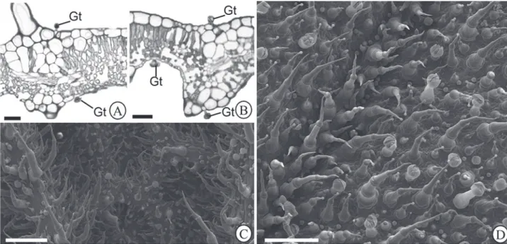
The leaf indument of H. villosa is comprised by glandular and non-glandular trichomes (Fig. 1A-D). Four morphotypes of glandular trichomes (I-IV) differing on size, shape, cell number, and histochemical and ultrastructural features were observed in both leaf surfaces (Fig. 1A-D).
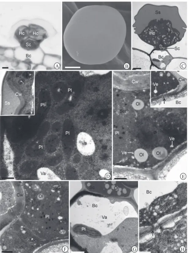
Morphotype I: peltate; comprised by a four-celled globular head, and body with a short unicellular stalk and a unicellular basis (Fig. 2A-C). The head cells are covered by smooth cuticle (Fig. 2B); secretion with homogenous and dense aspect fills the wide subcuticular space (Fig. 2C, 2D insert). The cell walls are thin with loose appearance (Fig. 2D insert). The cytoplasm is dense and abundant and with numerous plastids (Fig. 2D); the plastids are devoid of thylakoids and present tubular elements (Fig. 2D); oil drops occur free in the cytoplasm (Fig. 2D); the vacuoles are small, sparse and exhibits flocculate content (Fig. 2D). The stalk cell possesses periclinal walls with plasmodesmata and thick anticlinal walls (Fig. 2E-insert). The nucleus is voluminous and the cytoplasm is dense and abundant with smooth endoplasmic reticulum with dilated cistern, plastids, vesicles and scattered oil drops (Fig. 2E-F); the plastids are devoid of thylakoids and present osmiophilic globules (Fig. 2E-F); small vesicles occur in the cytoplasm (Fig. 2E, F). The basal cell has thin walls (Fig. 2G, H); numerous plasmodesmata occur in the periclinal walls (Fig. 2H). The
Morphology and histochemistry of glandular trichomes in Hyptis villosa Pohl ex Benth. (Lamiaceae) and differential labeling of cytoskeletal elements
cytoplasm is reduced due to the presence of a large central vacuole (Fig. 2G). Hyperactive dictyosomes, plastids with thylakoids, mitochondria, and polysomes characterize the basal cell (Fig. 2G-H).
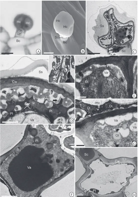
Morphotype II: capitate; composed by a bicellular rounded to oval head and a body with a unicellular short stalk and unicellular basis (Fig. 3A-C). The head cells are covered by smooth cuticle (Fig. 3B); secretion with flocculate aspect fills the wide subcuticular space (Fig. 3C); their walls are thick with loose appearance and form labyrinth projections (Fig. 3D and insert, 3E); plasmodesmata connect the head cell between themselves (Fig. 3D-insert) and to the stalk (Fig. 3F). The nucleus is voluminous with irregular contour (Fig. 3C); the cytoplasm is abundant and characterized by hyperactive dictyosomes, smooth and rough endoplasmic reticulum, mitochondria, small plastids devoid of thylakoids and polysomes (Fig. 3D-E); vacuoles of different size are present and contain fibrillar material (Fig. 3C-D); paramural bodies are observed in the periplasmic space (Fig. 3D). The stalk cell presents walls with loose aspect (Fig. 3F); the cytoplasm is abundant with mitochondria with dilated cristae, plastids without thylakoids, polysomes and scattered oil drops (Fig. 3F-G); a large central vacuole is filled with electron-dense content (Fig. 3F) Paramural bodies are observed in the stalk cell (Fig. 3F, G). The basal cell possesses moderately thick anticlinal walls and thin anticlinal walls (Fig. 3H). The cytoplasm is reduced and presents amorphous plastids (Fig. 3H), mitochondria, and free oil drops; a large central vacuole is observed (Fig. 3H). Morphotype III: capitate; constituted by a unicelular rounded head and body with a short neck cell, a bicellular long stalk (Fig. 4A-C), a large unicelular pedestal and a 6-8 celled basis (Fig. 4A-B). The head cell is covered by smooth cuticle (Fig. 4B); the walls are thin and present loose aspect (Fig. 4D). The nucleus is voluminous with irregular contour (Fig. 4C); the cytoplasm is abundant and contains hiperactive dictyosomes, rough endoplasmic reticulum, mitochondria, multivesicular bodies, polysomes and plastids devoid of thylakoids and containing voluminous electron-dense bodies (Fig. 4D, E and insert). Numerous vesicles occur scattered in the cytoplasm and next to the plasmalema (Fig. 4D, E-insert). Paramural bodies occur in the periplasmic space (Fig. 4D). The neck and stalk cells present similar features. Their walls are thin; the periplasmic space is narrow and contains paramural bodies (Fig. 4F). The nucleus is voluminous with evident nucleolus (Fig. 4C, F). The cytoplasm is abundant and rich in hyperactive dictyosomes, rough endoplasmic reticulum, polysomes, mitochondria with dilated cristae, oil drops, vesicles (Fig. 4F, G), and plastids with thylakoids containing voluminous electron dense bodies (Fig. 4F-insert). Plasmodesmata in the periclinal walls connect the stalk cells (Fig. 4G).The pedestal cell possesses thick walls and reduced cytoplasm (Fig. 4H) by the presence of a large central vacuole; plastids with starch
grains of different sizes occur in the pedestal cell (Fig. 4H). The basal cell possesses moderately thick anticlinal walls and thin anticlinal walls. The cytoplasm is reduced and presents mitochondria and rough endoplasmic reticulum (Fig. 4I); a large central vacuole is observed (Fig. 4I).
Morphotype IV: capitate; composed by a four-celled spherical glandular head and a body constituted by a long unicelular stalk and slightly elevated basis with 6-8 cells (Fig. 5A-C). The cuticle that covers the glandular head breaks via a horizontal line on the head top (Fig. 5C-insert). The description of the subcellular features of this morphotype was not possible because no sample analyzed on transmission electron microscopy exhibited this morphotype.
The distribution of each morphotype varied in the leaf surfaces (Tab. 1). The morphotype I and II were more abundant in the abaxial leaf surface while the morphotypes III and IV were the most plentiful on the adaxial leaf surface (Tab. 1). In generally, the adaxial leaf surface presented a higher density of glandular hairs in comparison to the abaxial leaf surface (Tab. 1).
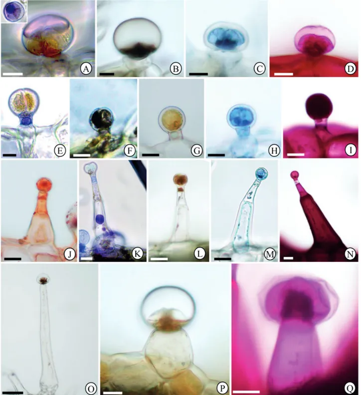
Histochemistry of the glandular trichomes
Different categories of chemical compounds were histochemically detected in the glandular trichomes of H. villosa (Tab. 2) (Fig. 6A-Q). Hydrophilic and lipophilic compounds were detected in all the glandular morphotypes. Different chemical compounds were histochemically detected along the parts of the trichome body and differences on the relative abundance of substances have been observed among the glandular morphotypes. (Tab. 2).
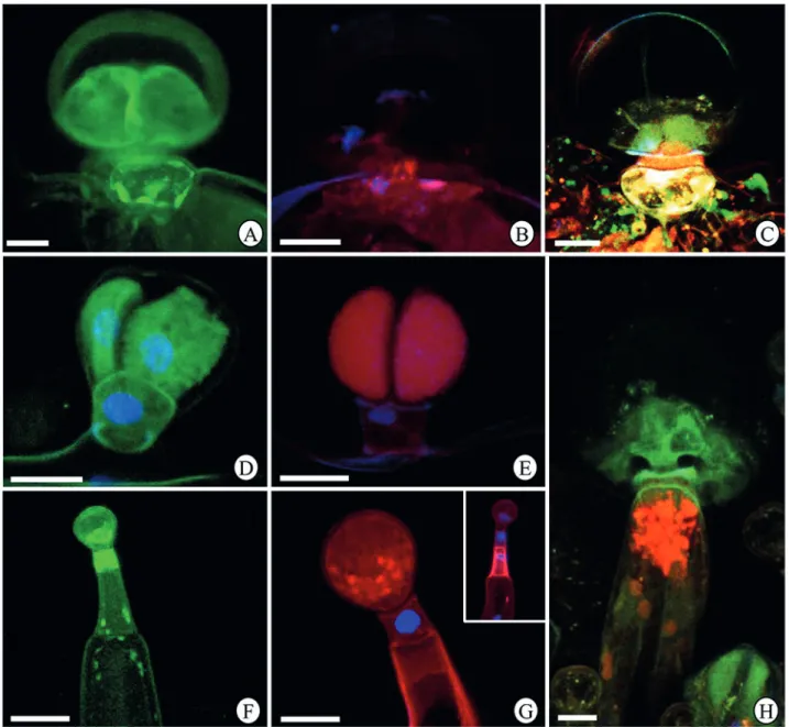
Distribution of the cytoskeletal elements
The distribution of microtubules and actin filaments was different in each portion of the glandular trichomes and among the different glandular morphotypes (Fig 7A-H).
In the morphotype I the head cells exhibited strong marking for microtubules (Fig. 7A, C); actin filaments were more abundant in the stalk cell (Fig. 7B-C) and the basal cell presented high abundance of actin filaments and microtubules (Fig. 7A-C).
Morphology and histochemistry of glandular trichomes in Hyptis villosa Pohl ex Benth. (Lamiaceae) and differential labeling of cytoskeletal elements
Morphology and histochemistry of glandular trichomes in Hyptis villosa Pohl ex Benth. (Lamiaceae) and differential labeling of cytoskeletal elements
Figure 5.Morphotype IV of glandular trichome in Hyptisvillosa leaf blade. Glandular trichome under (A-B) light and (C) scanning electron microscopy showing head cells (Hc), stalk cell (Sc) and basal cells (Bc). Observe the wide subcuticular space (Ss) in B. The insert in C present rupture of the cuticle on the head top. Bars = A, C, 50 μm; B, 25 μm.
Discussion
For our knowledge, this is the first report of the morphological, histochemical and subcellular features of glandular trichomes in a Hyptisspecies. The use of different convergent techniques of light and electron microscope enabled us to identify four glandular morphotypes of trichomes in both leaf surfaces of H. villosa, differing on shape, size, cell number, subcellular features and secretion composition. In addition, imunolabeling of actin filaments and microtubules produced differential results among the glandular morphotypes and along the trichome bodies, according to the main chemical classes of substances produced. A correlation between glandular morphotype and the composition of the secretion was possible to be established.
The presence of peltate and capitate glandular trichomes is a common feature to Lamiaceae species (Werker et al. 1993; Ascensão et al. 1999), and has been reported to species from different genera (Ascensão et al. 1995; 1999; Corsi & Bottega 1999; Rodrigues et al. 2006; Duarte & Lopes 2007; Gonçalves et al. 2010). As a rule, peltate trichomes present short stalk and a large secretory head, while in the capitate trichomes the stalk is more than half of the height of the head (Abu-Asab & Cantino 1987; Ascensão & Pais 1998; Werker 2000). According to this classification, the morphotypes I of H. villosa is peltate and the morphotypes II, III and IV are capitate.
In a general way, a higher density of glandular trichomes
was obtained to the adaxial leaf surface of H. villosa. This is a remarkable feature, since the most of the researches shows higher density of glandular hairs in the abaxial leaf surface in the studied plant species (Tozin et al. 2015b; Búfalo et al. 2016). In H. villosa the higher density of glandular trichomes in the adaxial leaf surface could represent an important strategy for dealing with the high light intensity that strikes on the plants in the campo cerrado (Tozin et al. 2015b), improving the reflection of the sunlight, minimizing the water loss and playing a chemical defense against biotic agents (Pérez-Estrada et al. 2000). In this sense, we can also suggest that the greatest need of protection against environmental factors, mainly the high luminosity, could also be related to the highest density of capitate trichomes (longer) in the adaxial leaf surface of H. villosa.
Hyptisspecies are mainly known by the production of essential oils with aromatic and medicinal potentials(Silva et al. 2013). In fact, terpenes were histochemically detected in all the glandular morphotypes analyzed, and can represent the major chemical category of substances responsible for the aromatic properties of H. villosa. However, hydrophilic compounds were also histochemically detected in different portions of the glandular trichomes. This evidences the mixed nature of the secretion in this species, although there are differences in the prevalence of lipophilic and hydrophilic fractions among the morphotypes and among the portions of a same trichome.
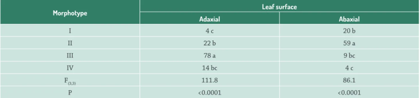
Table 1.Density of each glandular trichomes in both leaf surface of Hyptis villosa (Lamiaceae).
Morphotype Leaf surface
Adaxial Abaxial
I 4 c 20 b
II 22 b 59 a
III 78 a 9 bc
IV 14 bc 4 c
F(3,3) 111.8 86.1
P <0.0001 <0.0001
Table 2. Histochemistry of the leaf glandular trichomes in Hyptis villosa (Lamiaceae).
Metabolic group Reagent
Glandular morphotype
I II III IV
HC SC BC HC SC BC HC NC SC BC HC SC BC
Total lipids Sudan IV +++ ++ - + ++ + + ++ + - ++ +
-Terpenes Nadi reagent +++ - - + ++ - + ++ + - +++ +
-Phenolic compounds Ferric trichloride ++ - - ++ - - ++ - + + + -
-Alkaloids Dragendorff reagent + - - + - - ++ + + - + -
-Proteins Bromophenol blue + - + +++ + - ++ + + + + +
-Neutral polysaccharides PAS + ++ ++ +++ - - ++ - ++ + + ++
-Note: BC, basal cell; SC, stalk cell; NC, neck cell; HC, head cell; PAS, periodic acid-Schiff”s; +, ++, +++ abundance relative; -, absence.
(Harbone 1993; Langenheim 2003). Total lipids, terpenes and alkaloids in leaves are associated to the chemical defense against pathogens and herbivores (Langenheim 2003; Combrinck et al. 2007). Polysaccharides are important in the maintenance of the hydric potential of the cells protecting the organs against desiccation (Ascensão et al. 1999; Werker 2000), important role in environment with low humidity as in the Brazilian Cerrado. Phenolic compounds are also important in the defense against herbivores and pathogens and in the protection of the plants against high irradiance, like that in the Cerrado, that could cause photoinhibition and damage to the cell machinery (Liakoura et al. 1997; Tozin et al. 2015a; Silva et al. 2016).
Total lipids and terpenes were abundantly stained by the histochemical tests in the morphotype I, mainly in the head cells. In fact, the observation of several plastids lacking thylakoids in the head cells is associated with the production of oils (Fahn 1979; Rodrigues & Machado 2012); in addition, the presence of tubular inclusions in these plastids is common reported in cells secreting monoterpenes (Turner et al. 1999). In general way, lipids are released from the secretory cells via ecrine (Evert 2006), cross the cell wall driven by the mechanical pressure exercised by the protoplast (Paiva 2016) and accumulate in the subcuticular space; oil material can cross the cuticle and reach the outer side of the cells without requiring rupture of pore formation in the cuticle. In fact, the cuticle in the head cells of morpohtype I remained intact in all the samples analyzed. In the stalk cells, the presence of proliferative
smooth endoplasmic reticulum, plastids with osmiophilic inclusions and vesicles are indicative of production of lipophilic and hydrophilic substances (Fahn 1979; Evert 2006). Moreover, the occurrence of hyperactive dictyosomes characterizes the basal cells and indicates the intense production of hydrophilic substances (Fahn 1979; Evert 2006), corroborating our histochemical results.
Morphology and histochemistry of glandular trichomes in Hyptis villosa Pohl ex Benth. (Lamiaceae) and differential labeling of cytoskeletal elements
Figure 6. Histochemical tests of glandular trichomes in Hyptis villosa leaf blade. (A-D) Morphotype I. (E-I) Morphotype II. (J-N) Morphotype III. (O-Q) Morphotype IV. (A, E, K) Nadi reagent. The insert in A shows glandular head in overview. (B, F, O) Ferric Trichloride. (C, H, M) Bromophenol blue. (D, I, N, Q) PAS. (G, L, P) Dragendorff reagent. (J) Sudan IV. Bars = A-D, J-N, 15 μm; E-I, 10 μm; O, 100 μm; P-Q, 25 μm.
are the main sites of production of the total lipids and terpenes detected in these cells (Evert 2006; Tozin et al. 2015a). Similarly, the abundance of plastids and oil drops in the basal cells are indicatives of oil production in this portion of the trichome (Evert 2006; Tozin et al. 2015a).
Figure 7.Imunolabeling of cytoskeletal elements in the glandular trichomes in Hyptis villosa leaf blade under confocal microscopy.
(A-C) Morphotype I. (D-E) Morphotype II. (F-G) Morphotype III. (H) Morphotype IV. (A, D, F) Microtubule labeling by anti-β
-tubulin-FITC. (B, E, G) F-actin labeling by phalloidin-rhodamine. (C, H) Microtubule and F-actin labeling by anti-β-tubulin-FITC and
phalloidin-rhodamine.
lipids and polysaccharides in the whole trichome body (Evert 2006). However, the histochemical reactions to phenolic compounds, alkaloids, protein and polysaccharides were more intense in the head cells while the lipids were more intensely marked in the neck cells of the morphotype III. ). In the morphotype III, part of the secretion is release toward the apoplast by granulocrine process what is suggested by the presence of vesicles adjacent to the plasmalemma (Fahn 1979). In this morphotype, no subcuticular space was observed. Some secretion can accumulate as paramural bodies in the periplasmic space before to reach the outside of the cells. The presence of voluminous electron-dense bodies inside plastids as observed in the head, neck and stalk
Morphology and histochemistry of glandular trichomes in Hyptis villosa Pohl ex Benth. (Lamiaceae) and differential labeling of cytoskeletal elements
The widespread presence of plasmodesmata in morphotypes I, II and III connecting the cells of the trichomes among themselves and to the neighboring cells suggests the symplastic passage of substances from the lower cells toward the head cells of trichomes (Ascensão et al. 1999; Evert 2006; Argyropoulou et al. 2010; Silva et al. 2016). These substances may represent precursors of the secretion that will be synthetized in the head cells (Ascensão et al. 1999; Tozin et al. 2015a; Silva et al. 2016).
The rupture of the cuticle on the head cells via a horizontal line was a differential aspect in the morphotype IV, since no pores or rupture was observed in the other morphotypes. Although the analysis of the morphotype IV was not possible under tranwsmission electron microscopy, the rupture of the cuticle suggests the accumulation of secretion in a subcuticular space in a previous moment (Ascensão et al. 1995; Serrato-Valentini et al. 1997). In this morphotype, lipopholic and hydrophilic substances were histochemically detected in the head and stalk cells, with more intense reaction to lipids and terpenes in the head cells.
Our results for imunolabeling of the cytoskeleton elements in the glandular morphotypes of H. villosa corroborate the histochemical tests and the transmission electron microscopy data indicating a correlation between the main compounds produced in the cells and the predominance of actin filaments or microtubules. In general, microtubules were strongly marked in cells where the production of lipophilic substances was predominant, such as the head cells of morphotypes I, II and IV, the stalk cells of morphotype II and the neck cell of morphotype III. On the other hand, the imunolabeling for actin filaments was more intense in cells producing mainly lipophilic substances, such as the stalk cells of morphotypes I, III and IV and in the head cells of morphotype II. The involvement of microtubules in the transport of oil drops is well established in animal cells (Zehmer et al. 2009 and references therein). It has been showed that the ability of oil drops to form and grow in size (Andersson et al. 2006) and their rapid movement in the cytoplasm (Gross et al. 2000) is dependent on microtubules. So, we suggest that the more intense marking for microtubules in cells secreting abundant lipophilic compounds in H. villosa may be associated with the involvement of these elements in the intracellular transport of oil bodies. Concerning the actin filaments, their role in the conduction of the cytoplasmic current and in the vesicles movement is well studied in animal (Valderrama et al. 2001; Stamnes 2002) and plant cells (Evert 2006 and references therein), but not in secretory cells in plants. We purpose that the most intense labeling for actin filaments observed in cells producing secretion predominantly hydrophilic in glandular trichomes of H. villosa can be explained by being this cytoskeletal element the main responsible for the movement of vesicles produced by the dictyosomes throughout the cytoplasm (Evert 2006 and reference therein).
Our results showed that the four glandular morphotypes in the leaves of H. villosa are sites of production of bioactive compounds, occurring a correlation between the glandular morphotype and the composition of the secretion produced. Furthermore, we confirmed the existence of a differential distribution of actin filaments and microtubules between cells secreting mainly hydrophilic or lipophilic compounds. Additional studies with experimental approaches are being conducted to better understand the role of the cytoskeleton elements in the morphogenesis and functioning of the glandular hairs in Lamiaceae species.
Acknowledgements
We thank the CNPq (444205/2014-4) for the financial support; and the staff of the Centro de Microscopia Eletrônica (CME), IBB, UNESP, for helping in the sample preparation.
References
Abu-Asab M, Cantino P.D 1987. Phylogenetic implications of leaf anatomy in subtribe Melittidinae (Labiatae) and related taxa. Journal of Arnold Arboretum 68: 1-34.
Amaral LIV, Pereira MFDA, Cortelazzo AL. 2001. Formação das substâncias de reserva durante o desenvolvimento de sementes de Urucum (Bixa orellana L. - Bixaceae). Acta Botanica Brasilica 15: 125-132. Amrehn E, Heller A, Spring O. 2014. Capitate glandular trichomes of
Helianthus annuus Asteraceae): ultrastructure and cytological development. Protoplasma 251: 161-167
Andersson L, Bostrom P, Ericson J, et al. 2006. PLD1 and ERK2 regulate cytosolic lipid droplet formation. Journal of Cell Science 119: 2246-2257.
Appezzato-da-Glória B, Costa FB, Silva VC, Gobbo-Neto L, Rehder VLG, Hayashi AH. 2012. Glandular trichomes on aerial and underground organs in Chrysolaena species (Vernonieae – Asteraceae): structure, ultrastructure and chemical composition. Flora 207: 878-997. Argyropoulou C, Akoumianakiloannidou A, Christodoulakis SN, Fasseas
C. 2010. Leaf anatomy and histochemistry of Lippia citriodora
(Verbenaceae). Australian Journal of Botany 58: 398-409. Ascensão L, Marques N, Pais MS. 1995. Glandular trichomes on vegetative
and reproductive organs of Leonotis leonurus (Lamiaceae). Annals of Botany 75: 619-626.
Ascensão L, Mota L, Castro MM. 1999. Glandular trichomes on the leaves and flowers of Plectranthus ornatus: morphology, distribution and histochemistry. Annals of Botany 84: 437-447.
Ascensão L, Pais MS. 1998. The leaf capitate trichomes of Leonotis leonurus: histochemistry, ultrastructure and secretion. Annals of Botany 81: 263-271.
Baran P, Aktas K, Özdemir C. 2010. Structural investigation of the glandular of the glandular trichomes of endemic Salvia smyrnea L.. South African Journal of Botany 76: 572-578.
Búfalo J, Rodrigues TM, Almeida LFR, Tozin LRS, Marques MOM, Boaro CSF. 2016. PEG-induced osmotic stress in Mentha x piperita L.: structural features and metabolic responses. Plant Physiology and Biochemistry 105: 174-184.
Combrinck S, Plooy GWD, Mccrindle RI, Botha BM. 2007. Morphology and histochemistry of glandular trichomes of Lippia scaberrima
(Verbenaceae). Annals of Botany 99: 1111-1119.
Costa-Lotufo LV, Moraes MO, Araújo ECC, et al. 2004. Antiproliferative effects of abietane isolated from Hyptis martiusii (Labiatae). Pharmazie 59: 78-79.
David R, Carde JP. 1964. Coloration differentielle des inclusions lipidique et terpeniques des pseudophylles du Pin maritime au moyen du reactif Nadi. Comptes Rendus de l’ Academie dês Sciences Paris. Biologies 258: 1338-1340.
Duarte MR, Lopes JF. 2007. Stem and leaf anatomy of Plectranthus neochilus
Schltr., Lamiaceae. Brazilian Journal of Pharmacognosy 17: 549-556. Durigan G, Baitello JB, Franco GADC, Siqueira M F. 2004. Plantas do
Cerrado Paulista: Imagens de uma paisagem ameaçada. São Paulo, Páginas & Letras.
Evert RF. 2006. Esau’s plant anatomy. 3rd. edn. New Jersey, Wiley-Interscience.
Fahn A. 1979. Secretory tissues in plants. London, Academic Press. Falcão DQ, Menezes FS. 2003. Revisão etnofarmacológica, farmacológica
e química do gênero Hyptis. Revista Brasileira de Farmácia 84: 68-74. Figueiredo ACS, Barroso JMG, Pedro LMG, Ascensão L. 2007. Histoquímica e Citoquímica em Plantas: Princípios e Protocolos. 1st. edn. Lisboa, Faculdade de Ciências da Universidade de Lisboa, Centro de Biotecnologia Vegetal.
Gama TSS, Aguiar-Dias AC, Demarco D. 2016. Transfer cells in trichomatous nectary in Adenocalymma magnificum (Bignoniaceae). Anais da Academia Brasileira de Ciências 88: 527:537.
Gonçalves LA, Azevedo AA, Otoni WC. 2010. Characterization and ontogeny of the glandular trichomes of Ocimum selloi Benth. (Lamiaceae). Acta Botanica Brasilica 24: 909-915.
Gross SP, Welte MA, Block SM, Wieschaus EF. 2000. Dynein-mediated cargo transport in vivo. A switch controls travel distance. The Journal of Cell Biology 148: 945-956.
Gunning BES, Pate JS. 1969. “Transfer cells”: plant cells with wall ingrowths, specialized in relation to short distance transport of solutes – their occurrence, structure, and development. Protoplasma 68: 107-133. Harbone JB. 1993. Ecological biochemistry. 4th ed. London, Academic
Press.
Johansen DA. 1940. Plant michrotechnique. New York, McGraw-Hill. Langenheim JH. 2003. Plant resins: chemistry, evolution, ecology and
ethnobotany. Portland, Cambridge, Timber Press.
Liakoura V, Stepfanou M, Manetas Y, Cholevas C, Karabourniotis G. 1997. Trichome density and its UV-B protective potential are affected by shading and leaf position on the canopy. Environmental and Experimental Botany 38: 223-229.
Maggi F, Papa F, Cristalli G, Sagratini G, Vittori S, Giuliani C. 2010. Histochemical localization of secretion and composition of the essential oil in Melittismelissophyllum L. subsp. melissophyllum from Central Italy. Flavour and Fragrance Journal 25: 63-70.
Maroni BC, Stasi LC, Machado SR. 2006. Plantas medicinais do cerrado de Botucatu – guia ilustrado. São Paulo, Editora UNESP.
Martins MBG. 2002. Estudos de microscopia óptica e de microscopia eletrônica de varredura em folhas de Mentha spicata e de Mentha spicata × suaveolens (Lamiaceae). Bragantia 61: 205-218.
Mazia D, Brewer PA, Alfert M. 1953. The cytochemical staining and measurement of protein with mercuric bromphenol blue. The Biological Bulletin104: 57-67.
Mota L, Figueiredo AC, Pedro LG, Barroso JG, Ascensão L. 2013. Glandular trichomes, histochemical localization of secretion, and essential oil composition in Plectranthus grandidentatus growing in Portugal. Flavour and Fragrance Journal 28: 393-401.
Naidoo Y, Heneidak S, Gairola S, Nicholas A, Naidoo G. 2012. The leaf secretory scales of Combretum molle (Combretaceae): morphology, ultrastructure and histochemistry. Plant Systematic and Evolution 298: 25-32.
Nepi M, Ciampolini F, Pacini E. 1996. Development and ultrastructure of
Cucurbita pepo nectaries of male flowers. Annals of Botany 78: 95-104. O’Brien TP, Feder N, Mccully ME. 1964. Polychromatic staining of plant
cell walls by toluidine blue. Protoplasma 59: 368-373.
Oliveira CMA, Silva MDR, Kato L, Silva CC, Ferreira HD, Souza LKH. 2004. Chemical composition and antifungal activity of the essential oil of
Hyptis ovalifolia Benth. (Lamiaceae). Journal Brazilian of Chemical Society 15: 756-759.
Paiva EAS. 2016. How do secretory products cross the plant cell wall to be released? A new hypothesis involving cyclic mechanical actions of the protoplast. Annals of Botany 117: 533-540.
Paiva EAS, Machado SR. 2008. The floral nectary of Hymenaea stigonocarpa
(Fabaceae, Caesalpinioideae): Structural aspects during floral development. Annals of Botany 101: 125-133.
Paiva EAS, Martins LC. 2011. Calycinal trichomes in Ipomoea cairica
(Convolvulaceae): ontogenesis, structure and functional aspects. Australian Journal of Botany 59: 91-98.
Papini A, Tani G, Falco P, Brighigna L. 2010. The ultrastructure of the development of Tillandsia (Bromeliaceae) trichome. Flora 205: 94-100. Pate JS, Gunning B. 1972. Transfer cells. Annual Review of Plant Physiology
23: 173-196.
Pérez-Estrada LB, Cano-Santana Z, Oyama K. 2000. Variation in leaf trichomes of Wigandia urens: environmental factors and physiological consequences. Tree Physiology 20: 629-632.
Reynolds ES. 1963. The use of lead citrate at high pH as an electron-opaque stain in electron microscopy. The Journal of Cell Biology 17: 208-212. Robards AW. 1978. An introduction to techiniques for scanning electron
microscopy of plant cells. In: Hall JL. (ed.) Electron microscopy and cytochemistry of plant cells. New York, Elsevier. p. 343-403. Rodrigues LS, Monteiro P, Póvoa O, et al. 2006. Biodiversity studies on
Portuguese Thymbra capitata. Acta Horticulturae 723: 127-132. Rodrigues L, Póvoa O, Teixeira G, Figueiredo AC, Moldão M, Monteiro
A. 2013. Trichomes micromorphology and essential oil variation at different developmental stages of cultivated and wild growing
Mentha pulegium L. populations from Portugal. Industrial Crops and Products 43: 692-700.
Rodrigues TM, Machado SR. 2012. Oil glands in Pterodon pubescens benth. (Leguminosae-Papilionoideae): distribution, structure, and secretion mechanisms. International Journal of Plant Sciences 173: 984-992. Sacchetti G, Romagnoli C, Nicolleti M, Fabio A, Bruni A, Poli F. 1999.
Glandular trichomes of Calceolaria adscendens Lidl. (Scrophulariaceae): histochemistry, development and ultrastructure. Annals of Botany 83: 87-92.
Serrato-Velentini G, Bisio A, Cornara L, Ciarallo G. 1997. Structural and histochemical investigation of the glandular trichomes of Salvia aurea L. Leaves, and chemical analysis of the essential oil. Annals of Botany 79: 329-36.
Silva RS, Rezende CM, Santana HCD, Vieira RF, Bizzo HR. 2013. Scents from Brazilian Cerrado: chemical composition of the essential oil from the leaves of Hyptis villosa Pohl ex Benth (Lamiaceae). Journal of Essential Oil Research 25: 415-418.
Silva SCM, Tozin LRS, Rodrigues TM. 2016. Morphological and histochemical characterization of secretory sites of bioactive compounds in Lantana camara L. (Verbenaceae) leaves. Botany 94: 321-336.
Souza LKH, Oliveira CMA, Ferri PH, et al. 2003. Antimicrobial activity of Hyptis ovalifolia towards dermatophytes. Memórias do Instituto Oswaldo Cruz 98: 963-965.
Stamnes M. 2002. Regulating the actin cytoskeleton during vesicular transport. Current Opinion in Cell Biology 14: 428-433.
Svendsen AB, Verpoorte R 1983. Chromatography of alkaloids. New York, Elsevier Scientific Publishing Company.
Tozin LRS, Carvalho SF, Machado SR, Rodrigues TM. 2015a. Glandular trichome diversity on leaves of Lippia origanoides Kunth and Lippia stachyoides Cham. (Verbenaceae): morphology, histochemistry and ultrastructure. Botany 93: 297-306.
Tozin LRS, Marques MOM, Rodrigues TM. 2015b. Glandular trichome density and essential oil composition in leaves and inflorescences of Lippia origanoides Kunth (Verbenaceae) in the Brazilian Cerrado. Anais da Academia Brasileira de Ciências 87: 943-953.
Turner GW, Gershenzon J, Nielson EE, Froehlich JE, Croteau RB. 1999. Limonene synthase, the enzyme responsible for monoterpene biosynthesis in peppermint, is localized to leucoplasts of oil gland secretory cells. Plant Physiology 120: 879-886.
Morphology and histochemistry of glandular trichomes in Hyptis villosa Pohl ex Benth. (Lamiaceae) and differential labeling of cytoskeletal elements
Werker E. 2000. Trichome diversity and development. Advances in Botanical Research 31: 1-35.
Werker E. 1993. Function of essential oil-secreting glandular hairs in aromatic plants of Lamiaceae – A Review. Flavour and Fragrance Journal 8: 249-255.
Werker E, Putievsky E, Ravid U, Dudai N, Katzir I. 1993. Glandular hairs and essential oil in developing leaves of Ocimum basilicum L. (Lamiaceae). Annals of Botany 71: 43-50.