55 55 55 55 55 Mem Inst Oswaldo Cruz, Rio de Janeiro, Vol. 94(1): 55-66, Jan./Feb. 1999
Immunotherapy for Visceral Leishmaniasis: Ability of
Factors Produced during Anti-leishmania Responses of Skin
Test Positive Adults to Inhibit Peripheral Blood
Mononuclear Cell Activities Associated with Visceral
Leishmaniasis
Bettie J Holaday
Núcleo de Medicina Tropical, Universidade Federal do Ceará, Rua Alexandre Baraúna 949, 60430-160 Fortaleza, CE, Brasil
The course of human Leishmania chagasi infections appears to be determined by the balance be-tween type 1 (T1) CD4+ and CD8+ T suppressor (Ts) cell activities. Skin test positive adults living in hyperendemic areas who have no history of visceral leishmaniasis (VL) have T1 CD4+ T cell immunodominant responses against L. chagasi. The cytokines they secrete during anti-leishmania re-sponses are a probable source of cytokines which inhibit the CD8+ Ts cells associated with VL. The ability of supernatants generated from peripheral blood mononuclear cells derived from skin test posi-tive adults to reverse immune responses which appear to be mediated by CD8+ Ts cells was assessed in three sets of screening assays. The supernatants displayed three candidate factors. One, which could be explained by Leishmania antigens in the supernatant, decreased high endogenous IL-10 secretion acteristic of one class of VL patients. A second activity decreased high endogenous proliferation char-acteristic of the same class of patients without decreasing antigen specific proliferation. The third activity inhibited or killed CD8+ T cells but not CD4+ T cells. These activities might be useful in treating VL.
Key words: leishmaniasis - CD8+ T suppressor (Ts) - immunotherapy - interleukin (IL)-10 - IL-12 - antigens
Humans with a history of infection with Leish-mania chagasi display a continuum of immune re-sponses to the parasite. Subjects who have lived in hyperendemic areas for many years without clinically evident infections who have strong DTH+ responses against Leishmania are thought to have the strongest protective immune responses. They have the strongest Leishmania specific T1 CD4+ T cell responses (Holaday et al. 1993a). Conversely, CD4+ T cells are depleted in visceral leishmaniasis (VL) patients especially in the spleen (Cenini et al. 1993). CD8+ T cells, but not CD4+ T cells, isolated from patients with VL inhibit
Leishmania specific proliferation and IFN-g
secre-tion while increasing (interleukin) IL-10 secresecre-tion when mixed with autologous peripheral blood
mononuclear cells (PBMC) isolated after success-ful treatment (Holaday et al. 1993a). CD4+ T cell lines cannot be isolated from acute VL patients until CD8+ T cells are removed by cell sorting. Patients who have been successfully treated for VL gener-ally have more variable responses to L. chagasi
antigens ranging from responses characteristic of acute patients to those of DTH+ adults (Holaday et al. 1993b). Thus, the course of L. chagasi infec-tions appears to be due to the balance between T1 CD4+ T cells and CD8+ T suppressor (Ts) cells. These findings are in agreement with those of Uyemura et al. (1993) who found both T1 CD4+ and T2 CD8+ T cells in human cutaneous Leish-mania lesions.
Recent attempts to identify cytokines which might be useful in treating VL have focused on cytokines thought to maintain the balance between T1 and T2 CD4+ T cells in spite of the lack of evidence for a role for T2 CD4+ T cells in symp-tomatic human L. chagasi and L. donovani infec-tions (Hsieh et al. 1993, Manetti et al. 1993, Carvalho et al. 1994, Ghalib et al. 1995). The cur-rent study is the first to focus on detecting cytokines which might maintain a T1 CD4+ T cell immuno-dominant response by inhibiting the CD8+ Ts cells
This work was supported by United States National In-stitutes of Health grants 5PO1 AI-26512 and AI-26918 and TDR Strengthening Grant 880-225.
Present address: 170A Grattan St., San Francisco, CA 94117, USA. Fax: +1-415-566-9776.
56 56 56 56
56 Immunotherapy for Visceral Leishmaniasis Bettie J Holaday
associated with symptomatic human L. chagasi
infections. Cytokines with this property might re-store the ability of VL patients’ immune systems to eradicate Leishmania infections.
Three different types of screening assays were done. Each one provided different information. Since DTH+ adults have the strongest T1 CD4+ T cell response against L. chagasi the cytokines they secrete during anti-leishmania responses are the most probable source of cytokines which inhibit the CD8+ Ts cells associated with VL. In the first set of as-says supernatants generated by DTH+ adult PBMC in the presence (DS+A) or absence (DS-A) of L. chagasi antigens were tested for their ability to con-vert VL patient PBMC responses thought to be mediated by CD8+ Ts cells, that is, to decrease IL-10 secretion, increase IFN-g secretion, or increase Leishmania specific proliferation. The effects of DS+A were compared to those of IL-12 and anti-IL-10 which are thought to favor T1 CD4+ T cell activity over T2 CD4+ T cell activity (Hsieh et al. 1993, Manetti et al. 1993, Carvalho et al. 1994, Ghalib et al. 1995). The second assay used a recon-stitution assay to measure the ability of DS+A and IL-12 to prevent CD8+ Ts cell mediated changes in cytokine secretion and proliferation. The third set of assays directly tested the ability of a supernatant (CD4S) generated from Leishmania specific T1 CD4+ T cell lines derived from DTH+ adults to in-hibit the growth of CD4+ and CD8+ T cell lines.
The three sets of assays identified three candi-date activities, Leishmania antigens and two soluble factors. The assays also revealed the ex-istence of two categories of VL patients, those with high and low levels of endogenous IL-10 secre-tion, which responded differently to immuno-modulators.
MATERIALS AND METHODS
Human subjects - Subjects of the study were residents of the states of Ceará (CE), Piauí (PI), or Rio Grande do Norte (RN) in northeastern Brasil. DTH+ adults from a hyperendemic area near Itapipoca, CE, were selected as representative of people with strong protective immune responses against L. chagasi (Evans et al. 1992). PBMC from five DTH+ adults (ages 16-64, mean = 33 years) with indurations of 17-25 mm measured 48 hr af-ter intradermal injection of L. chagasi antigens were used to generate the DS-A and DS+A super-natants. Nineteen acute patients (ages 14-58, mean = 28 years) were self referred to Hospital São José or Hospital Walter Cantído in Fortaleza, CE; Santa Casa in Sobral, CE; Hospital Gizelda Trigueiro in Natal, RN and the Infectious Diseases Hospital of the Federal University of Piauí in Terezina; or were identified in field studies conducted by Fundação
Nacional de Saúde. All diagnoses were confirmed by the presence of amastigotes in bone marrow aspirates. Patients, except P14 and P23, were suc-cessfully treated with pentavalent antimony (20 mg/ kg/day) for 20 days. P14 had been symptomatic for eight months prior to seeking treatment. She showed no signs of clinical improvement after nine days of treatment with allopurinol, so she was then successfully treated with pentavalent antimony as described above. Blood for this study was drawn from P14 after allopurinol treatment was completed but before pentavalent antimonial treatment had begun. P23 had multiple relapses during a one and a half year period in spite of treatment with pen-tavalent antimony, allopurinol and splenectomy. The studies reported here were made during com-bined pentavalent antimony/allopurinol treatment of P23. All assays of acute VL patient PBMC were performed during the seven month period of April-October. P2 was successfully treated for VL in 1990 and has been free of kala azar symptoms since the end of treatment.
Parasite antigens - L. chagasi promastigotes were cultured in HOSMEM II medium containing 20% fetal calf serum, washed three times in PBS, harvested from stationary phase cultures by cen-trifugation, resuspended in PBS and lysed by soni-cation or repeated freeze thawing (Berens & Marr 1978). An aliquot of antigen suspension was as-sayed for protein content using a modification of the Lowry Rosebrough method (Dulley & Grieve 1975). Antigens prepared in this way are not mi-togenic to PBMC from non-immune subjects.
57 57 57 57 57 Mem Inst Oswaldo Cruz, Rio de Janeiro, Vol. 94(1), Jan./Feb. 1999
by venipuncture. Mononuclear cells were sepa-rated by centrifugation at 200 g for 20 min at 20oC over ficoll-hypaque. Cells were washed twice in Hank’s balanced salt solution containing 0.25% bovine serum albumin. The washed cells were cul-tured at 3 x 10⁶ cells/ml in Iscove’s modified Dulbecco’s medium supplemented with 10% fetal calf serum/10% human AB+ serum and contain-ing antibiotic/antimycotic in the absence or pres-ence of 20 mg/ml Leishmania antigens.
Superna-tants were collected on day 4, pooled, aliquotted and stored frozen prior to assay. Supernatants gen-erated from DTH+ adult PBMC with or without L. chagasi antigens were designated DS+A and DS-A, respectively. DS+A contained 1.9 ng/ml IFN-g
and 0.07 ng/ml IL-10. DS-A lacked measurable amounts of IFN-g but contained 0.07 ng/ml IL-10.
VL patients are anemic and leukopenic and the limited numbers of PBMC which could be collected from them prevented all assays from being per-formed with PBMC from each patient. PBMC from all 19 patients were assayed for proliferation but PBMC from only 12 patients were assayed for cytokine secretion. In addition, there were suffi-cient PBMC from only seven patients to allow test-ing of all the immunomodulators on proliferation and cytokine secretion. PBMC from P19 were col-lected and assayed twice, once three days after ini-tiation of treatment and a second time 10 days af-ter initiation of treatment (P19R).
Assays of the effects of immunomodulators on PBMC activities - In assays of PBMC from acute VL patients PBMC were cultured in triplicate, as described above. When immunomodulators were added to the cultures they were present at the fol-lowing concentrations: rIL-12 50 U/ml (Genetics Institute, Cambridge, MA), anti-IL-10 30 mg/ml
(DNAX, Palo Alto, CA) or 50% v/v DS-A or DS+A. When present, the CD8+ T cell lines were assayed at 2 x 10⁵/ml. In some assays DS+A was preincubated for 30 min at 37oC with 100 NU/ml of polyclonal rabbit anti-human IL-12 (Genetics Institute) or 20 mg/ml monoclonal anti-IL-12
(Hoffmann-La Roche, Nutley, NJ) prior to addi-tion to the PBMC. Supernatants were collected on day 4, pooled, aliquotted and stored frozen prior to assay. 3H-methyl thymidine (1.0 mCi/ml) was
added to PBMC cultures on day 5 and the cells were harvested on day 6. The small quantities of DS+A and DS-A available, as well as unavailabil-ity of long term cultured T cell lines, prevented measurement of the effects of DS+A, DS-A, rIL-12, and anti-IL-10 on the growth kinetics of the T cell lines used in CD4S assays.
Maintenance of long term T cell cultures - Long term P2 polyclonal CD8+ T cell lines, P2A/8/10/ 90 and P2A/8/1/91, polyclonal CD4+ T cell line,
P2A/4/10/90, and monoclonal CD4+ T cell line, P2A/4/1 were established from PBMC collected from P2 in 1990 during treatment for VL, as pre-viously described (Holaday et al. 1993a). Simi-larly, P2 monoclonal CD8+ T cell lines, P2R/8/52 and P2R/8/53 were established from PBMC col-lected from P2 after successful treatment. DTH+ adult Leishmania specific T1 CD4+ monoclonal T cell line, D4/46, and others used to generate CD4S (described below) were established from two DTH+ adults in 1990. All cell lines were stored frozen in liquid nitrogen. The T cells were thawed and expanded by stimulation with 5 mg/ml of PHA
(Sigma Chemical Co, St. Louis, MO) in the pres-ence of 25 U/ml rIL-2 (Cetus, Emeryville, CA), 106/ml inactivated allogeneic PBMC and 105/ml
inactivated JY cells once per three weeks (Yssel et al. 1984). Allogeneic PBMC and JY were inacti-vated by exposure to 50 mg/ml mitomycin c for 45
min, three washes to remove the mitomycin c, and exposure to 2000r g-irradiation.
IL-10 and IFN-g immunoenzymetric ELISA - The
levels of IL-10 in the supernatants were determined using commercially available monoclonal anti-hu-man IL-10, anti-huanti-hu-man IL-10-biotin, rhIL-10 (Pharmingen, San Diego, CA) and extravidin per-oxidase (Sigma) according to Pharmingen’s proce-dure. This ELISA is sensitive to 20 pg/ml. IFN-g
was measured using a commercially available hu-man IFN-g ELISA kit (Genzyme, Cambridge, MA).
The kit is sensitive to at least 100 pg/ml.
Production and assay of CD4S - Leishmania
specific CD4+ T1 T cell clones isolated from two DTH+ adults and tested for Leishmania specific-ity, as previously described, were stimulated with PHA, allogeneic PBMC, JY and rIL-2 as described above (Holaday et al. 1993a). Supernatants were collected 72 hr later, pooled and stored frozen prior to use. The levels of PHA and IL-2 remaining in CD4S were not mitogenic for PBMC. A small quantity of supernatant generated from the same CD4+ T cell clones by Leishmania antigen stimu-lation was also available for assay. The effects of CD4S on the kinetics of growth of CD4+ and CD8+ T cell lines was assessed by stimulating T cells with PHA in the presence or absence of 50% v/v CD4S. To eliminate the possibility that the effects of CD4S on T cell lines might be enhanced by insufficient IL-2 the effects of doubling the concentration of IL-2 in CD4S assays was tested. Since DS+A is known to contain IFN-g, lymphotoxin and tumor
necrosis factor-a, the effect of neutralizing
anti-bodies specific for IFN-g (30 mg/ml, DNAX, Palo
Alto, CA), lymphotoxin or tumor necrosis
factor-a (4000 NU/ml), (Genentech, South San Francisco,
58 58 58 58
58 Immunotherapy for Visceral Leishmaniasis Bettie J Holaday
added back to maintain the starting concentrations. After each week aliquots of the cells were counted and viability was assessed by trypan blue exclu-sion. Tests were carried out on a polyclonal CD4+ T cell line (P2A/4/10/90), a CD4+ clone derived from this line (P2A/4/1), a CD4+ clone from a DTH+ adult (D4/46), a polyclonal CD8+ T cell line derived from P2 during treatment (P2A/8/10/ 90) and two CD8+ clones (P2R/8/52 and P2R/8/ 53) derived after P2 had completed chemotherapy. CD4S assays were performed in the U.S. where PBMC from acute VL patients were unavailable and insufficient quantities of CD4S were available to measure the effects of CD4S on activities of acute VL patient PBMC.
Statistical analysis - The statistical significance of differences between means was determined by t tests. A probability < 0.05 was considered to be significant.
RESULTS
Assay 1 - Effects of immunomodulators on PBMC responses characteristic of VL
Existence of two categories of VL patients -PBMC from VL patients could be placed in two groups, based on their level of endogenous IL-10 secretion, which responded differently to immunomodulators. PBMC displaying low levels
(7 of 13 assays) had an IL-10 secretion mean of 0.07 ± 0.06 ng/ml and 0 ± 0 ng/ml of IFN-g (Table I).
PBMC displaying high levels of endogenous IL-10 secretion (6 of 13 assays) had an IL-10 secretion mean of 0.71 ± 0.21 ng/ml and 0 ± 0.01 ng/ml of IFN-g (Table II). The probability of the high
activ-ity IL-10 values being sampled from the same popu-lation as the low activity group was only 8 x 10⁻⁶.
PBMC from DTH+ adult controls had relatively low levels of endogenous IL-10 secretion (mean = 0.07 ± 0.04 ng/ml), IFN-g secretion (mean = 0 ± 0),
and proliferation (mean = 3,876 ± 1,904 cpm). En-dogenous IL-10 and proliferation values were posi-tively correlated (r = + 0.83). The y axis intercept of the regression line was 0.0 ng/ml IL-10 (Fig. 1). DTH+ PBMC secreted 1.9 ± 0.5 ng/ml IFN-g and
0.07 ± 0.04 ng/ml IL-10 in response to antigen and had a mean proliferation SI of 11.8 ± 4.5.
Since levels of endogenous IL-10 secretion and proliferation were positively correlated in the DTH+ controls, PBMC from patients for whom only proliferation data were available were classi-fied as having high or low activities by their level of endogenous proliferation. PBMC from patients with a mean endogenous 3H-thymidine incorpo-ration rate less than 3,650 cpm (mean value for the low activity group + 2 SD), P15, P18, P21, P26 and P31, were placed in the low activity group
TABLE I
Effects of Leishmania antigens and immunomodulators on IL-10 and IFN-g secretion by peripheral blood
mononuclear cells from low activity patients
IL-10 IFN-g
(ng/ml ± SDa) (ng/ml ± SD)
Patient Antigen Med DS+Ab IL-12 Med DS+A IL-12 Anti-IL-10
24 - .09 ± .03 .01 ± .01 .13 ± .03 0 ± 0 .06 ± .01 .18 ± .04 .21 ± .01
+ .19 ± .01 .27 ± .12 .07 ± .02 .08 ± .10 .06 ± 0 .16 ± .02 .24 ± .01
25 - .16 ± .0 .18 ± .01 .15 ± .06 0 ± 0 .06 ± .01 .18 ± .04 .20 ± .02
+ .11 ± .02 .19 ± .03 .19 ± .06 .08 ± .04 .15 ± .07 .18 ± .01 .20 ± .01
27 - .03 ± 0 .17 ± .03 .03 ± 0 0 ± 0 0 ± 0 0 ± 0 0 ± 0
+ .10 ± 0 .10 ± .03 .15 ± .10 0 ± 0 0 ± 0 0 ± 0 0 ± 0
28 - .11 ± .05 .17 ± .07 nd 0 ± 0 .08 ± .02 nd nd
+ .07 ± .02 .21 ± .02 0 ± 0 .05 ± 0
29 - .02 ± .01 .13 ± .01 nd 0 ± 0 .01 ± .01 nd nd
+ .06 ± .05 .18 ± 0 0 ± 0 .01 ± 0
30 - 0 ± 0 .18 ± .06 0 ± 0 0 ± 0 0 ± 0 .06 ± .02 .46 ± 0
+ 0 ± 0 .18 ± 0 .02 ± .02 0 ± 0 0 ± 0 .06 ± 0 .72 ± .34
32 - .05 ± .01 .19 ± .02 nd 0 ± 0 .04 ± .06 nd nd
+ .11 ± .01 .19 ± .01 0 ± 0 .08 ± 0
Mean - .07 ± .06 .15 ± .06 .08 ± .07 0 ± 0 .04 ± .03 .10 ± .09 .22 ± .19 + .09 ± .06 .19 ± .05 .11 ± .08 .02 ± .04 .05 ± .05 .10 ± .08 .29 ± .30
59 59 59 59 59 Mem Inst Oswaldo Cruz, Rio de Janeiro, Vol. 94(1), Jan./Feb. 1999
(Table III). PBMC from patients with a mean en-dogenous 3H-thymidine incorporation rate greater than 5,225 cpm (mean value for the low activity group + 4 SD), P14 and P16, were placed in the high activity group (Table IV). PBMC from the low activity group had a proliferation mean of 2,073 ± 788 cpm. These values were not corre-lated with endogenous IL-10 secretion (r = +0.15) but both variables were low compared to high ac-tivity group levels. PBMC in this group had a mean proliferation SI of 1.8 ± 1.1 (not including P18 PBMC which had an atypically high SI of 24). The endogenous proliferation mean for the high
activ-ity group was 12,922 ± 7,651; levels of endogenous IL-10 secretion and proliferation were correlated for this group (r = + 0.76). The y axis intercept of the regression line relating the two variables was 0.42 ng/ml IL-10 (Fig. 1).
Effects of immunomodulators on cytokine se-cretion by VL patient PBMC- The effects of Leish-mania antigens, by themselves or as part of DS+A, on IL-10 secretion by acute VL patient PBMC were different in assays of high and low activity patients. PBMC in the low activity group did not show sig-nificant changes in IL-10 or IFN-g secretion in
re-sponse to antigen alone. DS+A produced a small but significant increase in IL-10 secretion. IL-12 and anti-IL-10 caused small but significant in-creases in IFN-g secretion with or without antigen
present. In contrast, PBMC in the high activity group overall secreted less IL-10 in response to antigen. DS+A caused changes similar to antigen alone. IL-12 had no consistent effect on IL-10 se-cretion except to decrease the effect of antigen on secretion. Antigen alone or in DS+A caused a very small but significant increase in IFN-g secretion.
IL-12 caused a significant increase in IFN-g
secre-tion with or without antigen present. Anti-IL-10 had no significant effect on IFN-g secretion.
Effects of immunomodulators on proliferation by VL patient PBMC - The immunomodulators also had different effects on proliferation in assays of PBMC with high and low endogenous activities.
TABLE II
Effects of Leishmania antigens and immunomodulators on IL-10 and IFN-g secretion by peripheral blood
mononuclear cells from high activity patients
IL-10 IFN-g
(ng/ml ± SDa) (ng/ml ± SD)
Patient Antigen Med DS+Ab IL-12 Med DS+A IL-12 Anti-IL-10
17 - .46 ± .03 .13 ± .01 .37 ± .04 0 ± 0 .09 ± .01 .23 ± 0 nd
+ .11 ± .01 .10 ± .02 .33 ± .06 .03 ± 0 .12 ± .01 .21 ± .01
19 - .84 ± .01 .17 ± .01 .73 ± .01 0 ± 0 .04 ± .05 .23 ± 0 .05 ± .01
+ .19 ± .02 .11 ± .03 .34 ± .01 .11 ± .11 .10 ± .04 .21 ± 0 .16 ± .06 19R - .99 ± .19 .29 ± .02 .94 ± .05 .02 ± .04 .08 ± .02 .10 ± .14 .06 ± .04 + .18 ± .01 .20 ± .01 .42 ± .02 0 ± 0 .09 ± .01 0 ± 0 .10 ± .04
20 - .67 ± .01 0 ± 0 .70 ± .02 0 ± 0 .13 ± 0 .02 ± .02 nd
+ .14 ± .16 .08 ± .07 .44 ± .21 .18 ± 0 .15 ± 0 .24 ± .04
22 - .80 ± .11 .54 ± .11 .83 ± .03 0 ± 0 .10 ± 0 .26 ± .06 .05 ± .07
+ .25 ± .03 .28 ± .02 .74 ± .08 .12 ± .02 .12 ± .02 .25 ± 0 .18 ± .11
23 - .49 ± .08 .43 ± .13 .97 ± .16 0 ± 0 .12 ± .06 .04 ± .01 0 ± 0
+ 1.08 ± .03 .65 ± .06 .80 ± .10 .08 ± 0 .14 ± .02 .28 ± 0 .10 ± .01 Mean - .71 ± .21 .26 ± .20 .76 ± .22 0 ± .01 .09 ± .03 .15 ± .11 .04 ± .03 + .32 ± .37 .24 ± .22 .51 ± .20 .09 ± .06 .12 ± .02 .20 ± .10 .14 ± .04
a: mean ng per ml ± 1 standard deviation; b: supernatant derived by stimulation of peripheral blood mononuclear cells from skin test positive adults with Leishmania antigens.
60 60 60 60
60 Immunotherapy for Visceral Leishmaniasis Bettie J Holaday
None of the immunomodulators had a significant effect on proliferation in the low activity assays (Table III). DS+A alone decreased endogenous pro-liferation in assays of high activity PBMC produc-ing a mean SI of 0.7 ± 0.3, significantly less than
the SI for antigen alone, 1.4 ± 0.8 (p=0.04), or anti-gen plus DS+A, 2.3 ± 0.8, (p = 0.0001), (Table IV). Anti-IL-10 significantly increased proliferation in response to antigen (p = 0.04). Pretreatment of DS+A with 100 NU/ml polyclonal or 20 mg/ml TABLE III
Effects of Leishmania antigens and immunomodulators on proliferation by peripheral blood mononuclear cells from low activity patients
Patients Antigen Medium DS+Aa SIb IL-12 SI Anti-IL-10 SI
DS+A IL-12 anti-IL-10
15 - 1,038 ± 64c 2,731± 595 2.6 1,075 ± 269 1.0 nd
+ 2,401 ± 151 3,884 ± 476 3,573 ± 180
SI Antigen 2.3 1.4 3.3
18 - 1,406 ± 202 13,771 ± 634 9.8 nd nd
+ 33,384 ± 2,464 46,364 ± 2,331
SI Antigen 24 3.4
21 - 2,518 ± 414 8,489 ± 1,411 3.4 nd 4,968 ± 820 2.0
+ 5,254 ± 692 8,646 ± 1,034 7,686 ± 364
SI Antigen 2.1 1.0 1.6
24 - 2,160 ± 246 2,655 ± 599 1.2 7,257 ± 2,388 3.4 2,278 ± 488 1.0
+ 2,045 ± 534 4,703 ± 1,810 6,290 ± 365 5,341 ± 656
SI Antigen 1.0 1.8 0.9 2.3
25 - 2,183 ± 83 2,577 ± 463 1.2 6,228 ± 1,600 2.8 4,114 ± 1,228 1.9
+ 1,705 ± 132 3,511 ± 1,728 5,414 ± 986 2,505 ± 600
SI Antigen 0.8 1.4 0.9 0.6
26 - 3,225 ± 3,087 8,038 ± 524 2.5 nd nd
+ 2,614 ± 326 8,164 ± 1,503
SI Antigen 0.8 1.0
27 - 983 ± 60 1,998 ± 731 2.0 3,374 ± 650 3.4 7,396 ± 693 7.5
+ 1,179 ± 400 4,369 ± 488 3,644 ± 880 6,839 ± 4,540
SI Antigen 1.2 2.2 1.1 0.9
28 - 2,889 ± 1,700 11,996 ± 1,112 4.2 7,820 ± 2,084 2.8 nd
+ 9,922 ± 1,644 18,782 ± 4,522 21,621 ± 3,609
SI Antigen 3.4 1.6 2.8
29 - 2,136 ± 79 3,851 ± 632 1.8 2,927 ± 370 1.4 nd
+ 2,290 ± 493 8,330 ± 371 9,893 ± 573
SI Antigen 1.1 2.2 3.4
30 - 2,722 ± 1,086 5,069 ± 486 1.9 4,537 ± 2,007 1.7 5,852 ± 1,719 2.1
+ 3,978 ± 2,001 11,432 ± 2,248 6,050 ± 726 1,651 ± 319
SI Antigen 1.5 2.2 1.3 0.3
31 - 988 ± 356 1,888 ± 375 1.9 878 ± 372 0.9 nd
+ 1,202 ± 211 2,895 ± 565 1,819 ± 444
SI Antigen 1.2 1.5 2.1
32 - 2,627 ± 614 8,513 ± 742 3.2 6,028 ± 936 2.3 nd
+ 10,967 ± 1,918 12,203 ± 1,209 18,038 ± 2,447
SI Antigen 4.2 1.4 3.0
Mean SI 1.8 ± 1.1 1.6 ± 0.4 2.4 ± 0.9 2.1 ± 1.1 2.2 ± 1.0 1.1 ± 0.8 2.9 ± 2.6
valuesd antigen antigen + DS+A antigen IL-12 antigen anti-IL-10
only DS+A only + IL-12 only + anti-IL-10 only
61 61 61 61 61 Mem Inst Oswaldo Cruz, Rio de Janeiro, Vol. 94(1), Jan./Feb. 1999
monoclonal anti-IL-12 did not reverse the effects of DS+A (data not shown). IL-12 alone did not have a consistent effect on proliferation. Stimulation of DTH+ adult control PBMC with DS+A increased endogenous proliferation (SI = 11.5 ± 3.1) by the same amount as antigen only (11.8 ± 4.5).
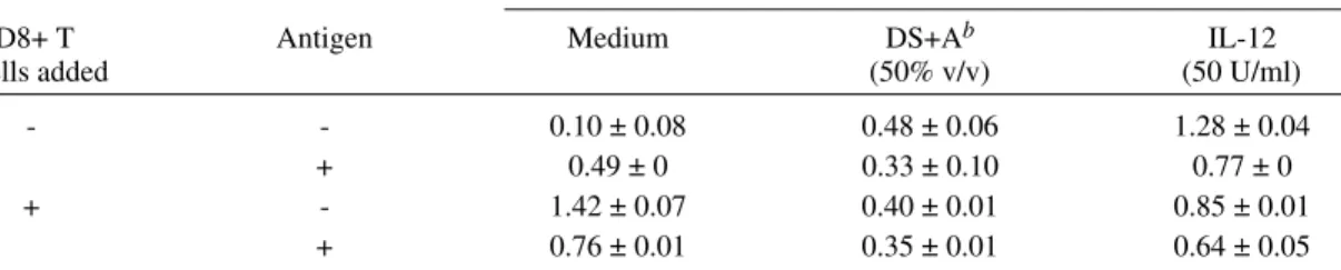
Assay 2 - Ability of immunomodulators to in-hibit CD8+ Ts mediated changes in cytokine se-cretion and proliferation - The addition of CD8+ T cells isolated during acute phase VL to autolo-gous PBMC collected after successful treatment has been shown to result in increased IL-10 secre-tion in the absence or presence of added Leishma-nia antigens (Holaday et al. 1993a). The ability of DS+A and IL-12 to reverse this increase was tested (Table V). PBMC collected from a VL patient (P2) three years after successful treatment secreted 0.10 ± 0.08 ng/ml IL-10 in the absence of antigen and
0.49 ± 0 ng/ml in the presence of antigen. Addi-tion of the autologous CD8+ T cell line, P2A/8/1/ 91 to these PBMC increased IL-10 secretion to 1.42 ± 0.07 ng/ml in the absence of antigen. Addition of antigen decreased this high level two-fold, simi-lar to the effect of antigen on endogenous IL-10 secretion by PBMC from high activity patients. Addition of DS+A alone increased PBMC IL-10 secretion by the same amount as antigen alone but prevented IL-10 secretion from increasing in re-sponse to the addition of CD8+ T cells twice as well as antigen alone. IL-12 increased IL-10 se-cretion in the absence of added antigen by about the same amount as the CD8+ T cells. The PBMC did not secrete IFN-g in any of the assays. CD8+
T cells inhibited antigen specific proliferation by about 50%. DS+A, but not IL-12, prevented the decrease (data not shown).
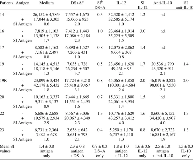
TABLE IV
Effects of Leishmania antigens and immunomodulators on proliferation by peripheral blood mononuclear cells from high activity visceral leishmaniasis patients
Patients Antigen Medium DS+Aa SIb IL-12 SI Anti-IL-10 SI
DS+A IL-12 anti-IL-10
14 - 26,152 ± 4,786c 7,557 ± 1,675 0.3 32,320 ± 4,412 1.2 nd + 17,044 ± 3,305 15,066 ± 925 32,585 ± 5,174
SI Antigen 0.6 2.0 1.0
16 - 7,819 ± 1,103 7,412 ± 1,443 1.0 23,464 ± 1,914 3.0 nd
+ 13,505 ± 1,178 17,086 ± 2,184 35,225 ± 5,509
SI Antigen 1.7 2.3 1.5
17 - 8,582 ± 1,162 6,890 ± 1,527 0.8 12,075 ± 2,862 1.4 nd
+ 7,161 ± 2,497 7,266 ± 431 9,664 ± 368
SI Antigen 0.8 1.0 0.8
19 - 14,145 ± 4,513 7,035 ± 728 0.5 23,456 ± 1,620 1.7 20,536 ± 790 1.4
+ 18,118 ± 3,146 26,234 ± 507 49,461 ± 95 43,320 ± 911
SI Antigen 1.3 3.7 2.1 2.1
19R - 23,099 ± 3,424 17,724 ± 3,218 0.8 45,065 ± 1,858 2.0 46,019 ± 3,822 2.0 + 42,178 ± 5,432 55,418 ± 9,457 110,001 ± 4,684 98,001 ± 7,530
SI Antigen 1.8 3.1 2.4 2.1
20 - 10,163 ± 3,337 7,444 ± 1,665 0.7 15,331 ± 1,800 1.5 nd
+ 9,311 ± 3,137 11,551 ± 2,495 22,061 ± 3,954
SI Antigen 0.9 1.6 1.4
22 - 6,686 ± 2,688 8,567 ± 3,036 1.3 10,776 ± 1,629 1.6 8,600 ± 5,152 1.3 + 19,579 ± 2,934 20,067 ± 4,349 43,257 ± 3,412 34,420 ± 3,907
SI Antigen 2.9 2.3 4.0 4.0
23 - 6,731 ± 2,364 2,638 ± 642 0.4 5,250 ± 1,170 0.8 8,670 ± 2,722 1.3
+ 7,021 ± 678 5,651 ± 793 6,737 ± 1,110 16,851 ± 2,167
SI Antigen 1.0 2.1 1.3 1.9
Mean SI 1.4 ± 0.8 2.3 ± 0.8 0.7 ± 0.3 1.8 ± 1.0 1.6 ± 0.6 2.5 ± 1.0 1.5 ± 0.3
values antigen antigen DS+A antigen IL-12 antigen IL-10
only + DS+A only + IL-12 only + anti-IL-10 only
62 62 62 62
62 Immunotherapy for Visceral Leishmaniasis Bettie J Holaday
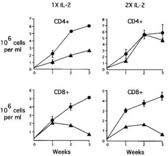
Assay 3 - The effects of CD4S on the growth of CD4+ and CD8+ T cell lines - Pooled superna-tant from Leishmania specific T1 CD4+ T cells (CD4S) had a differential effect on the growth of CD4+ and CD8+ T cell lines (Fig. 2). CD4S did not affect the growth of CD8+ cells during the first week in culture, but CD8+ cells began to die dur-ing the second week of culture with CD4S and nearly all of them were dead by four weeks. Dou-bling the concentration of rIL-2 or adding neutral-izing antibodies to INF-g, lymphotoxin or tumor
necrosis factor-a did not abrogate this effect. In
fact, extra rIL-2 inhibited CD8+ T cell prolifera-tion during the first week of culture (Fig. 3).
CD4S partially inhibited the growth of CD4+ T cell lines but did not result in cell death. The effect was completely abrogated by doubling the concentration of rIL-2 in the media to a final con-centration of 50 U/ml (Fig. 3). Experiments with a supernatant derived by antigen-stimulation of CD4+ T cell clones from the same two DTH+ adults produced similar changes in growth kinet-ics (data not shown).
DISCUSSION
An unexpected finding of this study was the discovery of two categories of VL patients, those with high and low levels of endogenous IL-10 se-cretion and proliferation, which responded differ-ently to immunomodulators. These groups may correspond to groups of patients with high and low levels of serum IL-4 (Zwingenberger et al. 1990, Holaday et al. 1993b). The existence of groups of VL patients with high and low levels of endog-enous IL-10 secretion has not been reported by other researchers who assayed PBMC at lower cell densities in less enriched media and at lower se-rum concentrations (Ghalib et al. 1993, 1995, Carvalho et al. 1994). Ghalib et al. (1995) assayed PBMC which had been frozen prior to culturing so that any PBMC activities occurring when the cells were first isolated may have been terminated before the assays were performed. Patients in this study were adults (14-58, mean = 28 years) living in both endemic and epidemic areas, rather than only adults in epidemic areas or small children. As explained below, subjects without previous exposure to Leishmania might rarely produce im-mune responses like those in the high endogenous activity group. High endogenous activity PBMC patients have been observed by members of the Núcleo de Medicina Tropical since 1989 using dif-ferent lots of sera. The most stringent conditions
TABLE V
Effects of Leishmania antigens, DS+A, and IL-12 on IL-10 secretion by an acute phase CD8+ T cell line mixed with autologous peripheral blood mononuclear cells from a recovered visceral leishmaniasis patient
IL-10 [ng/ml (mean ± SDa)] Immunomodulators added
CD8+ T Antigen Medium DS+Ab IL-12
cells added (50% v/v) (50 U/ml)
- - 0.10 ± 0.08 0.48 ± 0.06 1.28 ± 0.04
+ 0.49 ± 0 0.33 ± 0.10 0.77 ± 0
+ - 1.42 ± 0.07 0.40 ± 0.01 0.85 ± 0.01
+ 0.76 ± 0.01 0.35 ± 0.01 0.64 ± 0.05
a: standard deviation b: supernatant derived by stimulation of peripheral blood mononuclear cells from skin test positive adults with Leishmania antigens.
Fig. 2: growth of CD4+ T cells (D4/46) and CD8+ T cells (P2R/8/53) in the presence (triangles) or absence (circles) of CD4S (supernatant generated by PHA stimulation of pooled
63 63 63 63 63 Mem Inst Oswaldo Cruz, Rio de Janeiro, Vol. 94(1), Jan./Feb. 1999
were used in the assays reported here to prevent endotoxin contamination as described in Materi-als and Methods and the same sera were used in all the assays. In addition, high endogenous activities were not observed in any of ten assays of PBMC from DTH+ adults which were performed using the same media. Thus, it is safe to assume that the high endogenous levels were not the result of en-dotoxin contamination or differences in sera.
Elevated endogenous values could be explained in part by Leishmania independent immune re-sponses because poor, rural inhabitants of north-eastern Brazil are usually infected with intestinal helminths and other pathogens but the highest val-ues in this study were unique to VL patient PBMC. The high values may be correlated to disease pro-gression, similar to the positive correlation between level of endogenous IL-10 secretion by PBMC and alveolar macrophages from HIV+ subjects and CDC staging (Barcellini et al. 1994, Denis & Ghadirian 1994). Criteria for comparable staging of VL patients are not available but the fact that P14 and P23, who had been symptomatic the long-est and were not responding well to therapy, were among the high value patients supports this theory. Alternatively, more than one type of immuno-pathology may lead to delevopment of VL, simi-lar to the differences in immunopathology associ-ated with lepromatous and tuberculoid forms of leprosy (Salgame et al. 1991). For example, people
who have never been exposed to L. chagasi, such as those infected during epidemics, might develop clinical symptoms because they failed to develop protective immunity fast enough. All seven of the patients who lived in areas experiencing epidemic outbreaks (P18, P21, P26, P27, P29, P30, and P31) were in the low activity group. Adults who de-velop clinical symptoms of VL only after years of residence in an endemic area and those who have recovered from VL but relapse or are reinfected presumably already have protective immunity but may have developed symptomatic infections when their immunity was compromised by concurrent infections, allergies, malnutrition or other causes. There is evidence to support this theory. P2 had T1 or mixed T1/T2 responses to Leishmania anti-gens in assays performed one year after successful treatment for VL but had T2 responses to the anti-gens three years after treatment (Table V) (Holaday et al. 1993a). The T2 response occurred during a time when P2 suffered from undiagnosed gas-trointestinal symptoms of more than two months duration. P2 also had some asthma symptoms dur-ing this time. A DTH+ adult, D4, who had a strong T1 CD4+ T cell response to Leishmania antigens in 1990 produced PBMC which had a high endog-enous level of IL-10 secretion (0.41 ± 0 ng/ml) in 1992. Stimulation of the PBMC with Leishmania
antigens reduced IL-10 secretion (0 ± 0 ng/ml) but failed to cause a measureable increase in IFN-g
64 64 64 64
64 Immunotherapy for Visceral Leishmaniasis Bettie J Holaday
secretion. This atypical DTH+ adult response was similar to those of acute patients in the high activ-ity group and occurred at a time when the subject had been ill of undiagnosed causes for at least one month. If P2 or D4 had been infected with L. chagasi at the time they had T2 responses they might have developed symptomatic infections. The occurence of T2 responses in a treated patient and a DTH+ adult during illness suggests that immune responses to L. chagasi vary along a continuum when the immune system responds to changing demands.
Several lines of evidence suggest that an in-creased frequency or activity of a PBMC subset is responsible for the high levels of endogenous IL-10 secretion, and by correlation, proliferation also. A comparison of the y axis intercept values of high activity VL patients (0.42 ng/ml IL-10) and DTH+ adult controls (0.0 ng/ml IL-10) demonstrates that high activity patient PBMC must have had an in-creased frequency of a PBMC subset or a subset in a different activation state than the DTH+ adult controls. Acute phase CD8+ T cells, but not CD4+ T cells, have been shown to mediate increased IL-10 secretion when mixed with autologous PBMC collected after successful treatment of VL in the presence or absence of added antigen (Table V), (Holaday et al. 1993a). In addition, preliminary experiments of CD8+ T cell depletion of VL pa-tient PBMC have shown that depletion of CD8+ T cells ablates endogenous IL-10 secretion but not antigen specific IL-10 secretion (Holaday, unpub-lished observations). An increased frequency of T2 CD8+ T cells has also been observed in some CDC stage IV AIDS patients (Maggi et al. 1994).
Assays of the effects of immunomodulators on the activities of unfractionated PBMC detected two activities in DS+A which inhibited high endog-enous activities of PBMC from VL patients. Leish-mania antigens, by themselves or as part of DS+A, decreased high endogenous IL-10 secretion by VL patient PBMC. Similarly the antigens by them-selves or as part of DS+A prevented the CD8+ T cell mediated IL-10 secretion increase from occur-ring in the P2 reconstitution assay. The ability of
Leishmania antigens to lower IL-10 secretion is intriguing in light of reports of successful immu-notherapy of cutaneous leishmaniasis with Leish-mania antigens plus BCG (Convit et al. 1987, 1989). The means by which Leishmania antigens decreased IL-10 secretion are not known. IL-12 did not decrease IL-10 secretion and reduced the inhibitory effects of antigen on IL-10 secretion. INF-g has been shown to decrease IL-10 secretion
by monocytes and PBMC (Chomarat et al. 1993). However, PBMC secretion of IFN-g in response
to antigen stimulation does not appear to be
re-sponsible for the Leishmania antigens’ effect on IL-10 secretion because PBMC secretion of IFN-g
and IL-10 in response to antigen were not corre-lated in the high activity group of patients (r = +.06). Similarly, Karp et al found that levels of IL-10 and IFN-g mRNA in bone marrow of VL
patients were not inversely correlated (Karp et al. 1993).
A second DS+A activity decreased high endog-enous proliferation without decreasing antigen cific responses, effectively increasing antigen spe-cific proliferation. The effect of DS+A on prolif-eration cannot be explained by a “spent medium effect” because DS+A decreased endogenous pro-liferation only in assays of high activity VL pa-tient PBMC. It increased endogenous prolifera-tion in assays of low activity VL patient and DTH+ adult control PBMC. The identity of the factor(s) responsible for DS+A activity is not yet known but does not appear to include IL-12 or cytokines which act by reducing IL-10 secretion. If high endogenous proliferation was mediated by T2 CD4+ T cells the IFN-g present in DS+A might
explain the ability of DS+A to lower proliferation (Gajewsky et al. 1988). However, IFN-g does not
appear to have mediated the decrease in endog-enous proliferation since a constant amount of IFN-g was present in the DS+A added to each
culture but the amount of decrease in proliferation which occurred in response to DS+A varied. Also, the small but significant amount of IFN-g secreted
in response to antigen did not decrease prolifera-tion when antigen was added.
The data from the CD4S experiments show that a factor secreted by Leishmania specific T1 CD4+ T cells derived from DTH+ adults was toxic or inhibitory to CD8+ T cells but not to CD4+ T cells. The effect was not mediated by IFN-g, tumor
ne-crosis factor-a, or lymphotoxin. The factor in
CD4S is similar to that in DS+A in that it might decrease the number of CD8+ Ts cells relative to T1 CD4+ T cells. In fact the CD4S and DS+A factors might be the same. The CD4S data also suggest that the failure of DS+A to cause greater reduction in endogenous proliferation might be due to the fact that maximal inhibition or toxicity of the factors to CD8+ T cells occurred three-four weeks after exposure to the factors rather than dur-ing the first five days when the DS+A prolifera-tion assays were performed.
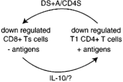
The data provide new insights into how anti-leishmania responses might be regulated in humans (Fig. 4). The model predicts that during L. chagasi
65 65 65 65 65 Mem Inst Oswaldo Cruz, Rio de Janeiro, Vol. 94(1), Jan./Feb. 1999
cells. Leishmania specific T1 CD4+ T cells can then eradicate the infection. The resulting lack of
Leishmania antigens allows CD8+ Ts cells to in-crease in number and secrete more IL-10. The CD8+ Ts cells can then prevent over-expansion and excess activity of T1 CD4+ T cells from damag-ing the body and preventdamag-ing other needed immune responses. Symptomatic infections occur when this immune regulatory cycle fails due to antagonistic demands on the immune system by concurrent in-fections, allergies, malnutrition, stress or other causes. The data suggest that Leishmania antigens and DS+A/CD4S factor(s) might be admistered to high activity patients to favor the development of T1 CD4+ T cell responses which could eradicate their infections.
antigens and the DS+A/CD4S factor(s) might be used to prevent these deaths. The same approach might be useful in treating other infections in which Ts or T2 CD8+ T cells are implicated in immuno-pathology, such as leprosy and AIDS.
ACKNOWLEDGMENTS
To AQ Sousa, SMB Jeronimo and CH Costa for sup-plying blood samples and clinical data from VL patients; TG Evans and MJ Teixeira for identifying the DTH+ adults in this study; R Pearson, R Locksley and A Vasconcelos for laboratory facilities. To Genetics Insti-tute for supplying rIL-12 and polyclonal anti-IL-12, to Hoffmann-La Roche for supplying monoclonal anti-IL-12, to Genentech for supplying anti-tumor necrosis fac-tor-a and lymphotoxin, to DNAX for supplying
anti-IL-10 and anti-IFN-g and to Cetus for supplying rIL-2.
REFERENCES
Aikat BK, Pathania AGS, Sehgal S, Bhattacharya PK, Dutta U, Pasricha N, Singh S, Parman RS, Sahaya S, Prasad LSN 1979. Immunological responses in Indian kala-azar. Indian J Med Res 70: 583-591. Barcellini W, Rizzardi GP, Borghi MO, Fain C, Lazzarin
A, Meroni PL 1994. TH1 and TH2 cytokine pro-duction by peripheral blood mononuclear cells from HIV-infected patients. AIDS 8: 757-762.
Berens R, Marr J 1978. An easily prepared defined medium for cultivation of Leishmania donovani promastigotes. J Parasitol 64: 160-162.
Carvalho E, Bacellar O, Brownell C, Regis T, Coffman RL, Reed SG 1994. Restoration of IFN-g
produc-tion and lymphocyte proliferaproduc-tion in visceral leish-maniasis. J Immunol 152: 5949-5956.
Cenini P, Berhe N, Hailu A, McGinnes K, Frommel D 1993. Mononuclear cell subpopulations and cytokine levels in human visceral leishmaniasis before and after chemotherapy. J Infect Dis 168: 986-993. Chomarat P, Rissoan M-C, Banchereau J, Miossec P
1993. Interferon-g inhibits interleukin 10 production
by monocytes. J Exp Med 177: 523-527.
Convit J, Castellanos P, Ulrich M, Castes M, Rondon A, Pinardi ME, Rodriguez N, Bloom BR, Formica S, Valecillos L, Bretana A 1989. Immunotherapy of lo-calized, intermediate, and diffuse forms of American cutaneous leishmaniasis. J Infect Dis 160: 104-115. Convit J, Rondon A, Ulrich M, Bloom B, Castellano
PL, Pinardi M E, Castes M, Garcia L 1987. Immu-notherapy versus chemotherapy in localized cuta-neous leishmaniasis. Lancet 1: 401-404.
Denis M, Ghadirian E 1994. Dysregulation of interleukin 8, interleukin 10, and interleukin 12 release by al-veolar macrophages from HIV type 1 infected sub-jects. AIDS Res Hum Retroviruses 10: 1619-1627. Dulley J, Grieve P 1975. A simple technique for
elimi-nating interference by detergents in the Lowry method of protein determination. Anal Biochem 64: 136-141.
Evans T, Teixeira M, McAuliffe I, Vasconcelos I, Vasconcelos AW, Sousa AQ, Lima JW, Pearson RD 1992. Epidemiology of visceral leishmaniasis in northeast Brazil. J Infect Dis 166:1124-1132.
Fig. 4: a new model of cross regulation of human anti-leishma-nia responses showing the role of DS+A (supernatant gener-ated from peripheral blood mononuclear cells from skin test positive adults by Leishmania antigen stimulation) and CD4S (supernatant derived by phytohemagglutinin stimulation of
Leishmania specific T1 CD4+ T cell clones derived from DTH+ adults) factor(s) in down regulating CD8+ Ts cells.
There is evidence to show that CD8+ Ts cells regulate T1 responses to other antigens in addition to L. chagasi antigens and that their activity is not always mediated by increased IL-10 secretion. Addition of acute phase CD8+ T cells to autolo-gous PBMC isolated from P2 one year after suc-cessful treatment decreased IFN-g secretion to other
antigens in addition to L. chagasi antigens. The CD8+ T cells decreased IFN-g secretion in response
66 66 66 66
66 Immunotherapy for Visceral Leishmaniasis Bettie J Holaday
Gajewski TF, Fitch FW 1988. Antiproliferative effect of IFN-g in immune regulation I. IFN-g inhibits
pro-liferation of Th2 but not Th1 murine helper T lym-phocyte clones. J Immunol 140: 4245.
Ghalib H, Piuvezam M, Skeiky Y 1993. Interleukin 10 production correlates with pathology in human Leish-mania donovani infections. J Clin Invest 92: 324-329.
Ghalib H, Whittle JA, Kubin M, Hashim FA, El-Hassan AM, Grabstein KH, Trinchieri B, Reed SG 1995. IL-12 enhances Th1-type responses in human Leish-mania donovani infections. J Immunol 154:4623-4629.
Ho M, Koech DK, Iha DW, Bryceson ADM 1983. Im-munosuppression in Kenyan visceral leishmaniasis. Clin Exp Immunol 51: 207-214.
Holaday BH, Pompeu MM, Jeronimo S, Texeira MJ, Sousa AQ, Vasconcelos AW, Pearson RD, Abrams JS, Locksley RM 1993. Potential role for interleukin-10 in the immunosuppression associated with kala azar. J Clin Invest 92: 2626-2632.
Holaday BH, Pompeu MM, Evans T, Braga DN, Texeira MJ, Sousa AQ, Sadick MD, Vasconcelos AW, Abrams JS, Pearson RD, Locksley RM 1993. Cor-relates of Leishmania-specific immunity in the clini-cal spectrum of infection with Leishmania chagasi. J Infec Dis 167: 411-417.
Hsieh C-S, Macatonia S, Tripp C, Wolf SF, O’Garra A, Murphy KM 1993. Development of Th1 CD4+ T cells through IL-12 produced by Listeria-induced macrophages. Science 260: 547-549.
Karp CL, El-Safi SH, Wynn TA, Satti MMH, Kordofani AM, Hashim FA, Hag-Ali M, Neva FA, Nutman TB, Sacks DL 1993. In vivo cytokine profiles in patients with kala-azar: Marked elevation of both interleukin-10 and interferon-gamma. J Clin Invest 91:
1644-1648.
Maggi E, Giudizi MG, Biagiotti R, Annunziato F, Manetti R, Piccinni M-P, Parronchi P, Sampognaro S, Giannarini L, Zuccati G, Romagnani S 1994. Th2-like CD8+ T cells showing B cell helper function and reduced cytolytic activity in human immunode-ficiency virus type 1 infection. J Exp Med 180: 489-495.
Manetti R, Parronchi P, Giudizi M, Piccinni M-P, Maggi E, Trinchieri G, Romagnani S 1993. Natural killer cell stimulatory factor [interleukin 12(IL-12)] in-duces T helper type 1 (Th1)-specific immune re-sponses and inhibits the development of IL-4-pro-ducing Th cells. J Exp Med 177:1199-1204. Salgame P, Abrams JS, Clayberger C, Goldstein H,
Convit J, Modlin RL, Bloom BR 1991. Differing lymphokine profiles of functional subsets of human CD4 and CD8 T cell clones. Science 254: 279-282. Uyemura K, Pirmez C, Sieling P, Kiene K, Paes-Oliveira M, Modlin RL 1993. CD4+ type 1 and CD8+ type 2 T cell subsets in human leishmaniasis have dis-tinct T cell receptor repertoires. J Immunol 151: 7095-7104.
Veress B, Omer A, Satir AA, El Hassan AM 1977. Mor-phology of the spleen and lymph nodes in fatal vis-ceral leishmaniasis. Immunology 33: 605-609. Yssel H, de Vries J, Koken M, Blitterswijk WV, Spits H
1984. Serum-free medium for generation and propa-gation of functional human cytotoxic and helper T cell clones. J Immunol Methods 72: 219-227. Zwingenberger K, Harms G, Pedrosa C, Omena S,
Sandkamp B, Neifer S 1990. Determinants of the immune response in visceral leishmaniasis: Evidence for predominance of endogenous interleukin 4 over interferon-g production. Clin Immunol