online | memorias.ioc.fiocruz.br
The genus Stenotrophomonas is widespread in the en-vironment and exists as a free-living and plant-associated organism as well as an opportunistic pathogen (Falagas et al. 2008, Nyc & Matejková 2010). Therefore, Stenotro-phomonas displays great metabolic versatility and in-traspecific heterogeneity (Ryan et al. 2009). The species Stenotrophomonas maltophilia is an important nosoco-mial pathogen (Nicodemo & Paez 2007) that is correlated with a high mortality rate among immunocompromised patients (Araoka et al. 2010). The impact of S. maltophilia as a multidrug-resistant pathogen has significantly in-creased (Hauben et al. 1999, Sanchez et al. 2009), which highlights the importance of rapid and reliable isolate identification. Moreover, endophytic Stenotrophomonas spp have an important role in plant development in a vari-ety of economically important agricultural species (Vega et al. 2005) by producing hormones, growth factors and N2 for their hosts. Some strains of this genus are used as biocontrol agents against pathogenic fungi and yeasts in plants (Minkwitz & Berg 2001). In addition, some Stenotrophomonas rhizophila strains can degrade xeno-biotic compounds and are used in the bioremediation of contaminated soils (Juhasz & Naidu 2000).
Ten species of Stenotrophomonas have been de-scribed as follows: S. maltophilia (Palleroni & Bradbury 1993), which was identified in soils, plants and in noso-comial infections (Ryan et al. 2009), Stenotrophomonas nitritireducens, which was isolated from ammonia-sup-plied biofilters (Finkmann et al. 2000), S. rhizophila, which was isolated from the rhizosphere of rape plants (Wolf et al. 2002), Stenotrophomonas acidaminiphila (Assih et al. 2002), which was originally found in a lab-scale methanogenic reactor that treated industrial waste-water, Stenotrophomonas koreensis (Yang et al. 2006), which was isolated from compost in Daejeon (South Ko-rea), and Stenotrophomonas terrae and Stenotrophomo-nas humi, which were described to encompass nitrate-reducing isolates that were obtained from soil samples in Ghent, Belgium (Heylen et al. 2007). The Stenotro- phomonas chelatiphaga strain was isolated from sewage sludge in Kazan City, Russian Federation (Kaparullina et al. 2009). The Stenotrophomonas pavanii strain was isolated from sugar cane cultivars in Brazil (Ramos et al. 2011). Recently, the Stenotrophomonas ginsengisoli strain was isolated from the soil of a ginseng field in South Korea (Kim et al. 2010).
The precise identification and classification of Stenotrophomonas remains a problem. Band pattern methods [e.g., amplified fragment length polymorphism (AFLP)] and DNA-DNA hybridisation (DDH) techniques have been used to underpin the taxonomy of this group. However, the data that are generated by these tools are often difficult to reproduce and are available only in a few laboratories (Coenye et al. 2004a). In addition, the data that are generated by these methods cannot be used Financial support: CNPq (300996/2006-7), FAPESP (2004/00814-9),
CNPq and IFS (to FLT), FAPESP (Biota Program) (to CAMF) + Corresponding author:[email protected]
Received 11 September 2010 Accepted 20 April 2011
An MLSA-based online scheme for the rapid
identification of Stenotrophomonas isolates
Patrícia Locosque Ramos1,2/+, Carlos Alberto Moreira-Filho1,2,
Stefanie Van Trappen3, Jean Swings3, Paul De Vos3, Heloiza Ramos Barbosa4,
Cristiane Carneiro Thompson5, Ana Tereza Ribeiro Vasconcelos6, Fabiano Lopes Thompson5
1Departamento de Pediatria, Faculdade de Medicina 2Centro de Pesquisas em Biotecnologia 4Departamento de Microbiologia,
Instituto de Ciências Biomédicas, Universidade de São Paulo, Av. Dr. Enéas Carvalho Aguiar 647, 01246-903 São Paulo, SP, Brasil
3Laboratory of Microbiology, Belgian Co-ordinated Collections of Micro-organisms, Bacteria Collection, Ghent University, Ghent, Belgium 5Departamento de Genética, Instituto de Biologia, Universidade Federal do Rio de Janeiro, Rio de Janeiro, RJ, Brasil
6Departamento de Matemática Aplicada e Computacional, Laboratório Nacional de Computação Científica, Petrópolis, RJ, Brasil
An online scheme to assign Stenotrophomonas isolates to genomic groups was developed using the multilocus se-quence analysis (MLSA), which is based on the DNA sequencing of selected fragments of the housekeeping genes ATP synthase alpha subunit (atpA), the recombination repair protein (recA), the RNA polymerase alpha subunit (rpoA) and the excision repair beta subunit (uvrB). This MLSA-based scheme was validated using eight of the 10 Stenotrophomo-nas species that have been previously described. The environmental and nosocomial StenotrophomoStenotrophomo-nas strains were characterised using MLSA, 16S rRNA sequencing and DNA-DNA hybridisation (DDH) analyses. Strains of the same species were found to have greater than 95% concatenated sequence similarity and specific strains formed cohesive readily recognisable phylogenetic groups. Therefore, MLSA appeared to be an effective alternative methodology to amplified fragment length polymorphism fingerprint and DDH techniques. Strains of Stenotrophomonas can be read-ily assigned through the open database resource that was developed in the current study (www.steno.lncc.br/).
to build an online electronic classification. These limita-tions can be overcome by the use of multilocus sequence typing (MLST), which uses the DNA sequence varia-tion in seven or more housekeeping genes to character-ise bacterial strains based on their unique allelic profiles (Maiden et al. 1998, Turner & Feil 2007). A simplified approach to MLST, which is widely applied in microor-ganism identification, uses only three-five housekeep-ing genes (Hanage et al. 2006, Brady et al. 2008) and is known as multilocus sequence analysis (MLSA). MLSA differs from MLST in the way the data are analysed. In MLSA, the sequence concatenation is used instead of the allelic profiles and the dataset includes multiple species collections. MLSA has been successfully used for species identification in several genera such as Vibrio (Thomp-son et al. 2005), Enterococcus (Naser et al. 2005) and the viridans group of Streptococcus (Bishop et al. 2009). The online scheme that is proposed in the current study
is based on the DNA sequencing of selected fragments of the genes coding for the ATP synthase alpha subunit (atpA), the recombination repair protein (recA), the RNA polymerase alpha subunit (rpoA) and the excision repair beta subunit (uvrB). This scheme allows for the rapid identification of Stenotrophomonas isolates through as-signment to the genomic groups of the genus (Hauben et al. 1999, Coenye et al. 2004b).
MATERIALS AND METHODS
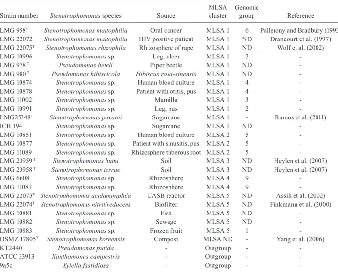
Bacterial strains and the isolation of DNA- All of the type strains of the genus Stenotrophomonas that were used for MLSA were deposited in the BCCMTM/LMG Bacteria Collection and in our own research collection at the University of São Paulo (Table I). The genomic bacte-rial DNA was isolated using the Wizard Genomic DNA Purification Kit (Promega, Madison, WI, USA: Cat. A 1120), according to the manufacturer’s instructions.
TABLE I Strains used in this study
Strain number Stenotrophomonas species Source
MLSA cluster
Genomic
group Reference
LMG 958T Stenotrophomonas maltophilia oral cancer MLSA 1 6 Pallerony and Bradbury (1993)
LMG 22072 Stenotrophomonas maltophilia HIV positive patient MLSA 1 ND Drancourt et al. (1997) LMG 22075T Stenotrophomonas rhizophila Rhizosphere of rape MLSA 1 ND Wolf et al. (2002)
LMG 10996 Stenotrophomonas sp. Leg, ulcer MLSA 1 2
-LMG 978 T Pseudomonas beteli Piper beetle MLSA 1 ND
-LMG 980 T Pseudomonas hibiscicola Hibiscus rosa-sinensis MLSA 1 ND
-LMG 10874 Stenotrophomonas sp. Human blood culture MLSA 1 4
-LMG 10878 Stenotrophomonas sp. Patient with otitis, pus MLSA 1 4
-LMG 11002 Stenotrophomonas sp. Mamilla MLSA 1 3
-LMG 10991 Stenotrophomonas sp. Leg, pus MLSA 1 2
-LMG25348T Stenotrophomonas pavanii Sugarcane MLSA 1 - Ramos et al. (2011)
ICB 194 Stenotrophomonas sp. Sugarcane MLSA 1 ND
-LMG 10851 Stenotrophomonas sp. Human blood culture MLSA 2 5
-LMG 10877 Stenotrophomonas sp. Patient with sinusitis, pus MLSA 2 5
-LMG 11089 Stenotrophomonas sp. Rhizosphere tuberous root MLSA 2 5
-LMG 23959 T Stenotrophomonas humi Soil MLSA 3 ND Heylen et al. (2007)
LMG 23958 T Stenotrophomonas terrae Soil MLSA 3 ND Heylen et al. (2007)
LMG 6608 Stenotrophomonas sp. Rhizosphere MLSA 4 9
-LMG 11087 Stenotrophomonas sp. Rhizosphere MLSA 4 9
-LMG 22073T Stenotrophomonas acidaminiphila UASB reactor MLSA 5 ND Assih et al. (2002)
LMG 22074T Stenotrophomonas nitritireducens Biofilter MLSA 5 ND Finkmann et al. (2000)
LMG 10881 Stenotrophomonas sp. Fish MLSA 5 ND
-LMG 10882 Stenotrophomonas sp. Sewage MLSA 5 ND
-LMG 10883 Stenotrophomonas sp. Frozen fruit MLSA 5 1
-DSMZ 17805T Stenotrophomonas koreensis Compost MLSA ND - Yang et al. (2006)
KT2440 Pseudomonas putida - outgroup -
-ATCC 33913 Xanthomonas campestris - outgroup -
-9a5c Xylella fastidiosa - outgroup -
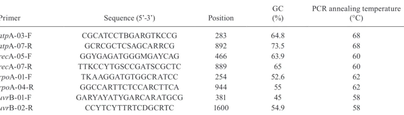
The amplification and sequencing of housekeeping and 16S rRNA genes-Approximately 50 ng of DNA were used as the PCR template for the amplification of select-ed fragments of the genes atpA, recA, rpoA and uvrB. The primers that were used in the current study and the respective annealing temperatures are listed in Table II. Because the complete Stenotrophomonas genome se-quences are only available for two of the S. maltophilia strains (NC 011071 and NC 010943) (Crossman et al. 2008), the primer design (using the software Kodon, Ap-plied Maths) included gene sequences of Xanthomonas axonopodis, Xanthomonas campestris, Xylella fastidi-osa, Pseudomonas aeruginosa, Pseudomonas putida, Pseudomonas syringae, Mesorhizobium loti, Sinorhizo-bium meliloti, Brucella melitensis, Brucella suis, Ralsto-nia solanacearum and Agrobacterium tumefaciens.
The polymerase chain reaction (PCR) for the house-keeping genes was composed of 38.2 µL of sterile MilliQ water, 1.5 µL of MgCl2 (1.5 mmol·µL-1), 5.0 µL of PCR buffer (10X), 0.4 µL of dNTPs (0.2 mmol·µL-1 each), 1.2 µL of the forward primer (20 µmol·µL-1), 1.2 µL of the reverse primer 20 µmol-1·µL, 0.4 µL of the Taq DNA Polymerase (2 U·µL-1) and 2.0 µL of the template DNA (0.05 µg·µL-1). The thermal program consisted of one cycle of 5 min at 95ºC, three cycles of 1 min at 95ºC, 2 min 15 s at 55ºC and 1 min 15 s at 72ºC, 30 cycles of 30 s at 95ºC, 1 min 15 s at 55ºC and 1 min 15 s at 72ºC and a final extension cycle of 7 min at 72ºC. The amplifica-tion of 16S rRNA was performed using 30-50 ng of DNA
in 50-μL reactions containing 2 mmol·µL-1 MgCl
2, 200
μmol·µL-1 dNTPs (each), 0.3 μmol·µL-1 of each of the uni-versal primers 27f (5’AGAGTTGATCCTGGCTCAG3’) and 1525r (5’AAGGAGGTGWTCCARCC3’) and 2U of the Taq DNA polymerase (Invitrogen) in the recom-mended buffer. The reaction mixtures were incubated in a thermal cycler Eppendorf Master Cycler Gradient (Eppendorf AG, Hamburg, Germany) at 94ºC for 2 min and then cycled 30 times at 94ºC for 1 min, 55ºC (an-nealing temperature) for 1 min and 72ºC for 3 min. A final extension at 72ºC for 10 min was used. All of the PCR products were purified using the GFX PCR DNA
and Gel Band Purification Kit (GE Healthcare, Uppsala, cat. 28-9034-70). Subsequently, 5.0 μL of the purified PCR product was mixed with 4.0 μL of the solution from the DYEnamicTM ET dye terminator kit MegaBACETM 1000 (GE Healthcare) and 1.0 μL of the sequencing primer (0.5 μmol·µL-1). The thermal program consisted of 30 cycles of 20 s at 95ºC, 15 s at 55º and 60 s at 60º. The sequencing products were purified according to the manufacturer’s instructions.
The comparison between the housekeeping genes and 16S rRNA and the phylogenetic analysis- Stenotro-phomonas bacteria were further characterised by partial sequencing of atpA (600 nt), recA (420 nt), rpoA (690 nt) and uvrB (1219 nt). The Chromas Pro 1.34 software was used to obtain the consensus sequences for the house-keeping genes and 16S rRNA. At least three reads were performed to obtain the consensus sequences. The con-sensus sequences were aligned using ClustalW (Altschul et al. 1990). The phylogenetic trees were created based on the maximum-parsimony (MP) and neighbour-joining (NJ) methods (Saitou & Nei 1987) using the software MEGA 4. We also separately analysed each of the house-keeping gene sequences and compared the resulting topologies with the topology that was obtained from the concatenated gene tree. The robustness of each topology was checked using 1,000 bootstrap replications. The type strain sequences of P. putida, X. campestris and X. fas-tidiosa were considered outliers because of their phyloge-netic similarities with those of Stenotrophomonas spp.
Split tree decomposition analysis and the Phi test were performed with SplitsTree4 (Huson 1998). The guanine-cytosine content, the ratio of the mean synony-content, the ratio of the mean synony-mous substitutions per the synonysynony-mous site to the mean non-synonymous substitutions per the non-synonymous site (ds/dn) and the recombination tests were calculated using the software package START (pubmlst.org/soft-ware/analysis/start/) (Jolley et al. 2001).
DDH-DDH experiments (Supplementary data) were performed to confirm the sequence data for the Steno-trophomonas isolates. A modification of the microplate
TABLE II
Amplification and sequencing primers used in this study
Primer Sequence (5’-3’) Position
GC (%)
PCR annealing temperature (°C)
atpA-03-F CGCATCCTBGARGTKCCG 283 64.8 68
atpA-07-R GCRCGCTCSAGCARRCG 892 73.5 68
recA-05-F GGYGAGATGGGMGAYCAG 466 63.9 60
recA-07-R TTKCCYTGSCCGATSCGCTC 889 65 60
rpoA-01-F TKAAGGATGTGGCRATCC 254 52.6 62
rpoA-04-R GGCCARTTCTCCARCTTCA 944 55 62
uvrB-01-F GARYAYATYGARCARATGCG 381 45 58
uvrB-02-R CCYTCYTTRTCDGCRTC 1600 54.9 58
method of Ezaki et al. (1989) that was described by Wil-lems et al. (2001) was used. The hybridisation temper-ature was 45ºC (calculated with the correction for the presence of 50% formamide).
Nucleotide sequence accession numbers- The gene sequences of the strains that were received from Labora- Labora-tory of Microbiology/Belgian Co-ordinated Collections of Micro-organisms, Pasteur Institute, German Collection of Microorganisms and Cell Cultures and the University of São Paulo Biomedical Sciences Institute collections were previously deposited in the GenBank. The sequence data are also available at our website www.steno.lncc.br.
RESULTS
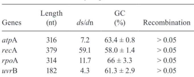
A broad collection of Stenotrophomonas strains was molecularly analysed to evaluate the usefulness of MLSA. The 16S rRNA results (data not shown) confirm that all of the strains and isolates that were used in the current work belong to the genus Stenotrophomonas, with the exception of the strains LMG 978T (Pseu-domonas beteli) and LMG 980T (Pseudomonas hibisci-cola). The genes that were analysed in the current study are typical housekeeping genes, which were shown by the relatively high ds/dn ratios and apparently have not undergone recombination (Table III). The NJ and MP methods were used to obtain the phylogenetic trees that were based on the concatenated sequences of atpA, recA, rpoA and uvrB. The strain clusters that were gen-erated were consistent with the former taxonomic stud-ies in Stenotrophomonas spp strains. overall, we found
five Stenotrophomonas genomic groups (Figure), which were named MLSA I-V, that correspond to the AFLP groups that were described by Hauben et al. (1999). The topological analysis for each of the housekeeping gene sequences yielded essentially the same strain clusters with a few differences in the relative position of some clusters (Supplementary data). The DNA-DNA related-ness experiments showed 70% DNA-DNA binding for three strains (Supplementary data), which may indicate the presence of new species or a single species that is composed of heterogenic strains. our results are clearly below the 70% DNA-DNA binding cut-off value for spe-cies delineation (Wayne et al. 1987).
TABLE III Summary of gene features
Genes
Length
(nt) ds/dn
GC
(%) Recombination
atpA 316 7.2 63.4 ± 0.8 > 0.05
recA 379 59.1 58.0 ± 1.4 > 0.05
rpoA 314 11.7 66 ± 3.3 > 0.05
uvrB 182 4.3 61.3 ± 2.9 > 0.05
ds/dn: the ratio of the mean synonymous substitutions per the synonymous site to the mean non-synonymous substitutions per the non-synonymous site; GC: guanine-cytosine content.
The MLSA I group encompassed three type strains: LMG 958T (S. maltophilia), LMG 22075T (S. rhizophila) and LMG 25348T (S. pavanii). These strains have been grouped based on the 16S rRNA sequence analysis (Hey-len et al. 2007, Ramos et al. 2011). The MLSA I group included nine other strains that were already described as members of the AFLP genomic groups 2, 3, 4 and 6 that were characterised by Hauben et al. (1999). The strains LMG 10996 and LMG 10991 also showed a high degree of similarity (84%) based on DDH results, consistent with previously reported levels (Hauben et al. 1999). The LMG 10996 strain in this group appears to be a putative new species considering our preliminary data of phenotypic tests (data not shown) and DDH results (Supplementary data). The MLSA II group contained three strains that belonged to Hauben’s AFLP group 5 (LMG 10877, LMG 10851 and LMG 11089). These strains share 98.8% of similarity in their 16S rRNA sequences (Hauben et al. 1999). The comparison of two of these strains (LMG 10877 and LMG 10851) using the DDH technique re-vealed only 58% homology (Hauben et al. 1999).
The MLSA III group harboured the two strains S. humi and S. terrae, which have 44.2% DDH similarity (Heylen et al. 2007). These strains displayed high similarity lev-els in the 16S rRNA sequences (Heylen et al. 2007). The MLSA IV group is formed by two strains (LMG 6608 and LMG 11087). These strains showed 74% and 99% homology based on the DDH and the 16S rRNA sequenc-ing, respectively. The MLSA IV group corresponded to Hauben’s genomic group 9 (Hauben et al. 1999).
The MLSA V group included the two closely related strains S. acidaminiphila (LMG 22073T) and S. nitritire-ducens (LMG 22074T), with nearly 65.8% DDH similar-ity between them (Assih et al. 2002). These two species have indistinguishable phenotypes. The MLSA V group also included the strains LMG 10883, which is a member of Hauben’s AFLP genomic group 1, and LMG 10881 and LMG 10882, which are not members of the Hauben’s AFLP genomic groups. The strains LMG 10883 and LMG 10882 were reported by Hauben et al. (1999) with a DNA-DNA binding value of 6% between both strains. However, Coenye et al. (2004b)repeated the experiment and obtained a value of 83%. These results confirm our data using the MLSA method.
DISCUSSION
The comparative genomic analysis of clinical and environmental isolates of Stenotrophomonas is of great interest. The analysis facilitates the rapid and reliable identification of these bacteria and contributes to our un-derstanding of the adaptation strategies of this genus to different niches (Ryan et al. 2009). However, the precise identification and classification of Stenotrophomonas re-mains a problem. The band pattern methods (e.g., AFLP) and DDH techniques have been used to underpin the taxonomy of Stenotrophomonas. The data that are gener-ated by these tools are often difficult to reproduce and are available only in relatively few laboratories (Coenye et al. 2004a). Moreover, this category of data cannot be used to build an online electronic classification.
Here, we proposed a rapid and cost-effective online scheme for molecular screening of the Stenotrophomo-nas isolates that was based on MLSA typing. our scheme allowed the assignment of bacterial isolates and strains into well-characterised genomic groups to estimate ge-netic distances, to measure the intraspecific diversity and to identify putative new species.
The results of the MLSA analysis confirm that there is significant diversity within Stenotrophomoas spp. More importantly, our MLSA groups were in close agreement with the genomic groups that were classified by Hauben et al. (1999). We used AFLP fingerprint, 16S rRNA se-quencing and DDH analyses. The results indicate that the partial genomic sequencing of housekeeping genes and ribosomal rRNAs provide sufficient information for the proper classification of these species.
Further studies that aim to refine and enhance the current MLSA scheme are currently under way in our laboratory. These studies will increase the number of loci and the length of sequence reads and will provide additional phenotypic information (e.g., multidrug re-sistance, adhesins, virulence factors) that are related to strains and isolates that are included in our database. Al-together, this new information should provide a reliable system of classification for this genus.
ACKNOWLEDGEMENTS
To Mrs. Thais Scudelletti, for revising and preparing our manuscript.
REFERENCES
Altschul SF, Gish W, Miller W, Myers EW, Lipman DJ 1990. Basic local alignment search tool. J Mol Biol 215: 403-410.
Araoka H, Baba M, Yoneyama A 2010. Risk factors for mortality among patients with Stenotrophomonasmaltophilia bacteremia in Tokyo, Japan, 1996-2009. Eur J Clin Microbiol Infect Dis 29: 605-608. Assih EA, ouattara AS, Thierry S, Cayol JL, Labat M, Macarie H
2002. Stenotrophomonas acidaminiphila sp. nov., a strictly aero-bic bacterium isolated from an upflow anaeroaero-bic sludge blanket (UASB) reactor. Int J Syst Evol Microbiol 52: 559-568.
Bishop CJ, Aanensen DM, Jordan GE, Kilian M, Hanage WP, Spratt BG 2009. Assigning strains to bacterial species via the internet.
BMC Biol 7: 3.
Brady C, Cleenwerck I, Venter S, Vancanneyt M, Swings J, Coutinho T 2008. Phylogeny and identification of Pantoea species associated with plants, humans and the natural environment based on multilo-cus sequence analysis (MLSA). Syst Appl Microbiol 31: 447-460. Coenye T, Vanlaere E, Falsen E, Vandamme P 2004a.
Stenotropho-monas africana Drancourt et al. 1997 is a later synonym of Ste- notrophomonas maltophilia (Hugh 1981) Palleroni and Bradbury 1993. Int J Syst Evol Microbiol 54: 1235-1237.
Coenye T, Vanlaere E, LiPuma JJ, Vandamme P 2004b. Identification of genomic groups in the genus Stenotrophomonas using gyrB RFLP analysis. FEMS Immunol Med Microbiol40: 181-185. Crossman LC, Gould VC, Dow JM, Vernikos GS, okazaki A,
Drancourt M, Bollet C, Raoult D 1997. Stenotrophomonas africana
sp. nov., an opportunistic human pathogen in Africa. IntJSyst
Bacteriol 47: 160-163.
Ezaki B, ogura T, Mori H, Niki H, Hiraga S 1989. Involvement of dnaK protein in mini-F plasmid replication: temperature-sensitive seg mu-tations are located in the dnaK gene. Mol Gen Genet 218: 183-189. Falagas ME, Valkimadi PE, Huang YT, Matthaiou DK, Hsueh PR
2008. Therapeutic options for Stenotrophomonas maltophilia
infections beyond co-trimoxazole: a systematic review. J Antimi-crob Chemother 62: 889-894.
Finkmann W, Altendorf K, Stackebrandt E, Lipski A 2000. Charac-terization of N2o-producing Xanthomonas-like isolates from bio-filters as Stenotrophomonas nitritireducens sp. nov., Luteimonas mephitis gen. nov., sp. nov. and Pseudoxanthomonas broegber- nensis gen. nov., sp. nov. Int J Syst Evol Microbiol 50: 273-282. Hanage WP, Fraser C, Spratt BG 2006. Sequences, sequence clusters and
bacterial species. Philos Trans R Soc Lond B Biol Sci 29: 1917-1927. Hauben L, Vauterin L, Moore ER, Hoste B, Swings J 1999. Genomic diversity of the genus Stenotrophomonas. Int J Syst Bacteriol 49: 1749-1760.
Heylen K, Vanparys B, Peirsegaele F, Lebbe L, De Vos P 2007.
Stenotrophomonas terrae sp. nov. and Stenotrophomonas humi
sp. nov., two nitrate-reducing bacteria isolated from soil. Int J Syst Evol Microbiol 57: 2056-2061.
Huson DH 1998. SplitsTree: analyzing and visualizing evolutionary data. Bioinformatics 14:68-73.
Jolley KA, Feil EJ, Chan MS, Maiden MC 2001. Sequence type analysis and recombinational tests (START). Bioinformatics 17: 1230-1231. Juhasz AL, Naidu R 2000. Enrichment and isolation of non-specific
aromatic degraders from unique uncontaminated (plant and fae-cal material) sources and contaminated soils. J Appl Microbiol 89: 642-650.
Kaparullina E, Doronina N, Chistyakova T, Trotsenko Y 2009.
Stenotrophomonas chelatiphaga sp. nov., a new aerobic EDTA-degrading bacterium. Syst Appl Microbiol 32: 157-162.
Kim HB, Srinivasan S, Sathiyaraj G, Quan LH, Kim SH, Bui TP, Liang ZQ, Kim YJ, Yang DC 2010. Stenotrophomonas ginsen-gisoli sp. nov., isolated from a ginseng field. Int J Syst Evol Mi-crobiol 60: 1522-1526.
Maiden MC, Bygraves JA, Feil E, Morelli G, Russell JE, Urwin R, Zhang Q, Zhou J, Zurth K, Caugant DA, Feavers IM, Achtman M, Spratt BG 1998. Multilocus sequence typing: a portable approach to the identification of clones within populations of pathogenic microorganisms. Proc Natl Acad Sci USA 95: 3140-3145. Minkwitz A, Berg G 2001. Comparison of antifungal activities and 16S
ribosomal DNA sequences of clinical and environmental isolates of Stenotrophomonas maltophilia. J Clin Microbiol 39: 139-145.
Naser SM, Thompson FL, Hoste B, Gevers D, Dawyndt P, Vancanneyt M, Swings J 2005. Application of multilocus sequence analysis (MLSA) for rapid identification of Enterococcus species based on rpoA and pheS genes. Microbiology 151: 2141-2150.
Nicodemo AC, Paez JI 2007. Antimicrobial therapy for Stenotro- phomonasmaltophilia infections. Eur J Clin Microbiol Infect Dis 26: 229-237.
Nyc o, Matejková J 2010. Stenotrophomonasmaltophilia: signifi-cant contemporary hospital pathogen. Folia Microbiol (Praha) 55: 286-294.
Palleroni NJ, Bradbury JF 1993. Stenotrophomonas, a new bacterial genus for Xanthomonas maltophilia (Hugh 1980) Swings et al. 1983. Int J Syst Bacteriol 43: 606-609.
Ramos PL, Van Trappen S, Thompson FL, Rocha RCS, Barbosa HR, De Vos P, Moreira-Filho CA 2011. Screening for endophytic nitrogen-fixing bacteria in Brazilian sugarcane varieties used in organic farming and description of Stenotrophomonas pavanii
sp. nov. Int J Syst Evol Microbiol61: 926-931.
Ryan RP, Monchy S, Cardinale M, Taghavi S, Crossman L, Avison MB, Berg G, van der Lelie D, Dow JM 2009. The versatility and adaptation of bacteria from the genus Stenotrophomonas. Nat Rev Microbiol 7: 514-525.
Saitou N, Nei M 1987. The neighbor-joining method: a new method for reconstructing phylogenetic trees. Mol Biol Evol 4: 406-425. Sanchez MB, Hernandez A, Martinez JL 2009. Stenotrophomonas
maltophilia drug resistance. Future Microbiol 4: 655-660. Thompson FL, Gevers D, Thompson CC, Dawyndt P, Naser S, Hoste
B, Munn CB, Swings J 2005. Phylogeny and molecular identi-fication of vibrios on the basis of multilocus sequence analysis.
Appl Environ Microbiol 71: 5107-5115.
Turner KM, Feil EJ 2007. The secret life of the multilocus sequence type. Int J Antimicrob Agents 29: 129-135.
Vega FE, Pava-Ripoll M, Posada F, Buyer JS 2005. Endophytic bacte-ria in Coffea arabica L. J Basic Microbiol45:371-380. Wayne LG, Brenner DJ, Colwell RR, Grimont PAD, Kandler o,
Krichevsky MI, Moore LH, Moore WEC, Murray RGE, Stack- Stack-ebrandt E, Starr MP, Truper HG 1987. Report of the ad hoc com-Report of the ad hoc com-mittee on reconciliation of approaches to bacterial systematics.
Int J Syst Bacteriol37: 463-464.
Willems A, Doignon-Bourcier F, Goris J, Coopman R, de Lajudie P, De Vos P, Gillis M 2001. DNA-DNA hybridization study of Bra-dyrhizobium strains. Int J Syst Evol Microbiol 51: 1315-1322. Wolf A, Fritze A, Hagemann M, Berg G 2002. Stenotrophomonas
rhizophila sp. nov., a novel plant-associated bacterium with anti-fungal properties. Int J Syst Evol Microbiol 52: 1937-1944. Yang HC, Im WT, Kang MS, Shin DY, Lee ST 2006.
TABLE
Percentage DNA-DNA hybridization between Stenotrophomonas strains
1a 2a 3a 4a 5a 6a 7a 8a 9a 10a 11a 12b 13b 14b 15b 16b 17b 18b 19b
1 LMG 10996 100
2 LMG 10991 84 100
3 ICB 194 64.8 - 100
4 LMG 25348TS. pavanii 58.1 - - 100
5 LMG 22073TS. acidaminiphila 17.7 - - - 100
6 LMG 23369TS. koreensis 12.3 - - - - 100
7 LMG 958TS. maltophilia 56.6 - 77.1 55.1 - - 100
8 LMG 22074TS. nitritireducens 26.3 - - 31.2 66.9 - - 100
9 LMG 23958TS. terrae 19.3 - - - - - - - 100
10 LMG 23959TS. humi 16 - - - - - - - - 100
11 LMG 24537TS. rhizophila 24.5 - - 30.7 - - - - - - 100
12 LMG 11004 65 89 - - - 100
13 LMG 11000 71 - - - 100
14 LMG 11111 - 76 - - - 100
15 LMG 978 - - - 54 - - - 100
16 LMG 980 53 - - - 55 - - - 100
17 LMG 10874 - - - 60 - - - 100
18 LMG 10878 - - - 49 - - - 100
19 LMG 10877 - - - 51 - - - 100
Neighbour-Joining phylogenetic tree based on the atpA gene sequences. Values of bootstrap after 1,000 repetitions are shown at the nodes. Pseudomonas putida, Xanthomonas campestris
and Xylella fastidiosa were used as out-groups.
Neighbour-Joining phylogenetic tree based on the recA gene sequences. Values of bootstrap after 1,000 repetitions are shown at the nodes. Pseudomonas putida and Xylella fastidiosa
Neighbour-Joining phylogenetic tree based on the uvrB gene sequences. Values of bootstrap after 1,000 repetitions are shown at the nodes. Pseudomonas putida, Xanthomonas campestris
and Xylella fastidiosa were used as out-groups.
Neighbour-Joining phylogenetic tree based on the rpoA gene sequences. Values of bootstrap after 1,000 repetitions are shown at the nodes. Pseudomonas putida, Xanthomonas campestris