Proteolytic release and partial
characterization of human
sperm-surface glycopeptides
1Laboratório de Fertilidade e Esterilidade Humana, Departamento de Bioquímica,
Instituto de Biociências, Universidade Federal do Rio Grande do Sul, 90050-170 Porto Alegre, RS, Brasil
2Departamento de Quimica Orgânica, Facultad de Ciências Exactas y Naturales,
Universidad de Buenos Aires, 1428 Buenos Aires, Argentina H. Tortorella1,
R.A. Konrath1,
M.N. Mazzini2
and A. Brandelli1
Abstract
Sperm-surface glycopeptides were obtained from intact sperm mem-branes after proteolytic release by different enzymatic treatments such as autoproteolysis, trypsin, papain and pronase. Glycopeptides were isolated, their properties and composition were examined, and their monosaccharide and amino acid constituents were characterized. The monosaccharides identified were fucose, mannose, galactose, N- ace-tylglucosamine, and N-acetylgalactosamine, which form part of more than one type of oligosaccharide units. Autoproteolytic treatment mainly provided O-glycosidic type oligosaccharides, while a mixture of O- and N-glycosidic oligosaccharides was obtained in variable proportions when treated with trypsin, papain or pronase. The highest degree of peptide cleavage was obtained with pronase. Despite the higher yields reached with trypsin, these glycopeptides contain the lowest percentage of oligosaccharide chains. Proteolytic treatment provides a simple, rapid procedure for the isolation of glycopeptides from the sperm surface.
Correspondence
A. Brandelli ICTA, UFRGS
Av. Bento Gonçalves, 9500 91501-970 Porto Alegre, RS Brasil
E-mail: [email protected]
Research supported by PROPLAN (UFRGS, Brasil), FAPERGS, UNESCO-Cono Sur, SECyT (Universidad de Buenos Aires, Argentina), and CNPq.
Received November 14, 1996 Accepted January 20, 1997
Key words •Carbohydrate •Glycoconjugate •Proteolysis •Human sperm •Peptide
Introduction
Oligosaccharides are involved in the im-munology of sperm and reproduction (1-7) and play a significant role in the sperm-oocyte interaction (8). Oligosaccharides con-stitute a singular source of biological rel-evant compounds for the knowledge of struc-tural features of sperm cell membrane or any cell membrane. Despite their involvement in reproduction and immunological infertility (1), few studies have been carried out regard-ing the chemical structure of the carbohy-drate constituents of the human sperm
mem-brane glycoproteins and their nature and com-position still remain unclear. Most of the efforts have been directed towards the iden-tification of sperm antigens by immunochemi-cal analysis of sperm cell membrane glyco-proteins (2-5).
Cytochemi-cal characterization and loCytochemi-calization of the
N-glycoside-linked oligosaccharide receptors indicated that some are cryptic ones that become exposed by the action of proteolytic enzymes, and most receptors on the acrosome are eliminated when digestion is carried out in the presence of seminal plasma (6).
In the present investigation, the glyco-peptides from human sperm membrane gly-coproteins were studied in order to deter-mine some of their chemical characteristics. These glycopeptides contain the carbohy-drate moiety of the glycoproteins, and were obtained by the action of proteolytic hydro-lases (autoproteolysis and extrinsic enzymes) on the sperm membrane.
Material and Methods
Reagents
Pronase (Streptomyces griseus protease, type XXV), papain (from papaya latex), and trypsin (from bovine pancreas, type III) were obtained from Sigma Chemical Co. All chemi-cals used were of analytical grade from Merck.
Isolation of sperm
Sperm were obtained from healthy do-nors and isolated according to the method of Saji et al. (9). Semen was diluted with five volumes of phosphate buffered saline (PBS) and centrifuged at 840 g for 10 min. The supernatant was discarded and the cells were washed three times with the same buffer.
Proteolytic release of glycopeptides from sperm membranes
Washed sperm were suspended in PBS (for autoproteolysis), trypsin (0.8 mg/ml PBS), activated papain (10) (0.8 mg/ml PBS), or pronase (160 µg/ml PBS containing 4 mM CaCl2), and then incubated at 37oC. Release of
glycopeptides was followed by periodic re-moval of aliquots (100 µl), centrifugation and
assays for changes in the protein content and liberation of free amino acids and low molec-ular weight peptides. After incubation, the total supernatant containing the glycopeptide mixture was centrifuged at 2,840 g for 10 min at 4oC, concentrated and freeze-dried.
Analytical methods
Protein analysis was carried out by the method of Lowry et al. (11). Total carbohy-drate was determined by the phenol-sulfuric acid method (12), and low molecular weight peptides by the ninhydrin method (13).
Gas-liquid chromatography (GLC)
Monosaccharide analysis was carried out as described by Reinhold (14), employing the trimethylsilyl derivatives of the corre-sponding methyl glycosides. A Hewlett-Packard 5840 gas chromatograph was used. A W-HP column was eluted with nitrogen at 29 ml/min. Monosaccharides were obtained by trifluoroacetic acid treatment for 2 h at 121oC, followed by methanolysis for 16 h at
65oC. The ratio O- versus N-glycosidic
link-ages was estimated presuming that all GalNAc is present in O-glycosidic type chains, and is reported as mol GalNAc rela-tive to Man = 3 (15,16), i.e., (O-/N-) = (GalNAc)/(Man/3).
Amino acid analysis
Amino acid analysis was performed as described elsewhere (17), using a Technicon Sequential Multi-Sample Amino Acids Ana-lyzer TM S2 and the amino acids obtained by peptide hydrolysis with 6 M HCl at 105oC
for 24 h. Amino acids were purified with Amberlite IR-120 before analysis.
Trypan blue incubation
blue in PBS, incubated at 37oC for 15 min
and centrifuged for 5 min at 900 g. The sperm pellet was washed in PBS until the supernatant became clear or pale blue (gen-erally 2 washes), and then fixed for 30 min in formaldehyde. Formaldehyde was removed by centrifuging the sperm and washing the pellet in deionized water. Fixed sperm were placed on slides and air-dried. The slides were examined under a bright-field micro-scope at 1000X magnification, and at least 100 cells/sample were evaluated for each of 2 replicates.
Gel filtration chromatography
Fractions from proteolytic treatments were dissolved in water and loaded on a column (40 x 2 cm) of Bio-Gel P-6DG, exclusion limit 6 kDa (Bio-Rad Laborato-ries), equilibrated with water. Elution was performed with water at room temperature and 5-ml fractions were collected and moni-tored for carbohydrate and protein. The pres-ence of salts was detected using the silver nitrate reagent.
Results
Sperm from normozoospermic donors were submitted to autoproteolysis and treat-ment with pronase, papain and trypsin to obtain glycopeptides from surface glycopro-teins. Proteolysis was monitored over time and aliquots were analyzed for free amino acids and low molecular weight peptides, proteins, and carbohydrates.
The production of free amino acids and low molecular weight peptides varied slightly with the type of enzyme and/or sample. The protein/peptide release showed a regular pat-tern with an increased rate during the first 4 h followed by a concomitant decrease in the production of amino acids and low molecu-lar weight peptides (see Figure 1, for trypsin-treated samples). Autoproteolysis produced larger amounts in some samples and
contin-ued without reaching a constant value as observed in the other treatments. Neverthe-less, all samples showed the expected be-havior with the enzymatic treatments, i.e., a decrease in protein content and an increase in amino acid amount against time.
The release of carbohydrate chains at different times is shown in Figure 2. Papain treatment caused the release of larger a-mounts of carbohydrates than trypsin or autoproteolysis at 4 h, although similar val-ues were reached by later incubation with trypsin. Treatment of sperm with pronase produced a release of 115 µg carbohydrates per mg total sperm protein after 1 h.
Figure 1 - Time course of pro-tein (
·
) and amino acid (à) re-lease by proteolytic treatments. The figure presents data for trypsin-treated normozoo-spermic samples as a typical pattern of peptide release. Sperm (90 x 106) wereincu-bated at 37oC in PBS containing
0.8 mg/ml trypsin for different times. Assays for amino acids/ low molecular weight peptides, and proteins were carried out by the ninhydrin and Lowry methods, as described in Mate-rial and Methods.
Protein (
·
) or amino acid
(
à
) release (mg/ml)
6
5
4
3
2
0 1
0 5 10 15 20 25
Time (h)
µg Carbohydrate/mg sperm protein
120
80
40
0
Autolysis Trypsin Papain Pronase
Figure 2 - Release of the sperm-surface glycopeptides by pro-teolytic treatments. Sperm were suspended in PBS (for autoproteolysis), trypsin (0.8 mg/ml PBS), activated papain (0.8 mg/ml PBS), or pronase (160 µg/ml PBS containing 4 mM CaCl2), and then incubated
at 37oC. Aliquots were removed
which caused lower levels of cell injury than the treatments with trypsin and papain (Fig-ure 3). After 1-h incubation with pronase, the percentage of Trypan blue-positive sperm increased by 18%. Digestion of sperm for a prolonged time with no disintegration of cells was previously observed by Mazzini et al. (6).
Samples obtained from the above treat-ments were isolated and studied. The glyco-peptide mixtures released at 4 h (treatments with trypsin, papain or autolysis) or at 1 h (pronase treatment) were analyzed by size exclusion chromatography. A similar profile of molecular weight distribution was ob-served within two main fractions: the glyco-peptide-containing fraction eluted with the void volume (exclusion limit 6 kDa) and a polydisperse fraction containing the free amino acids and low molecular weight pep-tides (data not shown). Some yields of the isolated sperm glycopeptide fractions are shown in Table 1. The carbohydrate content of the produced glycopeptides was moni-tored and calculated as shown in Table 1.
The carbohydrate composition of the oli-gosaccharide chains was analyzed. The monosaccharides found were fucose, man-nose, galactose, N-acetylglucosamine and
N-acetylgalactosamine (Table 2). Galactose was always the main constituent, with vari-able proportions of the other monosaccha-rides. Glucose was identified mainly in auto-lyzed and trypsin-treated samples.
O- versus N-linked oligosaccharide ra-tios were estimated as described in Material and Methods. As shown in Table 2, autopro-teolysis produced a molar ratio of O -glyco-sidic to N-glycosidic oligosaccharide chains of 10:1; trypsin and papain released elevated amounts of N-type oligosaccharides. When sperm were treated with pronase higher a-mounts of N-glycosidic chains were obtained (molar ratio O-/N- was 1:2).
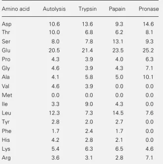
Amino acid analysis was carried out on representative samples only. Aspartate, threo-nine, serine and glutamate were the main
Figure 3 - Sperm viability after proteolytic treatments. Sperm were incubated as described in the legend to Figure 2 and then submitted to Trypan blue staining. (
·
) Autoproteolysis, (à) trypsin, (£) papain, (t) pronase.Table 1 - Release of glycopeptides from the surface of normal human sperm.
Sperm were incubated as described in the legend to Figure 2 and the glycopeptides released at 4 h (trypsin, papain or autoly-sis) or at 1 h (pronase) were analyzed. Yield is reported as mg dry weight/20 normospermic samples containing 80-140 x 106
cells. aNon-dialyzable material after autoproteolysis: 21% amino
acid/low molecular weight peptide content; small quantities of NeuNAc were also detected by the Warren assay (37). Glyco-peptides of mass higher than 6 kDa were not detected by size exclusion chromatography on Bio Gel P-6DG.
Treatment Yield Carbohydrate (%) Protein (%)
Autoproteolysis 3.5 27.2 67.0
Non-dialyzablea 3.2 >28.0 51.0
Trypsin 10.1 11.8 63.7
Papain 7.7 18.5 78.9
Pronase 5.9 25.0 71.4
% Trypan blue-positive sperm
100
80
60
40
20
0
0 5 10 15 20
Time (h)
constituents of the peptide moiety together with variable amounts of alanine, glycine, leucine, and lysine (Table 3). The percent-ages of the amino acids involved in the pro-teolysis breakdown, namely, tyrosine, lysine and arginine, were variable and usually low. Methionine was not detected.
Discussion
The sperm-surface glycopeptides from normospermic donors were examined. Sperm antigen preparations are difficult to deal with because of the somewhat rigid sperm cell membrane that resists the usual solubilization methods. For the isolation of glycopeptides we used autoproteolysis, a technique which has been previously demonstrated to be useful (6). Other types of proteolytic enzymatic degrada-tion were also investigated, such as trypsin and papain which have a more defined action, and pronase which has a wide specificity of action, cleaving glycosylated proteins of the mem-brane surface (18).
Acrosin, the sperm-specific protease in-volved in several aspects of fertilization (19,20), and trypsin possess similar pro-teolytic activity and specificity for peptide bonds which involve the basic amino acids lysine and arginine. Pronase is less specific (hydrolyzes arginine bonds and others) and differs from trypsin and other proteases since there are no known pronase inhibitors in the human sperm. Papain is commonly used for the release of ectoenzymes from membranes without incurring any loss of activity (21,22). Proteolytic enzymes other than acrosin have been extracted from sperm but are not well characterized, and activities of fibrinolytic enzymes (seminin), plasminogen activators, pepsin-like proteinase, aminopeptidase and dipeptidase have been described in seminal plasma (23). Treatments with extrinsic en-zymes also involve intrinsic proteases acti-vated by the hydrolytic conditions which characterize the autoproteolytic treatment.
The results of the enzymatic treatment,
Table 2 - Monosaccharide composition of human sperm-surface glycopeptides released by different proteolytic enzymes.
Sperm were incubated as described in the legend to Figure 2 and the glycopeptides released at 4 h (trypsin, papain or autoly-sis) or at 1 h (pronase) were analyzed. Results are reported as mol%. The molar ratio of O-glycosidic to N-glycosidic oligosac-charide chains was estimated as described in Material and Methods.
Monosaccharide Autolysis Trypsin Papain Pronase
Fucose 21.1 11.7 17.6 15.3
Mannose 4.9 16.9 9.3 26.2
Galactose 41.5 33.4 36.2 37.2
Glucose 8.2 8.4 7.9 0.8
N-Acetylgalactosamine 16.5 17.0 15.8 4.0
N-Acetylglucosamine 7.8 12.6 13.2 16.5
O-/N- ratio 10:1 3:1 5:1 1:2
Table 3 - Amino acid composition of human sperm glycopeptide fractions obtained by proteolytic treatment of normal sperm samples.
Data are reported as mol% after acid hydrolysis.
Amino acid Autolysis Trypsin Papain Pronase
Asp 10.6 13.6 9.3 14.6
Thr 10.0 6.8 6.2 8.1
Ser 8.0 7.8 13.1 9.3
Glu 20.5 21.4 23.5 25.2
Pro 4.3 3.9 4.0 6.3
Gly 4.6 3.9 4.3 7.1
Ala 4.1 5.8 5.0 10.1
Val 4.6 3.9 0.0 0.0
Met 0.0 0.0 0.0 0.0
Ile 3.3 9.0 4.3 0.0
Leu 12.3 7.3 14.5 7.6
Tyr 2.8 2.0 2.7 0.0
Phe 1.7 2.4 1.7 0.0
His 4.2 2.8 2.1 0.0
Lys 5.4 6.3 6.5 4.6
Arg 3.6 3.1 2.8 7.1
different enzymes. The differences observed in the monosaccharide composition of the sperm membrane glycopeptides released af-ter different enzymatic treatments indicate that important variations in the nature of the glycopeptides occur according to the mode of action of the enzyme(s).
The monosaccharides galactose, fucose, mannose, N-acetylglucosamine and
N-acetylgalactosamine, that are present in the sugar moiety of human sperm as indicated by lectin studies (24,25), were identified in the present study. Glucose found in consid-erable relative proportions in some cases is not a usual monosaccharide in glycoproteins (26), and therefore may indicate release of a glycolipid component from the membrane in the case of prolonged treatment (autolysis, trypsin and papain), but not in the case of degradation with pronase which is fast and effective (30-60 min) (27). The carbohy-drate composition of these glycopeptides is unlike that of either the N-glycosyl or the O -glycosyl type, since it contains GalNAc as well as Fuc and Glc, in addition to Man, Gal and GlcNAc residues. However, in the pep-tide moiety, a significant content of Asp and/ or Thr/Ser suggests a predominant propor-tion of both N- and O-glycosylated chains in the original glycoprotein. Under mild condi-tions, trypsin caused the removal of glyco-protein from the surface of Macaca radiata
sperm (28) and from human semen (29), albeit of higher molecular weight, results similar to those obtained in the present in-vestigation.
The Asp (Asp + Asn) and Ser + Thr contents of the autolysis products were 10.6 and 18.0 mol%, respectively. Hence, the O-/
N- ratio can only be 10:1 if the N- glycosyl-ated Asn is 1.8 mol%, or 17% of Asp content of the amino acid analysis. Extending the same rationale to the other treatments, the
N-glycosylated Asn is 36% for trypsin and 41.5% for papain. In the case of pronase treatment, the O-/N- ratio was estimated as 1:2, indicating a relative higher number of
N-linked carbohydrates.
The specific shape of complex oligosac-charide-protein structures located on the ex-tracellular side of membrane-bound peptides is thought to be responsible for their func-tion as carriers of significant biological in-formation and cellular recognition (30-32). The importance of these oligosaccharides in reproductive physiology has been demon-strated by several observations. The human sperm cell membrane antigen gp12, which is highly specific for sperm and seminal plasma, has been isolated from a plasma membrane fraction (9). The antigenic determinant of this molecule, recognized by the correspond-ing antibody, should be located in the sugar portion of the glycoprotein (33). On the other hand, antisperm monoclonal antibodies, re-acting to glycosylated epitopes, presented very strong properties of sperm agglutina-tion and/or immobilizaagglutina-tion (24). Finally, ga-mete interaction is mediated by protein-car-bohydrate recognition (34) and the cluster-ing of sperm receptors to the zona pellucida oligosaccharides has been proposed as a mechanism of acrosomal exocytosis induc-tion (35,36). In conclusion, the present study provides relevant information about the chemical properties of human sperm-sur-face glycopeptides.
Acknowledgments
References
1. Alexander NJ & Anderson DJ (1987). Im-munology of semen. Fertility and Sterility, 47: 192-205.
2. Czuppon AB, Mettler L, Schauer R & Pawassarat V (1981). Purification of a hu-man sperm antigen. Hoppe-Seylers Zeitschrift für Physiologische Chemie, 362: 963-968.
3. Olson GE & Gould KG (1981). Character-ization of sperm surface and seminal plasma glycoproteins of the chimpanzee.
Journal of Reproduction and Fertility, 62: 185-192.
4. Poulsen F (1983). The nature of an iso-antigen of the human sperm membrane.
Journal of Reproductive Immunology, 5: 49-54.
5. Lee CG, Lum V, Wong E, Menge AC & Huang Y (1983). Identification of human sperm antigens to antisperm antibodies.
American Journal of Reproductive Immu-nology, 3: 183-187.
6. Mazzini MN, Ceraci P, deCerezo JMS & Cerezo AS (1986). Carbohydrates of the surface of the normal human spermato-zoon. American Journal of Reproductive Immunology and Microbiology, 11: 107-111.
7. Tsuji Y, Clausen H, Nudelman E, Kaizo T, Hakomori SI & Isojima S (1988). Human sperm carbohydrate antigens defined by an antisperm human monoclonal antibody derived from an infertile woman bearing antisperm antibodies in her serum. Journal of Experimental Medicine, 168: 343-356. 8. Miller DJ & Ax RL (1990). Carbohydrates
and fertilization in animals. Molecular Re-production and Development, 26: 184-198. 9. Saji F, Minagawa Y, Negoro T, Nakamuro K & Tanizawa O (1985). A human sperm coating antigen isolated from sperm cell membrane. American Journal of Repro-ductive Immunology and Microbiology, 8: 132-136.
10. Butler PE (1990). Solubilization of mem-brane proteins by proteolysis. In: Beynon PJ & Bond JS (Editors), Proteolytic En-zymes. IRL Press, London, 193-200. 11. Lowry OH, Rosebrough NJ, Farr AL &
Randall RJ (1951). Protein measurement with the Folin phenol reagent. Journal of Biological Chemistry, 193: 267-275. 12. Dubois M, Giles KA, Hamilton KA, Rebers
PA & Smith F (1956). Colorimetric method for determination of sugars and related substances. Analytical Chemistry, 28: 350-356.
13. Moore S & Stein WH (1957). A modified ninhydrin reagent for the photometric de-termination of amino acids and related compounds. Journal of Biological Chem-istry, 211: 907-913.
14. Reinhold VN (1972). Gas liquid chromato-graphic analysis of constituent carbohy-drates in glycoproteins. Methods in Enzy-mology, 25: 244-249.
15. Montreuil J (1984). Spatial conformation of glycans and glycoproteins. Biology of the Cell, 51: 115-132.
16. Montreuil J, Bouquelet S, Debray H, Fournet B, Spik G & Strecker G (1986). Glycoproteins. In: Chaplin MF & Kennedy JF (Editors), Carbohydrate Analysis. IRL Press, Oxford, 143-204.
17. Mazzini MN, Di Giacomo MA & Cerezo AS (1987). Hydrophobic interaction chro-matography of autoproteolysis products of human seminal plasma. Biomedical Chromatography, 2: 152-155.
18. Cook GMW (1976). Techniques for the analysis of membrane carbohydrates. In: Maddy AH (Editor), Biochemical Analysis of Membranes. Chapman & Hall Ltd., Lon-don, 283-351.
19. Töpfer-Petersen E, Steinberger M, Von Eschenbach CE & Zucker A (1990). Zona pellucida-binding of boar sperm acrosin is associated with theN-terminal peptide of the acrosin ß-chain (heavy chain). FEBS Letters, 265: 51-54.
20. Eberspaecher U, Gerwien J, Habenicht UF, Schleuning WD & Donner P (1991). Activation and subsequent degradation of proacrosin is mediated by zona pellucida glycoproteins, negatively charged poly-saccharides, and DNA. Molecular Repro-duction and Development, 30: 164-170. 21. Louvard D, Maroux S, Vannier C &
Desnuelle P (1975). Topological studies on the hydrolases bound to intestinal brush border membrane. Solubilization by papain and Triton X-100. Biochimica et Biophysica Acta, 375: 236-248.
22. Maestracci D (1976). Enzymatic solubili-zation of the human intestinal brush bor-der enzymes. Biochimica et Biophysica Acta, 433: 469-481.
23. Morton DB (1977). The occurrence and function of proteolytic enzymes in the re-productive tract of mammals. In: Barrett AJ (Editor), Proteinases in Mammalian Cells and Tissues. Elsevier/North Holland, Amsterdam, 445-500.
24. Kurpisz M & Alexander NJ (1995). Carbo-hydrate moieties on sperm surface: physi-ological relevance. Fertility and Sterility, 63: 158-165.
25. Cross NL & Overstreet JW (1987). Glycoconjugates of the human sperm sur-face: distribution and alterations that ac-company capacitation in vitro. Gamete Re-search, 16: 23-35.
26. Montreuil J (1980). Primary structure of glycoprotein glycans: basis for the molec-ular biology of glycoproteins. Advances in Carbohydrate Chemistry and Biochemis-try, 37: 157-223.
27. Harrison R, Higginbotham JD & Newman R (1975). Sialoglycopeptides from bovine milk fat globule membrane. Biochimica et Biophysica Acta, 389: 449-463.
28. Nasir UD, Walker-Nasir E, Jeanloz RW & Shalev M (1980). Isolation and identifica-tion of spermatozoon-surface glycopro-teins from Macaca radiata. Carbohydrate Research, 85: C7-C9.
29. Nasir UD, Walker-Nasir E, Ajaz Z & Malghani MA (1984). Sperm-surface gly-coproteins: isolation, purification and par-tial characterization. In: Vliegerithart JGF, Karuberlinig JP & Veldirek GA (Editors),
Abstracts of the XIIth International Carbo-hydrate Symposium. Vorik Publishers, Utrecht, The Netherlands, 249.
30. Taylor ME & Drickamer K (1993). Struc-tural requirements for high affinity bind-ing of complex ligands by the macrophage mannose receptor. Journal of Biological Chemistry, 268: 399-404.
31. Zanetta JP, Kuchler S, Lehmann S, Badache A, Maschke S, Thomas D, Dufourcq P & Vicendon G (1992). Glyco-proteins and lectins in cell adhesion and cell recognition processes. Histochemical Journal, 24: 791-804.
32. Weston AS & Parish CR (1992). Evidence that mannose recognition by splenic sinu-soidal cells plays a role in the splenic en-try of lymphocytes. European Journal of Immunology, 22: 1975-1981.
33. Saji F, Minagawa Y, Ohashi K, Negoro T & Tanizawa O (1986). Further characteriza-tion of a human sperm coating antigen.
American Journal of Reproductive Immu-nology and Microbiology, 12: 13-16. 34. Macek MB & Shur BD (1988).
Protein-carbohydrate complementarity in mam-malian gamete recognition. Gamete Re-search, 20: 93-109.
35. Leyton L & Saling PM (1989). Evidence that aggregation of sperm receptors by ZP3 triggers the acrosome reaction. Jour-nal of Cell Biology, 108: 2163-2168. 36. Brandelli A, Miranda PM & Tezon JG
(1994). Participation of glycosylated resi-dues in the human sperm acrosome reac-tion: possible role of N-acetylhexosamini-dase. Biochimica et Biophysica Acta, 1120: 299-304.