online | memorias.ioc.fiocruz.br
Assessment of immunological changes in Epstein-Barr
virus co-infection in Egyptian chronic HCV patients
Sahar Shoman1/+, Mohamed Nabil1, Ashraf Tabl2, Hussam Ghanem1, Sherif El kafrawy3,4
1Department of Microbiology, Faculty of Science, Ain Shams University, Cairo, Egypt
2Department of Microbial Biotechnology, National Research Centre, Giza, Egypt 3National Liver Institute, Menufia, Egypt 4King Fahd Medical Research Canter-King Abdulaziz University, Jada, Kingdom of Saudi Arabia
Epstein-Barr virus (EBV) plays a major role in liver pathology. Similar to other members of the herpesvirus family, EBV establishes a persistent infection in more than 90% of adults. The aim of this study was to evaluate the impact of EBV and chronic hepatitis C co-infection (HCV) on biochemical and immunological responses in patients. The study was conducted in 62 patients and 33 apparently healthy controls. Patients were divided into three groups: group I, consisting of 31 patients with chronic hepatitis C infection (CHC), group II, consisting of eight patients with EBV infection and without HCV infection and group III, consisting of 23 patients with EBV and chronic HCV. The percentage of CD3+ cells, helper CD4+ cells and CD19+ B-cells was measured by flow cytometry. Human interferon-γ (IFN-γ) and interleukin (IL)-15 levels were measured by an ELISA. The levels of liver alanine aminotransferase and aspartate aminotransferase enzymes were higher in EBV/HCV patients compared to that in EBV and HCV mono-infected patients. EBV/HCV patients had significantly reduced percentages of CD3+ and CD4+ cells compared to EBV patients. Serum IFN-γ levels were significantly reduced in EBV/HCV patients (3.86 pg/mL) compared to CHC patients (6.76 pg/mL) and normal controls (4.69 pg/mL). A significant increase in serum IL-15 levels was observed in EBV/HCV patients (67.7 pg/mL) compared to EBV patients (29.3 pg/mL). Taken together, these observations suggest that HCV and EBV co-infection can potentiate immune response dampening in patients.
Key words: Epstein-Barr virus - hepatitis C virus - IFN-γ - IL-15
Epstein-Barr virus (EBV) [human herpes virus (HHV)-4)] infects a narrow range of hosts and replicates slowly. It persists in a latent state in B-lymphocytes and is thought to lead to their immortalisation and malignant
transformation (Henry et al. 2013). Similar to other her -pes viruses, EBV tends to become latent (Petrovaet al.
2010). Primary infection by EBV results in transitional
viraemia followed by a powerful T-cell adaptive immune response, which, in immunocompetent subjects, main-tains the infection in its latent state (Cohen et al. 2009).
In immunocompetent individuals, the immortalisa-tion of B-lymphocytes is associated with EBV reactiva-tion and the process is conducted by cytotoxic T lym-phocytes specific for lytic and latent antigens (Petrova
et al. 2010). EBV may also cause illness in immuno -competent people, such as viral hepatitis patients, in whom hepatitis C is the agent accountable for the ma-jority of transfusion-associated hepatitis (Yeung et al. 2007). EBV and hepatitis C co-infection (HCV) leads to higher HCV production than HCV infection alone. It has
been reported that the EBV-encoded nuclear antigen 1 (EBNA1) protein of EBV is responsible for higher HCV replication (Sugawara et al. 1999, Palma et al. 2010).
The aim of this study was (i) to investigate the cel-lular and humoral immune responses to EBV infection
doi: 10.1590/0074-0276140049
+ Corresponding author: [email protected] Received 7 February 2014
Accepted 2 June 2014
in chronic HCV patients by measuring the changes in
se-rum levels of interleukin (IL)-15 and interferon (IFN)-γ
and (ii) to study the changes in cellular immunity in these patients through the measurement of the
percent-age of total T lymphocytes CD3+, CD4+ helper cells and
CD19+ B lymphocytes.
SUBJECTS, MATERIALS AND METHODS
Study population - This study (approved by the
Ethi-cal Committee of Ain Shams University) included 99
cases collected from Cairo, Egypt (El Demerdash and El Bakri hospitals), Al Qalubia (Banha hospital), Al
Menia (El Menia hospital) and Al Monofia (Sheben El Kom hospital) from August 2011-February 2012. The 95 cases included 36 females and 59 males between 18-68
years of age. The subjects were divided into four groups:
EBV patients with HCV infection (n = 23), EBV patients without HCV (n = 8), patients with chronic hepatitis C infection (CHC) infection (n = 31) and healthy controls
[individuals negative for HCV, human immunodeficien-cy virus and hepatitis B virus antibodies, as determined
from each hospital visit (n = 33)]. Ethics approval was
obtained for the study and informed consent forms were signed by patients and healthy controls. Blood samples were drawn from all study participants and serum
sam-ples were separated and stored at -80ºC until further
testing. Blood samples for lymphocyte subset staining (immunophenotyping) were processed the same day.
Detection of HCV IgG antibodies (Abs) - Abs were as-sayed in serum samples from all studied subjects using the
using the acid guanidium
thiocyanate-phenol-chloro-form method (Chomczynski & Sacchi 1992). Primers used in the detection of HCV RNA were as follow. P1: 5’GGTGCACGGTCTACGAGACCTC3’, P2 forward primer: 5’AACTACTGTCTTCACGCAGAA3’, P3 reverse primer: 5’TGCTCATGGTGCACGGTCTA3’, nested re
-verse primer P4: 5’ACTCGGCTAGCAGTCTCGCG3’ and nested forward primer P5: 5’GTGCAGCCTCCAG-GACCC3’. All primers were purchased from Promega (Madison, USA). cDNA was synthesised by incubating 10 µL of RNA at 37ºC for 60 min with 20 U of cloned Avian myloblastosis virus reverse transcriptase, 1 × RT-buffer (Qbiogene, USA), 40 units of RNAsin (Clonetech, USA), 0.2 mmol/L each dNTP (Promega, USA) and 10 pmol primer (P1). First round amplification was performed in a total volume of 50 µL using 10 µL of cDNA, 10 pmol of each of the primers P2 and P3, 0.2 mmol/L of each dNTP
(Promega), two units of Taq DNA polymerase (Promega)
and 1 × Taq buffer. The second round of amplification was
similar to the first, except for using the nested primers P4
and P5 and 10 µL of the first round PCR product as tem -plate. PCR cycling conditions for both rounds consisted
of 30 cycles of 1 min at 94ºC, 1 min at 55ºC and 1 min at 72ºC. The nested PCR products were separated by elec -trophoresis on a 2% ethidium bromide stained agarose gel and visualised under ultraviolet light.
Serological analysis of EBV infection - Human EBV IgM antibodies were detected in all samples by the
qual-itative ELISA test using commercially available EBV kits (Diagnostic Automation, USA). EBV-IgG antibod -ies were detected using commercially available kits
(AT-LAS Medical EBV-IgG Kit, UK) according to the manu
-facturer’s instructions. The results of EBV IgM and IgG
measurements were expressed as optical density units.
Detection of EBV-DNA - Viral nucleic acid DNA was
extracted from 300 μL of serum using the Wizard® DNA
purification mini kit (Promega) following the
manufac-turer’s instructions. For the detection of EBV DNA, nest -ed PCR of the serum samples was perform-ed according to
previously established protocols (Kapranos et al. 2003). The 25-μL qualitative PCR reaction mixture contained 2.5 μL of 10x buffer (10 mM Tris-HCl pH 8.0, 50 mM KCl, 25 mM MgCl2), 0.5 μL of 50 mM dNTP mix, 10 pmol of
primers E2P1 (5’ATCCTTGCACTTAGCCAAGC3’) and E2P2 (5’TCCAGATGTGTCTCCCTTCT3’) (Bioneer, USA) for the amplification of a 556-bp fragment in the EBNA-2 gene, 5 μL of DNA solution (DNA template), 14.15 μL of distilled water and 0.1 μL (2U) of Taq DNA
polymerase (Bioneer). Nested PCR was performed ac-cording to the following thermal cycling protocol:
pre-denaturation at 94ºC for 5 min, followed by 35 cycles of 94ºC for 30 s, annealing at 57ºC for 30 s and extension at 72ºC for 60 s and final extension at 72ºC for 4 min. The
second round of the nested PCR was conducted using the same thermal cycling conditions as described above and
Nested amplification products were visualised by 2% aga-rose gel electrophoresis and ethidium bromide staining.
Liver enzyme levels - Alanine aminotransferase
(ALT) (normal range is up to 40 U/L) and aspartate ami
-notransferase (AST) (normal range is up to 38 U/L) lev -els were measured in all samples using commercial kits
(Siemens Healthcare Diagnostic Inc, USA) according to
the manufacturer instructions.
Measurement of serum cytokine levels - Human IFN-γ and IL-15 were measured using commercially available ELISA kits (Human IFN-γ ELISA kit, Bender MedSys
-tems, Austria, and Human IL-15 ELISA kit, Ray Bio -tech®, USA) according to the manufacturer’s instructions.
The results are presented as the concentration (pg/mL). Measurement of CD4+, CD3+ and CD19+ percentages in whole blood by flow cytometry - The percentage of
to-tal CD3+ T lymphocytes, CD4+ helper T cells and CD19+
B lymphocytes in whole blood was determined by direct staining with the following conjugated monoclonal
an-tibodies: anti-CD3+ fluorescein isothiocyanate (FITC),
anti-CD4+ FITC and anti-CD19+ phycoerythrin. The
percentages were determined using a flow cytometer (Coulter® Epics® XLTM, USA) and the data were analy
-sed using system IITM software (Flow cytometry Core
Laboratory, El-Demerdash Hospital, Cairo)
Statistical analysis - All statistical analyses were
per-formed using GraphPad InStat statistical software program. Differences were considered significant when p ≤ 0.05.
RESULTS
Based on the HCV-RNA RT-PCR (Fig. 1) and
EBV-DNA PCR (Fig. 2) results, the study population was divided into four groups. The first group, representing
32.6% of the total, included 31 HCV infected patients (15 males and 16 females) between 17-63 years of age, with a mean age of 40.03 ± 11.1 years. The second group, rep
-resenting 8.4% of the total, included eight EBV-infected patients (4 males and 4 females) between 18-58 years of age, with a mean age of 36.8 ± 15.1 years. The third group, representing 24.2% of the total, included 23 cases (18 males and 5 females) with both EBV and HCV infec
-tion in the age range of 22-68 years, with a mean age of 50.5 ± 15.1 years. The fourth group, representing 34.7% of the total, included 33 individuals (22 males and 11
females) negative for EBV and HCV infection (normal
healthy control group) whose age range was between 18-46 years, with a mean age of 31.1 ± 8.2 years.
increase in AST levels was highly significant (p < 0.001) in the EBV/HCV (93.9 ± 11.39 U/mL), HCV (75.4 ± 16.5 U/mL) and EBV (51.5 ± 8.3 U/mL) patients compared to the NC (29.5 ± 14.2) (Fig. 3).
Serum levels of human IL-15 - Serum human IL-15 levels were significantly higher (p < 0.001) in the EBV/ HCV (67.8 ± 21.1 pg/mL) and HCV-infected (47.2 ± 7.7 pg/mL) patients compared to the NC group (9.5 ± 11.1 pg/ mL). In addition, IL-15 levels were significantly higher in the EBV/HCV patients than in the EBV-infected pa
-tients (29.3 ± 17.2 pg/mL). No significant difference was
observed between the EBV-infected and HCV-infected patients (Fig. 4).
Serum level of human IFN-γ - Human IFN-γ levels in the serum were significantly (p < 0.001) elevated in HCV patients (6.5 ± 2.3 pg/mL) compared to the HCV/ EBV patients (3.4 ± 1.01 pg/mL), the EBV patients (2.9 ± 0.8 pg/mL) and the NC (4.9 ± 1.2 pg/mL). IFN-γ se
-rum levels were also significantly lower (p < 0.05) in the EBV and EBV/HCV patients compared to the NC.
No significant difference was detected between the EBV
and EBV/HCV groups (Fig. 5).
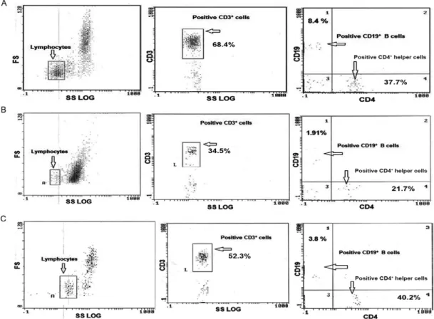
Flow cytometry analysis - The percentage of CD3+,
CD4+ and CD19+ lymphocytes was measured by flow
cytometry and found to be lower by a statistically
sig-nificant amount in the EBV/HCV patients compared to
the healthy individuals. The mean percentage of total T
lymphocytes (CD3+ cells) was significantly lower in the
HCV/EBV patients (27.8 ± 11.07) compared to the chronic HCV patients (46.7 ± 4.6), the EBV patients (50.5 ± 4.3) and the NC (65.2 ± 8.1). The percentage of T-helper cells
(CD4+ cells) was also significantly lower in the HCV/
EBV (15.75 ± 1.3), chronic HCV (25.08 ± 2.4) and EBV patients (32.01 ± 3.01) compared to the NC (43.8 ± 3.6). Additionally, CD19+ B lymphocytes were significantly
reduced in number in the HCV/EBV (2.41 ± 1.2), EBV (2.5 ± 1.3) and chronic HCV patients (4.7 ± 1.19) com
-pared to the NC group (8.2 ± 1.5), as shown in Figs 6, 7.
DISCUSSION
Virus-to-virus interactions are reported to modify the progression of viral infections in humans (Palma et
al. 2010). The classic hepatotropic viruses, hepatitis A
through E, are not the only viral agents capable of infect-ing the liver and causinfect-ing hepatitis as a part of an organ-specific or systemic involvement with hepatic injury ranging from elevations in aminotransferases to acute hepatitis with or without acute liver failure and fulminant hepatitis. Their ability to cause chronic liver disease has not been fully proven. Cytomegalovirus (CMV), EBV, herpes simplex virus, varicella-zoster virus and adeno-viruses have also been shown to be hepatotropic (Galle-gos-Orozco et al. 2010). Epstein-Barr viral infection is characterised by alternating periods of latency and re-activation. The reactivation of the virus is observed dur-ing periods of immune system down-regulation, such as
Fig. 1: nestedreverse transcription-polymerase chain reaction re-sults of serum samples. Lanes 2, 3, 5, 7: positive for hepatitis C virus (HCV) RNA; 1, 4, 6: negative for HCV RNA.
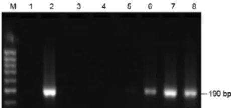
Fig. 2: nested polymerase chain reaction results of serum samples. Lanes 2, 6-8: positive for Epstein-Barr virus (EBV) DNA; 1, 3-5: negative for EBV DNA.
Fig. 3: alanine aminotransferase (ALT) and aspartate aminotrans -ferase (AST) activity levels among study subjects. EBV: Epstein-Barr virus; HCV: hepatitis C virus; NC: normal controls.
Fig. 4: interleukin (IL)-15 activity levels among study subjects. EBV: Epstein-Barr virus; HCV: hepatitis C virus; NC: normal controls.
during drug treatment and illness-related stress or dur-ing co-infection with various pathogens (Gandhi et al. 2004, Gredmark et al. 2007).
Consistent with previous reports, our results showed
that the activity of liver enzymes (ALT and AST) was sig
-nificantly increased in the chronic HCV patients (p < 0.001) and in the EBV-infected group (p < 0.01) compared to the control group (Herrine 2002, Hinedi & Koff 2003, Mis
-siha et al. 2008). The observed higher serum ALT and AST levels in the EBV/HCV patients compared to the chronic HCV or EBV mono-infected patients (p < 0.001) suggest
a synergistic effect of EBV co-infection on liver cell
dam-age. Similar results have also been reported for chronic
HCV patients infected with other HHVs, such as CMV and
HHV-6 (Claudio et al. 1999, Petrova et al. 2010).
IL-15 plays an important role in the immune system
and is especially important in the activation of the in-nate and tissue-associated immune responses because it promotes the activation, proliferation and survival of natural killer and CD8+ memory T-cells (Golden-Mason
& Rosen 2006). In this study, the highly significant (p < 0.001) increase in serum IL-15 levels in chronic HCV
patients compared to the NC indicated the degree of liver tissue damage caused by HCV, which is in agreement
with previous reports (Budhu & Wang 2006). These re -sults also indicate the synergetic effect of double
infec-tion on IL-15 levels due to increased liver damage (Xu et al. 2000, Ohga et al. 2001, Kimura et al. 2005).
in IFN-γ in CHC cases may be due to HCV viral replica -tion and disease progression, as observed in previous
re-ports (Lechmann et al. 1999, Budhu & Wang 2006, Posta
et al. 2009). Other studies have reported a reduction in
IFN-γ production in EBV-infected cases (Hislop et al. 2007, Saghafian et al. 2013); this reduction in produc
-tion can account for the gradual decrease in IFN-γ levels
upon the introduction of EBV infection. This observa-tion is consistent with our results demonstrating that the
highest level of IFN-γ was observed in the CHC group followed by the CHC/EBV co-infected group; the low
-est IFN-γ level was observed in the EBV mono-infected
cases. EBV infection has evolved multiple mechanisms for disrupting IFN-stimulated Janus kinases (JAK)
sig-nal transduction. EBV infection inhibits the IFN-γ re
-sponse by decreasing the JAK1 protein, which is a nec
-essary component of IFN-γ signalling. Using a variety
of such mechanisms, EBV is able to escape antiviral immune responses throughout the primary infection
pe-riod, as demonstrated by Morrison et al. 2001, Ramana et al. 2002 and Ressing et al. 2008.
In this study, total T lymphocytes (CD3+ cells) were
significantly lower (p < 0.05) in CHC patients than in
controls. These findings are in agreement with Iken et
al. (2006) who observed an unusually low frequency of
HCV-specific T-cells in the liver and peripheral blood of CHC patients. Moreover, Ciccaglione et al. (2007) de-duced that HCV persistence in most infected individu-als is associated with the ability of the virus to evade the host immune response at local and systemic levels and that the virus is capable of replicating in immune cells, effector cells and hepatocytes, which are the main
target of HCV replication. Shete et al. (2010) previously reported that the CD3+ T-cell count was lower in
EBV-infected patients compared to control individuals. Our results also showed a significant decrease in the percent-age of CD4+ cells in the CHC, HCV/EBV, HCV and EBV
patients compared to the NC group. These results were
consistent with another study by Gonzalez et al. (2008),
showing that the CD4+ count and percentage were
sig-nificantly decreased in HCV infected patients compared
to healthy individuals. Mozer-Lisewska et al. (2006) and Harcourt et al. (2006) reported that the percentage
of CD4+ cells was decreased in HCV cases compared to
control groups, while other studies reported a reduction in CD4+ T cells in patients with EBV-associated
Hodg-kin’s disease (Karcheva et al. 2008, Shete et al. 2010).
The enumeration of B-cells in this study indicated a significant difference in the percentage of B-cells
(CD19+) between the CHC and control groups. Durand
et al. (2010) suggested that CD19+ B-cells were
signifi-cantly reduced in cirrhotic patients with or without hepa-tocellular carcinoma (HCC). However, HCV induces a powerful antibody response to its envelope glycoprotein E2. Because E2 binds to B-cells via CD81, it is possible
that antibodies to E2 block the binding of HCV to cells,
thus protecting against HCV infection in some cases
(Merani et al. 2011). Once E2 binds to B-cells via CD81, it associates with CD19 and CD21, forming a complex that lowers the activation threshold (Ito et al. 2011). In this study, the percentage of CD19+ cells in
HCV/EBV-infected cases was lower than that in the EBV-HCV/EBV-infected cases, the CHC patients and the controls. The differences
in CD19+ counts between these cases were statistically
significant. Yang et al. (2005) demonstrated a visible de
-crease in CD19+ expression in EBV-infected cases
com-pared to normal B lymphocytes. Karcheva et al. (2008)
showed that acute EBV infection is characterised by a
decrease in CD19 (B-cell) counts.
Previous studies have reported that EBV is responsi-ble for the increased replication of HCV in chronic HCV
patients through EBNA1 (Sugawara et al. 1999, Morrison et al. 2001), suggesting that EBV could be involved in the development of HCC (Sugawara et al. 2000, Petrova et al. 2010). This observation suggests an urgent need for a management protocol for EBV/HCV co-infected pa -tients and especially in the context of the study by Bader
El-Din et al. (2011), who reported that co-infection with
CMV, which is a member of the same family as EBV, complicates the effectiveness of antiviral therapy in the majority of chronic HCV cases.
In conclusion, we recommend introducing EBV treatment in chronic HCV patients to prevent rapid de-terioration and to slow the progression to liver
cirrho-sis and HCC. It is important to measure the efficiency
of immunological parameters (CD3+, CD4+ and CD19+
cells) and their related cytokines (IFN-γ and IL-15) in
the peripheral blood and serum of HCV-infected patients before, during and after full-term antiviral therapy to as-sess their prognostic significance.
ACKNOWLEDGEMENTS
To the managers of the hospitals reported in this paper, for their permission to collect blood samples, and to the Depart-ment of Microbial Biotechnology, National Research Center, for support to execute this research work.
REFERENCES
Bader El-Din NG, El-Meguid MA, Tabll AA, Anany MA, Esmat G, Zayed, N, Helmy A, El-Zayady AR, Barakat A, El-Awady MK 2011. Human cytomegalovirus infection inhibits response of chronic hepatitis C virus infected patients to interferon-based therapy. J Gastroentero Hepat26: 55-62.
Budhu A, Wang XW 2006. The role of cytokines in hepatocellular carcinoma. J Leukoc Biol80: 1197-1213.
Chomczynski P, Sacchi N 1992. Single-step method of RNA isolation by acid guanidium-thiocyanatephenol-chloroform extraction.
Anal Biochem162: 156-159.
Ciccaglione AR, Srellacci E, Marcantonio C, Muto V, Equestre M, Mar -silli G, Rapicetta M, Battistini A 2007. Repression of interferon reg-ulatory factor 1 by hepatitis C virus core protein result in inhibition of antiviral and immunomodulatory genes. J Virol81: 202-214.
tional meeting, 8-9 September 2008. Ann Oncol 20: 1472-1482. Durand TG, Di-Liberto H, Colman E 2010. Occult infection of periph
-eral B cells by hepatitis C variants which have low translational efficiency in cultured hepatocytes. Gut 59: 934-942.
Gallegos-Orozco JF, Rakela-Broder J 2010. Hepatitis viruses: not al -ways what it seems to be. Rev Med Chile138: 1302-1311.
Gandhi MK, Tellam JT, Khanna R 2004. Epstein-Barr virus-associat-ed Hodgkin’s lymphoma. Br J Haematol125: 267-281.
Golden-Mason L, Rosen HR 2006. Natural killer cells: primary target for hepatitis C virus immune evasion strategies? Liver Transplant
12: 363-372.
Gonzalez VD, Falconer K, Michaelsson J, Moll M, Rechard O, Alaeus A, Sandberg JK 2008. Expansion of CD56- NK cells in chronic
HCV/HIV-1 co-infection: reversion by antiviral treatment with pe -gylated interferon-gamma and ribavirin. Clin Immunol128: 46-56.
Gredmark S, Jonasson L, Van Gosliga D, Ernerudh J, Soderberg-Nau -cler C 2007. Active cytomegalovirus replication in patients with coronary disease. Scand Cardiovasc J41: 230-234.
Harcourt G, Gomperts E, Donfield S, Klenerman P 2006. Profound loss of HCV-specific interferon-gamma secreting CD4+ T-cells in
HIV/HCV co-infected patients. Gut 55: 1484-1487.
Henry H, Balfour JR, Priya V 2013. Primary Epstein–Barr virus in -fection: impact of age at acquisition, co-infection and viral load.
J Infect Dis207: 1787-1789.
Herrine SK 2002. Approach to the patient with chronic hepatitis C virus infection. Ann Intern Med136: 747-757.
Hinedi TB, Koff RS 2003. Cholestatic hepatitis induced by Epstein-Barr virus infection in an adult. Dig Dis Sci 48: 539-541. Hislop AD, Taylor G, Sauce D, Rickinson AB 2007. Cellular responses
to viral infection in humans: lessons from Epstein-Barr virus.
Annu Rev Immunol25: 587-617.
Iken K, Huang L, Bekele H, Schmidt EV, Koziel MJ 2006. Apoptosis of activated CD4+ and CD8+ T-cells in enhanced by co-culture
with hepatocytes expressing hepatitis C virus structural proteins through FasL induction. Virol346: 363-372.
Ito M, Kusunoki H, Mochida K, Yamaguchi K, Mizuochi T 2011. HCV infection and B-cell lymphomagenesis. Adv in Hematol
2011: 835314.
Kapranos N, Petrakou E, Anastasiadou C, Kotronias D 2003. Detec -tion of herpes simplex virus, cytomegalovirus and Epstein-Barr virus in the semen of men attending an infertility clinic. Fertil Steril79 (Suppl.): 1566-1570.
Karcheva M, Lukanov Tz, Gecheva S, Slavcheva V, Veleva G, Nachev R 2008. Infectious mononucleosis - Diagnostic potentials. J Imab 93: 431-438.
Kimura H, Hoshino Y, Hara S, Sugaya N, Kawada J, Shibata Y, Kojima S, Nagasaka T, Kuzushima K, Morishima T 2005. Dif -ferences between T cell-type and natural killer cell-type chronic active Epstein-Barr virus infection. J Infec Dis 191: 531-539.
Lechmann M, Woitas RI, Langhans B, Kaiser R, Ihlenfeldt HG, Jung G, Sauerbruch T, Spengler U 1999. Decreased frequency of HCV
Missiha SB, Ostrowski M, Heathcote EJ 2008. Disease progression in chronic hepatitis C: modifiable and non-modifiable factors. Gas-troenterol134: 1699-1714.
Morrison TS, Mauser A, Wong A 2001. Inhibition of IFN-γ signaling by EBV immediate-early protein. Immunity15: 787-797.
Mozer-Lisewska I, Dworackiy G, Kaczmarekz E, Sluzewski W, Kacz -marky M, Wozniakz A, Zeromskiy J 2006. Significance of altera -tions in PBMC immunophenotype of children with chronic viral hepatitis C - the role of dendritic cells. Scand J Immun63: 311-319.
Ohga S, Nomura A, Takada H 2001. Epstein-Barr virus (EBV) load and cytokine gene expression in activated T cells of chronic ac-tive EBV infection. J Infect Dis183: 1-7.
Palma T, Doonan PB, Trager NN, Kasman LM 2010. A systematic approach to virus-virus interactions. Virus Res 149: 1-9.
Petrova M, Kamburov V, Nikolovska D, Kosseva O, Nikolova M, Krastev Z 2010. Epstein-Barr virus: is there any contribution to chronic hepatitis B and C? Liver Int30: 488-489.
Posta JJ, Ratnarajah S, Lloyd AR 2009. Immunological determinants of the outcomes from primary hepatitis C infection. Cell Mol Life
Sci66: 733-756.
Ramana CV, Gil MP, Schreiber RD, Stark GR 2002. Stat1-dependent and independent pathways in IFN-gamma-dependent signaling.
Trends Immunol 23:96-101.
Ressing ME, Horst D, Griffin BD, Tellam J, Zuo J, Khanna R, Rowe M, Wiertz EJ 2008. Epstein-Barr virus evasion of CD8(+) and
CD4(+) T cell immunity via concerted actions of multiple gene
products. Semin Cancer Biol18: 397-408.
Saghafian HS, Sundstro Y, Sohlberg E, Nilsson C, Linde A, Troye BM, Berg L, Sverremark EE 2013. Herpesvirus seropositivity in childhood associates with decreased monocyte-induced NK cell IFN-γ production. J Immunol182: 2511-2517.
Shete A, Thakar M, Abraham PR, Paranjape R 2010. A review on peripheral blood CD4+ T lymphocyte counts in healthy adult
In-dians. Ind J Med Res132: 667-675.
Sugawara Y, Makuuchi M, Kato N, Shimotohno K, Takada K 1999. Enhancement of hepatitis C virus replication by Epstein-Barr virus-encoded nuclear antigen 1. EMBO18: 5755-5760
Sugawara Y, Makuuchi M, Takada K 2000. Detection of Epstein-Barr virus DNA in hepatocellular carcinoma tissues from hepatitis C-positive patients. Scand J Gastroenterol35: 981-984.
Xu J, Ahmad A, Jones JF 2000. Elevated serum transforming growth factor b1 levels in Epstein-Barr virus-associated diseases and their correlation with virus-specific immunoglobulin A (IgA) and IgM. J Virol 74: 2443-2446.
Yang W, Agrawal N, Patel J, Edinger A, Osei E, Thut D, Powers J, Meyerson H 2005. Diminished expression of CD19 in B-cell lym -phomas. Cytometry - Part B. Clin Cyto63 B: 28-35.