Decreased Interleukin-10 Responses in
Children with Severe
Mycoplasma
pneumoniae
Pneumonia
Shenggang Ding1☯, Xiaowu Wang2,4☯, Wei Chen2, Yuan Fang2, Boyu Liu2, Yan Liu2, Guanghe Fei3*, Linding Wang2*
1Department of Pediatrics, the First Affiliated Hospital of Anhui Medical University, Hefei, China,
2Department of Microbiology, Anhui Medical University, Hefei, Anhui, China,3Department of Respiratory Medicine, First Affiliated Hospital of Anhui Medical University, Hefei, Anhui, China,4Department of Clinical Laboratory, Fuyang Second People’s Hospital, Fuyang, Anhui, China
☯These authors contributed equally to this work.
*[email protected](LW);[email protected](GF)
Abstract
Several cytokines may play roles in the immunological pathogenesis of mycoplasmal pneu-monia caused byMycoplasma pneumoniae. In this study, we investigated serum cytokine profiles in children with mycoplasmal pneumonia. The serum levels of interleukin (IL)-8, IL-10, and IL-18 were examined using ELISA kits in 34 patients withM.pneumoniaeinfection (Group 1, 11 with severe mycoplasmal pneumonia; Group 2, 13 with mild mycoplasmal pneumonia; Group 3, 10 with asthma) and 32 age-matched, non-infected controls. The serum levels of IL-8, IL-10, and IL-18 increased significantly in patients with mycoplasmal pneumonia compared with those in controls (P<0.01). The serum levels of IL-10 decreased significantly in Group 1 compared with those in Group 2 (P<0.01). The serum levels of IL-18 increased significantly in Group 1 compared with those in Group 2 (P<0.01). The serum lev-els of IL-10 and IL-18 decreased significantly in 10M.pneumoniae-infected patients with asthma compared with those in 24M.pneumoniae-infected patients without asthma (P<0.01). We examined the level of interleukins (IL-8, IL-10 and IL-18) after the patients
started therapy. The data showed that IL-18 were lower after therapy (P<0.01). Collectively,
our data suggested that these cytokines may be involved in the pathogenesis of mycoplas-mal pneumonia.
Introduction
Mycoplasma pneumoniaeis recognized as one of the most important pathogens in
commu-nity-acquired pneumonia (CAP) in young adults and children [1].Mycoplasma pneumoniae
gives rise to mycoplasmal pneumonia, also called atypical pneumonia, which has been shown
by many physicians to have different clinical features. It accounts for 7–20% of CAP [2]. Many
studies showed that atypical pneumonia might have a higher incidence in patients with mild
a11111
OPEN ACCESS
Citation:Ding S, Wang X, Chen W, Fang Y, Liu B, Liu Y, et al. (2016) Decreased Interleukin-10 Responses in Children with SevereMycoplasma pneumoniaePneumonia. PLoS ONE 11(1): e0146397. doi:10.1371/journal.pone.0146397
Editor:Xue-jie Yu, University of Texas Medical Branch, UNITED STATES
Received:September 15, 2015
Accepted:December 16, 2015
Published:January 11, 2016
Copyright:© 2016 Ding et al. This is an open access article distributed under the terms of the
Creative Commons Attribution License, which permits unrestricted use, distribution, and reproduction in any medium, provided the original author and source are credited.
Data Availability Statement:All relevant data are within the paper and its Supporting Information files.
Funding:This work was supported by National Science Foundation of China (Grant No. 81271837, 81571963) and Natural Science Foundation Key project of Anhui Education Department (KJ2012A161, KJ2015A020). The funders had no role in study design, data collection and analysis, decision to publish, or preparation of the manuscript.
symptoms, and atypical pneumonia is not only considered to cause respiratory disease, but also severe diseases including neurological, dermatological, hematological, renal,
gastrointesti-nal, and musculoskeletal diseases [3]. Further reports showed that the clinical parameters of
atypical pneumonia were different in patients withoutM.pneumoniapneumonia, and
symp-toms include fevers, crackles, thrombocytosis, consolidation, sore throat, headache, skin rash, ear infection. Although the pathogenesis of mycoplasmal pneumonia is not clear at present, these manifestations may result from the immune response to infection and/or the ability ofM.pneumoniaeto directly invade the ciliated airway epithelial cells in the respiratory tract [2–4].
Researchers have found that the cell-mediated immune response of the host plays an
impor-tant role in the mechanism of mycoplasmal pathogenicity, specifically Th1-type cytokines [5].
IL-8, IL-18 and IL-10 level in serum were higher in people with mycoplasmal pneumonia
[6–13]. Measurement of their levels can help understand conditions of the patients. Recent
investigations have shown thatM.pneumoniaelysates may increase interleukin-8 (IL-8) levels
in human airway epithelial cells, may contribute to neutrophilic asthma [5,6,8]. Several studies
showed thatM.pneumoniaeinfection in children induced IL-18, which may be a central factor
in the immunopathologic response [10,11,13]. IL-18, a Th1-type cytokine, was originally
des-ignated interferon gamma (IFN-γ)-inducing factor. This cytokine was first reported to be
pro-duced by Kupffer cells and activated macrophages, and it was shown to be a critical factor in
inducing liver injury in mice [9]. IL-18 serums level in adult with severe mycoplasmal
pneumo-nia was significantly higher than those in patients with mild cases, suggesting that pulmonary
lesions may be modulated inM.pneumoniaeinfected humans [13]. Chung et al. studied 75
children with mycoplasmal pneumonia and divided them into asthmatic and non-asthmatic groups. They found that children with asthma exhibited a deficient IL-18 response and had
more severe pneumonia [6]. Oishi et al. measured serum IL-18 level in 23 acute or convalescent
patients and demonstrated that there was a significant correlation between the level of IL-18
and lactate dehydrogenate (LDH) in serum [11]. They thought that the LDH level can be used
to estimate the IL-18 level. A gnotobiotic mouse model ofM.pneumoniaepneumonia was
established, which showed that cytokines, including IL-4, IL-10 and IFN-γ, were significantly
increased in mice that were repeatedly infected withM.pneumonia[7,12]. However, few
researchers have examined IL-10 level in serum in severeM.pneumoniaeinfections in children
patients. The aim of this study was to investigate IL-10 level in the serum of children infected withM.pneumoniaepneumonia. At the same time, the correlation between the cytokines (IL-8, IL-10 and IL-18) and the other clinical factors was analyzed. We found that the serum levels of IL-10 decreased significantly in patients with severe mycoplasmal pneumonia compared
with those with mild mycoplasmal pneumonia (P<0.01). The serum levels of IL-10 and IL-18
decreased significantly in 10M.pneumoniae-infected patients with asthma compared with
those in 24M.pneumoniae-infected patients without asthma (P<0.01). Our data showed that
both IL-8 and IL-18 levels were positively correlated with CRP levels. Our findings suggest that CRP level can be used to estimate the levels of IL-8 and IL-18. The levels of IL-10 and IL-18
could help diagnose the severity of pediatricM.pneumoniaepneumonia.
Materials and Methods
Study Subjects
We recruited 34 children with mycoplasmal pneumonia, including 20 girls and 14 boys, aged
1–12 years, (median age 5 years), who were admitted to the First Affiliated Hospital of Anhui
Medical University between August 2013 and April 2014. We enrolled 32 age-matched,
The diagnosis of pneumonia in all patients was judged on both clinical and radiological
find-ings.M.pneumniaeinfection was confirmed by a serologic test (IgM titer1:160) using the
Serodia Myco II particle agglutination test (Fujirebio, Tokyo, Japan,). Acute blood sample was taken from each patient within 7 days from the onset of the symptoms. Serum samples were stored at -70°C until they were assayed for IL-8, IL-10, and IL-18. The disease severity was defined as sores from 0 to 5 based on the following clinical findings according to the previous
studies with modification [14,15]: rapid breathing or lower chest wall in drawing, fever
(>38.5°C), more than 2 affected pulmonary lobes on chest X-rays, more than 7 days of hospital
stay.
The patients with severity score3 was defined as severe pneumonia group and2 as mild
pneumonia group. Asthma was defined with the physician’s diagnosis of bronchial
hyperre-sponsiveness on at least 12% of reversibility of EFV1 afterβ2 agonist inhalation or
methacho-line challenge [6]. The patients were divided into three groups: those who had severe cases of
M.pneumoniaepneumonia without asthma (Group 1, n = 11), those who had mild cases of
M.pneumoniaepneumonia without asthma (Group 2, n = 13), and those who were diagnosed withM.pneumoniaepneumonia with asthma (Group 3, n = 10). Informed written consents were obtained from all parents and permission to conduct the study was approved by the Ethics Committee of Anhui Medical University.
Cytokine Assays
Serum cytokine levels of IL-8, IL-10, and IL-18 were determined using Human IL-8, IL-10 and IL-18 ELISA Kit (Shanghai Yuanye Bio-Technology, Shanghai, China) following the
manufac-turer’s instruction. The minimal significant level detected was 12.5 pg/ml for IL-8 and IL-18,
and 1.0 pg/ml for IL-10, as set by the manufacturer.
Clinical Laboratory Data
White blood cell (WBC) counts, neutrophil (NEU) counts, C-reactive protein (CRP) levels, lac-tate dehydrogenate (LDH) levels, lymphocyte counts, and red blood cell (RBC) counts were
performed by routine methods. Demographic data were obtained from the children’s parents.
Statistical Analysis
Data on serum IL-8, IL-10, and IL-18 levels were expressed as means ± standard deviation (SD). Sixty-six children were classified into 34 patients with mycoplasmal pneumonia and 32 patients without mycoplasmal pneumonia. The 34 patients with mycoplasmal pneumonia were classified into three groups. IL-8, IL-10, and IL-18 levels were compared among patients
with mycoplasmal pneumonia and control subjects using a Student’st-test, as the data were
normally distributed. RBC counts, WBC counts were compared using chi-squared tests, and their counts were also shown with means ± SD. Neutrophil counts, CRP levels, LDH levels, and lymphocyte counts, which were not normally distributed, were compared using Mann-Whitney U tests, and their levels were shown with interquartile range (IQR). The relationship
between IL-8, IL-10, IL-18, and laboratory data were analyzed using Pearson’s product
moment correlation coefficient. A two-sided P value<0.05 was considered significant. The
results of the analysis were obtained using SPSS for windows.
Results
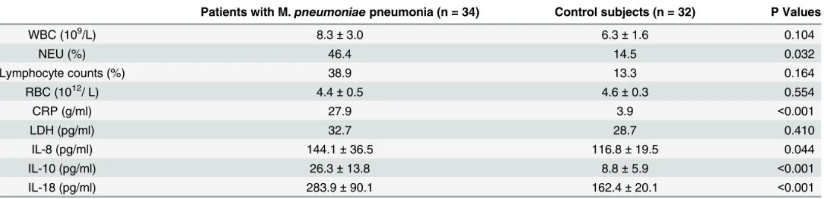
The characteristics, demographic data, serum IL-8, IL-10, and IL-18 levels of patients with
differences in WBC counts, LDH levels, lymphocyte counts, and RBC counts between patients with mycoplasmal pneumonia and control subjects. IL-8, IL-10, and IL-18 levels of patients
before therapy and after therapy are shown inTable 2. Patients with mycoplasmal pneumonia
had significantly higher CRP, IL-8, IL-10, and IL-18 levels than control subjects. However, con-trol subjects had significantly higher NEU counts than patients with mycoplasmal pneumonia. The IL-8, IL-10, and IL-18 levels of the 34 patients with mycoplasmal pneumonia are shown in
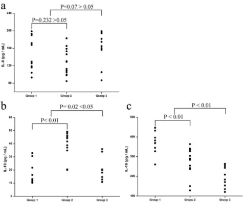
Fig 1. The levels of IL-8 in Group 1 (mean ± SD: 146.0 ± 36.9 pg/ml) and Group 2 (mean ± SD: 128.7 ± 29.0 pg/ml) were not significantly different (P = 0.232). IL-8 levels did not significantly
differ between Group 3 (mean ± SD: 161.9 ± 36.1 pg/ml; P = 0.07) and the 24M.pneumoniae
-infected patients without asthma (mean ± SD: 136.6 ± 33.9 pg/ml) (Fig 1a). There were
signifi-cantly lower levels of IL-10 in Group 1 (mean ± SD: 16.8 ± 7.8 pg/ml; P<0.0001) compared
with Group 2 (mean ± SD: 39.9 ± 9.4 pg/ml). The study also found lower levels of IL-10 in
Group 3 (mean ± SD: 19.0 ± 8.6 pg/ml) compared with the 24M.pneumoniae-infected patients
without asthma (mean ± SD: 29.4 ± 14.4 pg/ml), which was significantly different (P = 0.02) (Fig 1b). The IL-18 level of Group 1 (mean ± SD: 374.4 ± 48.9 pg/ml) was significantly different from that of Group 2 (mean ± SD: 272.5 ± 67.9 pg/ml; P = 0.001). The IL-18 level of Group 3
(mean ± SD: 199.1 ± 51.2 pg/ml; P<0.01) was significant lower than that of the 24M.
pneumo-niae-infected patients without asthma (mean ± SD: 319.2 ± 78.5 pg/ml) (Fig 1c).
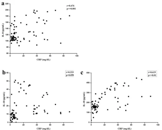
The correlations between IL-8, IL-10, and IL-18 levels, and clinical parameters CRP levels
are shown inFig 2. Meanwhile, the correlation between the levels of IL-8 and CRP (r = 0.476,
P<= 0.001), as well as the correlation between the levels of IL-18 and CRP (r = 0.619,
P<= 0.001), were also statistically significant, although the degree of the relationships was not
Table 1. Clinical factures, laboratory findings, and serum levels of IL-8, IL-10 and IL-18 of patients withM.pneumoniaepneumonia and control subjects.
Patients with M.pneumoniaepneumonia (n = 34) Control subjects (n = 32) P Values
WBC (109/L) 8.3±3.0 6.3±1.6 0.104
NEU (%) 46.4 14.5 0.032
Lymphocyte counts (%) 38.9 13.3 0.164
RBC (1012/ L) 4.4±0.5 4.6±0.3 0.554
CRP (g/ml) 27.9 3.9 <0.001
LDH (pg/ml) 32.7 28.7 0.410
IL-8 (pg/ml) 144.1±36.5 116.8±19.5 0.044
IL-10 (pg/ml) 26.3±13.8 8.8±5.9 <0.001
IL-18 (pg/ml) 283.9±90.1 162.4±20.1 <0.001
Data on serum IL-8, IL-10, IL-18, WBC, and RBC are presented as means±SD; Date on Neutrophil counts, CRP levels, LDH levels, and lymphocyte
counts are shown with IQR.
doi:10.1371/journal.pone.0146397.t001
Table 2. Serum levels of IL-8, IL-10 and IL-18 of patients before therapy and after therapy.
Before therapy After therapy P Values
IL-8 (pg/ml) 139.1±31.5 110.8±19.5 0.576
IL-10 (pg/ml) 30.3±13.7 10.8±4.3 0.514
IL-18 (pg/ml) 249.9±40.9 97.5±19.1 0.020
CRP (g/ml) 48.4±21.3 4.5±3.2 0.001
Data are presented as means±SD.
very strong (Fig 2a and 2c). The correlation between IL-10 and CRP was not significant (Fig
2b). However, significant correlations were not found between IL-8, IL-10, and IL-18 levels, and
WBC counts, lymphocyte counts, RBC counts, NEU counts, and LDH levels (S1–S4Figs).
Discussion
M.pneumoniae, which lacks a peptidoglycan cell wall, is one of the extracellular pathogens that
cause community-acquired respiratory infections. PCR methods can be used to detectM.
pneu-moniaein sputum, throat swabs, or bronchoalveolar lavage fluid, asM.pneumoniaeattaches to ciliated airway epithelial cells of the respiratory tract. PCR methods have a high sensitivity to detect microbial nucleic acids. However, positive PCR results cannot confirm reliably that the
organism is alive or infectious [16,17]. Some studies showed that levels of secreted cytokines,
such as IL-2 and IL-5, were significantly different in patients infected byM.pneumoniae
com-pared with other patients [18,19]. Pang et al. reported that the TNF-α, IL-6 and IL-10 levels in
children with M. pneumoniae pneumonia were higher at acute stage than those in the control group, suggesting the cytokines maybe involved in the pathogenesis of M. pneumoniae
pneu-monia [20]. Delayed-type hypersensitivity skin reaction responses toM.pneumoniaeantigens
appear to be correlated with the severity of pneumonia. Serum IL-18 and sIL-2R levels in
patients with severeM.pneumoniaeinfection group were higher than those in patients with
mild infections, and there was a significant relationship between them [13]. Oishi et al. pointed
out that most hospitals and clinics cannot usually measure IL-18 secretion levels quickly, so it
is important to search for correlations between IL-18 levels and other clinical features [11]. But
Fig 1. IL-8 (a), IL-10 (b) and IL-18 (c) levels in non-asthmatic patients with severeM.pneumoniae pneumonia (group 1) and mildM.pneumoniaepneumonia (group 2), and in asthmatic patients with M.pneumoniaepneumonia (group 3).Serum IL-10 and IL-18 levels were significantly higher in patients infected byM.pneumoniaewithout asthma than those in group 3. However, IL-8 levels were not significantly different. Serum IL-10 levels were significantly lower in group 1 than in group 2, while IL-18 levels were significantly higher in group 1; IL-8 levels were not significantly different.
if the condition permits, cytokines detection is necessary. Transgenic IL-10-deficient mice can spontaneously acquire a chronic inflammatory bowel disease. The finding indicated that endogenous IL-10 is important to inhibit the inflammatory response, while IL-10 has a dual
role in the inflammatory bowel disease [21]. IL-10 is expressed in the lungs ofM.pneumoniae
-infected BALB/c mice, and it is believed that IL-10 serves to turn off the specific T-cell response
[10,12]. In our study, serum IL-10 levels were higher in patients with mycoplasmal
pneumo-nia, which is in accordance with the findings of previous studies [16,22]. Mizukane et al.
reported an extremely rare case of a 76-year-old Japanese who developed hemophagocytic
syn-drome due to fulminantM.pneumoniaepneumonia, and they found that elevated s2R,
IL-6, and IL-10 levels may be caused by severe mycoplasmal pneumonia [9]. We examined three
groups of patients with mycoplasmal pneumonia, including severe and mild patients without asthma and patients with mycoplasmal pneumonia with asthma. In severe cases, IL-10 levels were lower than in mild cases. This result can be explained by the fact that the ability of IL-10 to inhibit inflammation may be decreased by some factors. Recently, researchers have
sug-gested that the onset of asthma may be preceded byM.pneumoniaeinfections, or that asthma
symptoms may be aggravated by infectingM.pneumonia[23]. In cases with asthma, IL-10
lev-els were lower, which suggests that some asthmatic children have deficient IL-10 responses
when infected byM.pneumoniae. Seo YH et al reported the levels of interleukin-18 in
naso-pharyngeal aspirate were significantly higher in patients infected withMycoplasma
pneumo-niaewho did not respond to macrolide treatment[24]. We also detected other chemokines,
including IL-8, IL-18, and other clinical factors. Serum IL-8 levels in patients withM.
pneumo-niaewere significantly higher than those in control subjects, and IL-8 was induced in airway
Fig 2. Serum levels of IL-8 (a), IL-10 (b), and IL-18 (c) relationship with CRP.Significant relationships were found between IL-8 levels and CRP levels, as well as between IL-18 levels and CRP levels, although no significant relationship was found between IL-10 and CRP levels.
epithelial cell line A549 by aM.pneumoniaelysate (MPL), which may have increased
neutro-phil chemotaxis and ERK activity [8]. Kyung et al. reported that MPL stimulated the
produc-tion of IL-8 in human airway epithelial cells [23]. Narita et al. detected IL-8 levels in pleural
fluid samples and serum samples from patients withM.pneumoniaeinfections, and the data
showed that IL-8 levels were significantly elevated in pleural fluid and serum [25]. Our results
are in agreement with the view that serum IL-8 may be involved in the pathogenesis of the
pul-monary disease features ofM.pneumoniaepneumonia. Our data showed that IL-8 levels were
not significantly different in patients with or without asthma, which indicates that IL-8 may not play a pivotal role in asthma. IL-18 levels in patients with mycoplasmal pneumonia were significantly higher than those in control subjects, and they were lower in patients with asthma compared with patients without asthma. Serum IL-18 levels in children in our study were
simi-lar to those of previous reports in adults and children [6,13]. These results suggest that these
chemokines play an important role in the pathogenesis of respiratory infections.
Seo YH et al also found that the levels of CRP in nasopharyngeal aspirate were significantly
higher in patients infected withMycoplasma pneumoniaewho did not respond to macrolide
treatment. They suggested that serum CRP could be a useful marker for predicting the efficacy of macrolides and helping clinicians make better clinical decisions in children with
macrolide-resistantMycoplasma pneumoniae[24]. We also analyzed the clinical characteristics of the
study subjects, including CRPTable 1shows that CRP levels were higher and NEU counts
were lower in patients withM.pneumoniainfection. We analyzed the correlation between
IL-8, IL-10, and IL-1IL-8, and the clinical factors. The cause of this result may be due to different cell-mediated immune responses of the host. Our data showed that both IL-8 and IL-18 levels were positively correlated with CRP levels. An association of IL-10 levels and clinical factors
was not found.Table 2shows that IL-18 levels were lower after therapy (P = 0.020), CRP levels
were also significant lower after therapy (P = 0.001). But after therapy, the correlation between
the levels of IL-8, IL-18 and CRP was not significant. (S5 Fig) In summary, our data indicate
that IL-8, IL-10, and IL-18 might modulate pulmonary lesions in mycoplamal pneumonia.
Conclusions
Our findings suggest that CRP level can be used to estimate the levels of IL-8 and IL-18. If nec-essary, the levels of IL-10 and IL-18 should be measured, as they can help diagnose the severity
of pediatricM.pneumoniaepneumonia.
Supporting Information
S1 Fig. Serum levels of IL-8 (a), IL-10 (b), and IL-18 (c) relationship with RBC counts. (TIF)
S2 Fig. Serum levels of IL-8 (a), IL-10 (b), and IL-18 (c) relationship with NEU.Significant relationships were not found between them and NEU, respectively.
(TIF)
S3 Fig. Serum levels of IL-8 (a), IL-10 (b), and IL-18 (c) relationship with Lymphocyte counts.
(TIF)
S4 Fig. Serum levels of IL-8 (a), IL-10 (b), and IL-18 (c) relationship with WBC counts. (TIF)
Acknowledgments
This study was supported by the National Science Foundation of China (Grant No. 81271837, 81571963) and Natural Science Foundation Key project of Anhui Education Department (KJ2012A161, KJ2015A020).
Author Contributions
Conceived and designed the experiments: LW GF. Performed the experiments: SD XW. Ana-lyzed the data: WC YF. Contributed reagents/materials/analysis tools: BL YL. Wrote the paper: SD XW GF LW.
References
1. Clyde WA Jr. Clinical overview of typical Mycoplasma pneumoniae infections. Clinical infectious dis-eases: an official publication of the Infectious Diseases Society of America. 1993; 17 Suppl 1:S32–6. PMID:8399935.
2. Izumikawa K, Izumikawa K, Takazono T, Kosai K, Morinaga Y, Nakamura S, et al. Clinical features, risk factors and treatment of fulminant Mycoplasma pneumoniae pneumonia: a review of the Japanese liter-ature. Journal of infection and chemotherapy: official journal of the Japan Society of Chemotherapy. 2014; 20(3):181–5. doi:10.1016/j.jiac.2013.09.009PMID:24462437.
3. Othman N, Isaacs D, Daley AJ, Kesson AM. Mycoplasma pneumoniae infection in a clinical setting. Pediatrics international: official journal of the Japan Pediatric Society. 2008; 50(5):662–6. doi:10.1111/ j.1442-200X.2008.02644.xPMID:19261116.
4. Chmura K, Bai X, Nakamura M, Kandasamy P, McGibney M, Kuronuma K, et al. Induction of IL-8 by Mycoplasma pneumoniae membrane in BEAS-2B cells. American journal of physiology Lung cellular and molecular physiology. 2008; 295(1):L220–30. doi:10.1152/ajplung.90204.2008PMID:18487355; PubMed Central PMCID: PMC2494795.
5. Arae K, Hirata M, Kurata S, Kamiya S, Taguchi H. Mycoplasma pneumoniae induces interleukin-8 pro-duction via the epidermal growth factor receptor pathway. Microbiology and immunology. 2011; 55(10): 748–50. doi:10.1111/j.1348-0421.2011.00375.xPMID:21831204.
6. Chung HL, Shin JY, Ju M, Kim WT, Kim SG. Decreased interleukin-18 response in asthmatic children with severe Mycoplasma pneumoniae pneumonia. Cytokine. 2011; 54(2):218–21. doi:10.1016/j.cyto. 2011.02.008PMID:21356600.
7. Hayakawa M, Taguchi H, Kamiya S, Fujioka Y, Watanabe H, Kawai S, et al. Animal model of Myco-plasma pneumoniae infection using germfree mice. Clinical and diagnostic laboratory immunology. 2002; 9(3):669–76. PMID:11986276; PubMed Central PMCID: PMC119980.
8. Lee KE, Kim KW, Hong JY, Kim KE, Sohn MH. Modulation of IL-8 boosted by Mycoplasma pneumoniae lysate in human airway epithelial cells. Journal of clinical immunology. 2013; 33(6):1117–25. doi:10. 1007/s10875-013-9909-yPMID:23779254.
9. Mizukane R, Kadota Ji J, Yamaguchi T, Kiya T, Fukushima H, Nakatomi M, et al. An elderly patient with hemophagocytic syndrome due to severe mycoplasma pneumonia with marked hypercytokinemia. Respiration; international review of thoracic diseases. 2002; 69(1):87–91. 49377. PMID:11844970. 10. Narita M, Tanaka H, Abe S, Yamada S, Kubota M, Togashi T. Close association between pulmonary
disease manifestation in Mycoplasma pneumoniae infection and enhanced local production of interleu-kin-18 in the lung, independent of gamma interferon. Clinical and diagnostic laboratory immunology. 2000; 7(6):909–14. PMID:11063497; PubMed Central PMCID: PMC95984.
11. Oishi T, Narita M, Matsui K, Shirai T, Matsuo M, Negishi J, et al. Clinical implications of interleukin-18 levels in pediatric patients with Mycoplasma pneumoniae pneumonia. Journal of infection and chemo-therapy: official journal of the Japan Society of Chemotherapy. 2011; 17(6):803–6. doi:10.1007/ s10156-011-0265-7PMID:21681500.
12. Pietsch K, Ehlers S, Jacobs E. Cytokine gene expression in the lungs of BALB/c mice during primary and secondary intranasal infection with Mycoplasma pneumoniae. Microbiology. 1994; 140 (Pt 8): 2043–8. doi:10.1099/13500872-140-8-2043PMID:7921254.
14. Kin Key N, Araujo-Neto CA, Nascimento-Carvalho CM. Severity of childhood community-acquired pneumonia and chest radiographic findings. Pediatric pulmonology. 2009; 44(3):249–52. doi:10.1002/ ppul.20988PMID:19205052.
15. Mathisen M, Strand TA, Sharma BN, Chandyo RK, Valentiner-Branth P, Basnet S, et al. Clinical pre-sentation and severity of viral community-acquired pneumonia in young Nepalese children. The Pediat-ric infectious disease journal. 2010; 29(1):e1–6. doi:10.1097/INF.0b013e3181c2a1b9PMID:
19935451.
16. Hassan J, Irwin F, Dooley S, Connell J. Mycoplasma pneumoniae infection in a pediatric population: analysis of soluble immune markers as risk factors for asthma. Human immunology. 2008; 69(12): 851–5. doi:10.1016/j.humimm.2008.09.003PMID:18835573.
17. Huijskens EG, Biesmans RC, Buiting AG, Obihara CC, Rossen JW. Diagnostic value of respiratory virus detection in symptomatic children using real-time PCR. Virology journal. 2012; 9:276. doi:10. 1186/1743-422X-9-276PMID:23164039; PubMed Central PMCID: PMC3511061.
18. Chen ZR, Zhang GB, Wang YQ, Yan YD, Zhou WF, Zhu CH, et al. Soluble B7-H3 elevations in hospital-ized children with Mycoplasma pneumoniae pneumonia. Diagnostic microbiology and infectious dis-ease. 2013; 77(4):362–6. doi:10.1016/j.diagmicrobio.2013.09.006PMID:24139879.
19. Esposito S, Droghetti R, Bosis S, Claut L, Marchisio P, Principi N. Cytokine secretion in children with acute Mycoplasma pneumoniae infection and wheeze. Pediatric pulmonology. 2002; 34(2):122–7. doi: 10.1002/ppul.10139PMID:12112778.
20. Pang HX, Qiao HM, Cheng HJ, Zhang YF, Liu XJ, Li JZ. [Levels of TNF-alpha, IL-6 and IL-10 in bronch-oalveolar lavage fluid in children with Mycoplasma pneumoniae pneumonia]. Zhongguo dang dai er ke za zhi = Chinese journal of contemporary pediatrics. 2011; 13(10):808–10. PMID:22000436. 21. Glehen O, Mithieux F, Traverse-Glehen A, Isaac S, Bienvenu J, Francois Y, et al. Enteral
immunother-apy in the treatment of chronic enterocolitis in interleukin-10-deficient mice. Hepato-gastroenterology. 2003; 50(51):670–5. PMID:12828057.
22. Salvatore CM, Fonseca-Aten M, Katz-Gaynor K, Gomez AM, Hardy RD. Intranasal interleukin-12 ther-apy inhibits Mycoplasma pneumoniae clearance and sustains airway obstruction in murine pneumonia. Infection and immunity. 2008; 76(2):732–8. doi:10.1128/IAI.00878-07PMID:18039833; PubMed Cen-tral PMCID: PMC2223484.
23. Sohn MH, Lee KE, Choi SY, Kwon BC, Chang MW, Kim KE. Effect of Mycoplasma pneumoniae lysate on interleukin-8 gene expression in human respiratory epithelial cells. Chest. 2005; 128(1):322–6. doi: 10.1378/chest.128.1.322PMID:16002953.
24. Seo YH, Kim JS, Seo SC, Seo WH, Yoo Y, Song DJ, et al. Predictive value of C-reactive protein in response to macrolides in children with macrolide-resistant Mycoplasma pneumoniae pneumonia. Korean journal of pediatrics. 2014; 57(4):186–92. doi:10.3345/kjp.2014.57.4.186PMID:24868216; PubMed Central PMCID: PMC4030120.