*Correspondence: A. M. Pedrosa. Laboratório de Hematologia, Facul-dade de Farmácia, UniversiFacul-dade Federal do Ceará. Rua Capitão Francisco Pedro, 1210 - Rodolfo Teóilo, 60370-430 - Fortaleza – CE, Brasil. E-mail: [email protected]
A
vol. 50, n. 2, apr./jun., 2014 http://dx.doi.org/10.1590/S1984-82502014000200020
Cytotoxicity and DNA damage in the neutrophils of patients with
sickle cell anaemia treated with hydroxyurea
Alano Martins Pedrosa
1,*, Maritza Cavalcante Barbosa
1, Thayna Nogueira dos Santos
1,
Luzia Kalyne Almeida Moreira Leal
1, Amanda de Araújo Lopes
1, Darcielle Bruna Dias Elias
1,
Greyce Luri Sasahara
1, Bruno Coêlho Cavalcanti
2, Romélia Pinheiro Gonçalves
11Pharmacy Department, Faculty of Pharmacy, Odontology and Nursing, Federal University of Ceará, Fortaleza, CE, Brazil, 2Physiology and Pharmacology Department, Faculty of Medicine, Federal University of Ceará, Fortaleza, CE, Brazil
Hydroxyurea (HU) is the most important advance in the treatment of sickle cell anaemia (SCA) for preventing complications and improving quality of life for patients. However, some aspects of treatment with HU remain unclear, including their effect on and potential toxicity to other blood cells such as neutrophils. This study used the measurement of Lactate Dehydrogenase (LDH) and Methyl
ThiazolTetrazolium (MTT) and the comet assay to investigate the cytotoxicity and damage index (DI) of the DNA in the neutrophils of patients with SCA using HU.In the LDH and MTT assays, a cytoprotective
effect was observed in the group of patients treated, as well as an absence of toxicity. When compared to
patients without the treatment, the SS group (n=20, 13 women and 07 men, aged 18-69 years), and the group of healthy individuals (AA) used as a control group (n=52, 28 women and 24 men, aged 19-60 years), The SSHU group (n=21, 11 women and 10 men, aged 19-63 years) showed a signiicant reduction
(p<0.001) in LDH activity and an increase in the percentage of viable cells by the MTT (p<0.001).
However, the SSHU group presented signiicantly higher DI values (49.57±6.0 U/A) when compared to the AA group (7.43 ± 0,94U/A) and the SS group (22.73 ±5.58 U/A) (p<0.0001), especially when
treated for longer periods (>20 months), demonstrating that despite the cytoprotective effects in terms of cell viability, the use of HU can induce DNA damage in neutrophils.
Uniterms: Hydroxyurea/cytotoxicity. Sickle cell anaemia/treatment. Neutrophils/DNA damage.
Deoxyribonucleic acid/damage.
A hidroxiuréia (HU) constitui o avanço mais importante no tratamento da anemia falciforme (AF) por
prevenir complicações e aumentar a qualidade de vida dos pacientes. Entretanto, alguns aspectos do
tratamento com HU permanecem obscuros, incluindo a sua ação e potencial toxicidade em outras células
sanguíneas, tais como neutróilos. Este estudo utilizou a mensuração da lactato desidrogenase (LDH) e do metil tiazoltetrazólio (MTT) e o ensaio do cometa para investigar a citotoxicidade e índice de dano (ID) ao DNA em neutróilos de pacientes com AF em uso do medicamento. Nos ensaios de LDH e MTT,
observou-se além de ausência de toxicidade, uma ação citoprotetora no grupo de pacientes tratados,
Grupo SSHU (n=21, 11 mulheres e 10 homens, com idades entre 19-63 anos), quando comparados aos pacientes sem tratamento, Grupo SS (n=20, 13 mulheres e 07 homens, 18-69 anos), e grupo de indivíduos saudáveis (AA) usado como controle (n=52, 28 mulheres e 24 homens, 19-60 anos), com redução signiicativa (p<0,001) na atividade de LDH e aumento no percentual de células viáveis pelo MTT (p<0,001). Entretanto, o grupo SSHU apresentou valores de ID signiicativamente elevados (49,57±6,0 U/A), quando comparados ao grupo AA (7,43 ± 0,94U/A) e grupo SS (22,73 ±5,58 U/A) (p<0,0001),
especialmente quando tratados por períodos mais longos (>20 meses), demonstrando que apesar dos
efeitos citoprotetores quanto à viabilidade celular, o uso da HU pode induzir lesão ao DNA de neutróilos.
INTRODUCTION
Sickle cell anaemia (SCA) is a hereditary disease caused by the substitution of glutamic acid with valine in the sixth codon of the β-globin leading to the formation of haemoglobin S (HbS). The sickle red blood cell is rigid
and inlexible, favoring the occurrence of vaso-occlusive
and haemolytic events and starting a chain reaction that culminates with the generation of reactive oxygen species (ROS), reduced bioavailability of nitric oxide (NO) and
the chronic inlammatory process, with direct involvement
of neutrophils in the early stages and propagation of these
mechanisms (Okpala, 2004; Conran et al., 2007; Rees, Gibson, 2011; Chirico, Pialoux, 2012).
The leukocytes play an important role in the pathophysiology of SCA. The vaso-occlusive process and endothelial injury result in a chronic inflammatory
response characterized by high levels of proinlammatory
cytokines, which are capable of making the activated endothelium. The endothelial activation results in increased expression of adhesion molecules on neutrophils, such as
E-selectin, P-selectin and ICAM-1 (Intercellular Adhesion
Molecule-1), inducing the chemotaxis of neutrophils and its interaction with adhesion molecules on sickle red blood cells, other leukocytes and platelets, leading to pancellular
activation resulting in the release of most proinlammatory
cytokines. Thus, there is a vicious circle between production of inflammatory mediators and cellular adhesion to the
endothelium, leading to a state of chronic inlammation,
fundamental to the process of vaso-occlusion (Okpala,
2006; Canalli et al., 2008; Conran, Franco-Penteado, Costa, 2009; Segel, Halterman, Lichtman, 2011).
The high count of neutrophils, often seen in patients with SCA, even in the absence of infection, has been associated with the occurrence of painful crises, acute chest syndrome (ACS), cerebrovascular accident (CVA)
and early death; however, the mechanisms responsible
for this are not completely understood (Platt et al., 1994;
Castro et al., 1994; Ohene-Frempong et al., 1998). Some
factors that may cause the interruption of apoptosis, such
as proinlammatory cytokines, cell adhesion, hypoxia,
and transmigration of bacterial lipopolysaccharide (LPS) may be related to the increased number of neutrophils and the survival time of these cells, suggesting an important relevance to the pathogenesis of SCA, since changes in the processes of apoptosis may affect cellular function and increase the damage potential (Ohene-Frempong et al., 1998; Moulding et al., 1998; Cross et al., 2005; Cross et al., 2006; Conran et al., 2007).
Hydroxyurea (HU) is the only drug approved by the
U.S. Food and Drug Administration (1999) and European
Medicines Agency (2008) for the treatment of SCA. HU inhibits the enzyme ribonucleotide reductase (RNR),
causes cell-cycle arrest, and allows globin genes to be
more actively expressed. By killing cycling cells, HU
changes the kinetics of erythroid proliferation, forcing more F cells to be produced from primitive progenitors and directly stimulating HbF production (Franco et al.,
2006).Furthermore, HU therapy increases haemoglobin
concentration, reduces the expression of adhesion molecules on erythrocytes, platelets and neutrophils, decreases the production of granulocytes and contributes to the improvement of clinical events, reducing the number of hospital admissions, the frequency of painful episodes, the need for transfusion and the occurrence of CVA and ACS (Platt et al., 1984; Steinberg et al., 2003; Zago, Pinto, 2007; Cartron, Elion, 2008; Orah, Platt, 2008; Conran, Franco-Penteado, Costa, 2009; Lou et al., 2009).
Despite these benefits the HU therapy may have clastogenic, mutagenic and teratogenic effects (Murphy,
Chaube, 1964; Oppenheim, Fishbein, 1965; Aliverti, Bonanomi, Giavini, 1980; Ware, Aygun, 2009). In the
literature, the HU ability to cause cancer is controversial
and the long-term eficacy and safety of HU in treating
patients with SCA remains inconclusive (Steinberg
et al., 2010). Some studies have shown that HU is genotoxic while other studies suggest that HU has low mutagenicity in vivo (Hanft et al., 2000; Friedrisch et al.,
2008). Furthermore, some reports relate that HU acts as
a competitive inhibitor of catalase-mediated hydrogen peroxide decomposition and this effect could be related to in vivo toxicity (Juul et al., 2010).
The long term safety of using HU remains an important issue, especially regarding genotoxicity and cell damage, and impact on the function of different organs (spleen kidneys, brain, lings) (Cançado et al., 2009).
Thus, some aspects regarding the treatment with HU and the safety of its long term use are still not fully elucidated, especially its effect on other blood cells other than erythrocytes. Therefore, it is necessary to evaluate the effect of HU regarding the genotoxicity and cytotoxicity in neutrophils isolated from patients with sickle cell anaemia at baseline, seeking evidence about the effect of HU on these cells and its influence on the modulation of the
chronic inlammatory process in these patients.
MATERIAL AND METHODS
Criteria for inclusion
Subjects and samples
(24 women and 17 men, aged 18-69 years) with clinical
and laboratory diagnosis (confirmed by haemoglobin electrophoresis and molecular biology) of sickle cell
anaemia (homozygotic SS form), representing 50% of the
patients attending the outpatient unit of the haematology
service of a reference hospital in Fortaleza, CE, Brazil (Table I presents the characteristics and clinical details of all
individuals participating in the study). The study included SCA patients in a stable condition, according to the criteria
of Ballas: absence of painful episodes and/or intercurrent illnesses, such as infections and inlammations in the four weeks preceding the study; no hospital admissions in the 2-3 days preceding the study and no blood transfusions in the four months preceding the study (Ballas, 2012).
In Brazil, the Ministry of Health degree nº 872 of 6th
November 2002 approved the use of HU for patients with sickle cell disease. The drug is recommended for patients with a laboratory diagnosis of sickle cell disease (HbSS,
S/β Thalassemia, HbSC, Hb SD), including children with three or more episodes of vaso-occlusive crises requiring
hospital medical care; a recurring acute chest crises; one or more strokes; recurrent priapism and severe and
persistent anaemia in the past 12 months,Hb concentration
consistently less than 7 g/dL, concentration of fetal Hb<8% after 2 years of age, WBC count 20x109/L in the
absence of infection. Patients with hypersensitivity to HU will be excluded from this protocol, as well as patients with presence of at least one of the following items related
to bone marrow dysfunction: neutrophils <2.5x10⁹/L, platelets <95x109/L,Hb concentration <4.5 g/dL or reticulocyte count <95 x 109/L, pregnant women and HIV-infected patients (Brazil, 2002; Cançado et al., 2009).
The selected patients were divided into two groups
according to the use of HU: SS (without HU treatment) (n=20, 13 women and 07 men, aged 18-69 years)
and SSHU (receiving oral HU dose between 15 and
25mg/kg/day) (n=21, 11 women and 10 men, aged 19-63 years). A control group (AA) was made up of 52 healthy blood donors (28 women and 24 men, aged 19-60 years) from a blood center in Fortaleza, CE, Brazil. Exclusion criteria for all groups were: presence of infectious diseases (HIV-1 and 2, HBsAg, HCV and
HTLV-1 & 2), pregnancy, presence of renal failure or liver disease, smoking, alcohol consumption, use of chelated
iron, anti-inlammatory drugs, antioxidant vitamins, or
any immunosuppressant. The survey was approved by
the Ethics Committee of the Federal University of Ceará (COMEP/Protocol number 101/12), and all subjects signed
an informed consent form. Samples (10 mL) of peripheral
blood were collected with EDTA for molecular biology. Samples (10 mL) of heparinized peripheral blood were
collected for isolation of polymorphonuclear leukocytes, predominantly neutrophils.
Molecular biological analysis
Molecular diagnosis of patients was based on a Polymerase Chain Reaction for mediated Restriction Fragment Length Polymorphism (PCR-RFLP), by
digestion with DdeI restriction enzyme, according to the
methods of Saiki et al. (1985).
Isolation of polymorphonuclear leukocytes
The isolation of polymorphonuclear leukocytes was carried from whole blood by differential gradient
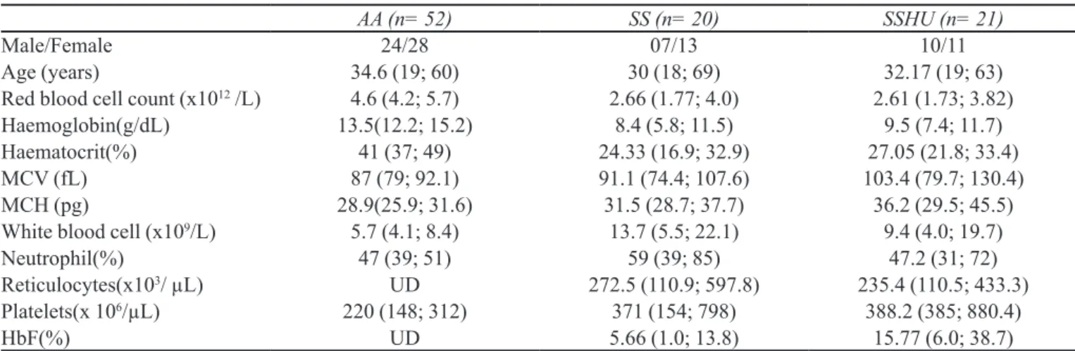
TABLE I -Characteristics and clinical details of the control group and patients with sickle cell anemia participating in the study
AA (n= 52) SS (n= 20) SSHU (n= 21)
Male/Female 24/28 07/13 10/11
Age (years) 34.6 (19; 60) 30 (18; 69) 32.17 (19; 63)
Red blood cell count (x1012 /L) 4.6 (4.2; 5.7) 2.66 (1.77; 4.0) 2.61 (1.73; 3.82)
Haemoglobin(g/dL) 13.5(12.2; 15.2) 8.4 (5.8; 11.5) 9.5 (7.4; 11.7)
Haematocrit(%) 41 (37; 49) 24.33 (16.9; 32.9) 27.05 (21.8; 33.4)
MCV (fL) 87 (79; 92.1) 91.1 (74.4; 107.6) 103.4 (79.7; 130.4)
MCH (pg) 28.9(25.9; 31.6) 31.5 (28.7; 37.7) 36.2 (29.5; 45.5)
White blood cell (x109/L) 5.7 (4.1; 8.4) 13.7 (5.5; 22.1) 9.4 (4.0; 19.7)
Neutrophil(%) 47 (39; 51) 59 (39; 85) 47.2 (31; 72)
Reticulocytes(x103/ µL) UD 272.5 (110.9; 597.8) 235.4 (110.5; 433.3)
Platelets(x 106/µL) 220 (148; 312) 371 (154; 798) 388.2 (385; 880.4)
HbF(%) UD 5.66 (1.0; 13.8) 15.77 (6.0; 38.7)
using a gelatin solution, following the methodology
proposed by Henson (1971) and modiied by Lucisano and Mantovani (1984) in which the cells were suspended in
buffered Hank’s balanced solution, containing calcium and magnesium. The preparations contained predominantly
neutrophils (85.0 ± 2.8%).
Cell viability assay
Cell viability was determined by a Trypan Blue
dye exclusion assay, in which the neutrophil suspension (2.5x10⁶/mL) was mixed with an equal volume of Trypan
Blue dye, 0.1%, and transferred to a Neubauer chamber. The Trypan Blue dye incorporated only non-viable cells due to membrane lesions (Lucisano, Mantovani, 1984).
The proportion of viable cells was estimated by counting 200 cells in an optical microscope.
Cytotoxicity assays
The cytotoxicity was analyzed by
lactatedehydro-genase (LDH) release, which assesses cell death by
necrosis, and by the MethylThiazolTetrazolium (MTT)
assay, measuring cellular metabolic activity through the
pathway succinate-tetrazolium redutase, enzyme active
only in cells with intact respiratory metabolism, which
reduces the tetrazolium salt (yellow coloration) to a salt purple colour (formazan) (Renzi, Valtolina, Forster, 1993).
After isolation, a suspension of cells (2.5 × 10⁶/mL) was incubated with buffered Hank’s balanced solution (test
group) or 0.2% Triton X-100 (known to cause cell lysis and used as a positive control), for 30 min at 37°C. The activity
of LDH was determined in the supernatant according to the manufacturer’s instructions (LDH liquiform of Labtest
Diagnosis, MG, Brazil) in which the consumption of NADH
was monitored by conversion of pyruvate to lactate at
340 nm. A solution of MTT at a concentration of 10 mg/mL
was added to a cell suspension of neutrophils (5.0x10⁶/mL)
for the colorimetric determination of the formazan crystals at 540 nm, according to the protocol described by Mosmann (1983).
Comet assay
The standard alkaline protocol for comet assay was used, as reported by Singh et al. (1988). The assay was
performed in accordance with general guidelines for in vivo use of the comet assay (Tice et al., 2000; Hartmann et al., 2003). Briely, the neutrophil suspension (2.5x10⁶ cells/mL) was mixed with low-melting-point agarose
and spread onto glass slides pre-coated with agarose, and cover slips were gently placed over their content. Once the samples solidified, the cover slips were removed and the slides were soaked in freshly made, chilled lysis
solution (2.5M NaCl, 100 mM EDTA, 10 mM Tris, pH 10.2, to which 1% Triton X-100 and 10% DMSO had been added) for 1-2 days under refrigeration. Excess liquid was blotted away from each slide’s back and edges; the
slides were then transferred to an electrophoresis tank,
and an alkaline solution (300 mM NaOH, 1 mM EDTA, pH>13) was added. The slides were exposed to the alkaline
solution for 20 min to allow for DNA unwinding and for the expression of alkali-labile sites as single strand breaks.
DNA was then electrophoresed for 20 min (25 V; 300 mA; 0.9 V/cm). Slides were removed from the electrophoresis
tank, cleaned, washed three times (5 min each time) with
neutralization buffer (0.4M Tris, pH 7.5), washed three
times with distilled water, and allowed to air dry. All steps of the assay were conducted under dim light.
Slides were then ixed and silver-stained according
to the methods of Nadin, Vargas-Roig and Ciocca (2001). For evaluation of DNA damage, 100 cells per subject were
analyzed at 200x magniication under a light microscope.
Cells were assessed visually and received scores from 0
(no migration) to 4 (maximum migration) according to tail intensity (size and shape). Therefore, the total scores (damage index or DI) for 100 cells ranged from 0 (all cells with no migration) to 400 (all cells with maximum
migration). Slides from patients and controls were processed, coded, mixed, and evaluated together.
Statistical analyses
The Statistical Package for the Social Sciences (SPSS) 10.0 for Windows was used for all data analyses,
and graphs were made with the GraphPad Prism 4.0 for Windows. The level of signiicance was 0.05 for all tests, and the data is presented as mean ± S.E. of the mean (SEM). The Kolmogorov-Smirnov test was used to
check for normal distribution of the data. The Analysis of variance (ANOVA) followed by the Tukey post-test was used to determine statistical differences between groups AA, SS and SSHU. The parametric T-test was used to test
the inluence of the mean dose of HU and HU Length of treatment on LDH release, MTT reduction and DI values.
The mean dose of HU and the treatment length were separated into two categories according to their medians.
RESULTS
Influence of treatment with HU on cell viability evaluated by exclusion assay with Trypan Blue dye in neutrophils from patients with sickle cell anaemia
in the AA group (95.8±0.29%) (p<0.05). The number of viable cells in the SSHU group (95.0±0.42%) was similar to the SS and control groups, i.e., there was no signiicant
difference.
Influence of treatment with HU on LDH release and MTT reduction in neutrophils from patients with sickle cell anaemia
Table III shows that the SS group (10.25 ± 1.21U/L)
showed a greater LDH release compared to the AA group
(6.71±0.68 U/L) and the SSHU group (5.59±0.37U/L) (p=0.0003). The LDH release did not differ between the SSHU and the AA groups (p>0.05).In the experimental model MTT reduction, the SS group (35.14±2.32%)
showed a lower number of viable cells when compared to
the AA group (56.8±3.2%) and the SSHU group (50.59 ± 3.64%) (p<0.0001). There was no signiicant difference
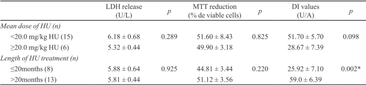
between the number of viable cells in the SSHU and the AA groups (p> 0.05). The mean dose of HU and the Length of HU treatment did not affect the LDH release and MTT
reduction (Table IV).
Influence of treatment with HU in the DI values in neutrophils from patients with sickle cell anaemia
Figure 1 shows an association between DI values and treatment with HU. DI values for the SSHU group were greater (49.57 ± 6.0U/A) when compared to the AA group (7.43± 0.94 U/A) and the SS group (22.73 ± 5.58 U/A) (p<0.0001). The SS group showed DI values higher than in the AA group, but the difference was not signiicant
(p> 0.05). The mean dose of HU and the Length of HU
treatment did not affect the DI values (Table IV).
DISCUSSION
The clinical and laboratory eficacy of HU in the TABLE II - Influence of treatment with HU on cell viability
evaluated by exclusion assay with Trypan Blue dye in
neutrophils from patients with sickle cell anaemia
Numberofcells (%)
Viable Non-viable p
AA (n=52) 95.8±0.29 4.2±0.33 p>0.05
SS (n=20) 93.7 ±0.34a 6.2 ±0.35 p<0.05
SSHU (n=21) 95.0 ±0.42 5.0 ±0.42 p>0.05
AA: Control group (healthy individuals), SS: patients with sickle cell anaemia not treated with HU; SSHU: patients with sickle
cell anaemia treated with HU. Results were expressed as mean
± SEM. ANOVA followed by Tukeypost test. a p<0.05 versus
AA group.
TABLE III - Inluence of treatment with HU in the parameters of cytotoxicity in neutrophils from patients with sickle cell anaemia
Parameters AA (n=52) SS (n=20) SSHU (n=21) p
LDH release (U/L) 6.71 ± 0.68 10.25 ± 1.21 a 5.59 ± 0.37 b 0.0003
MTT reduction (% de viable cells) 56.80 ± 3.20 35.14 ± 2.32 a 50.59 ± 3.64 b <0.0001
AA: Control group (healthy individuals), SS: patients with sickle cell anaemia not treated with HU; SSHU: patients with sickle
cell anaemia treated with HU. Results were expressed asmean ± SEM. ANOVA followed by Tukeypost test. a p<0.05 versus AA
group, b p<0.05 versus SS group
TABLE IV - LDH release, MTT reduction and DNA damage according to length of HU treatment and mean HU dose (SSHU group) (n=21)
LDH release
(U/L) p (% de viable cells)MTT reduction p DI values (U/A) p
Mean dose of HU (n)
<20.0 mg/kg HU (15) 6.18 ± 0.68 0.289 51.60 ± 8.43 0.825 51.70 ± 5.70 0.098
≥20.0 mg/kg HU (6) 5.32 ± 0.44 49.90 ± 3.18 28.67 ± 7.39
Length of HU treatment (n)
≤20months (8) 5.88 ± 0.64 0.925 44.81 ± 3.44 0.220 25.92 ± 7.10 0.002*
>20months (13) 5.81 ± 0.44 51.12 ± 3.56 59.0 ± 6.39
treatment of SCA has been convincingly demonstrated, with a decrease in morbidity and mortality with HU use,
coupled with the modest short-term toxicity proile and
ease of once daily oral administration, HU is an ideal treatment option (Ware, 2010).These consistent results leave little question as to the clinical benefits of HU therapy for adults with SCA. However, the long-term efficacy and safety of HU use for patients with SCA remains one of the most critical unanswered questions
(Brawley et al., 2008), and not much is known about
the effects of this medicament on neutrophils and on the functionality of these cells, since the majority of the studies found in the literature are related to red blood cells.
The results of this study showed that patients with
SCA not treated with HU showed signiicantly lower cell viability, both the cellular viability assay by Trypan Blue
dye as well as the MTT assay, and increased LDH release compared to the control group (AA). Similar results are found in studies with erythrocytes of SCA patients, revealing an increase in plasma levels of LDH of patients
when compared to healthy individuals (Kato et al., 2006;
Cançado et al., 2009), and this increase in enzymes is
positively associated with an increased risk that patients will suffer from priapism, pulmonary hypertension, cutaneous ulcers in the lower limbs, resistance to NO or even early
death (Kato et al., 2006; Steinberg, 2008; Rees, Gibson, 2011). There was no signiicant difference of these variables
between the patients treated with HU and the control group (AA). These results suggest that neutrophils from patients with SCA not treated with HU do have damage of
cellular integrity and respiratory metabolism, and that HU did not have any cytotoxic effect on patients’ neutrophils when investigated using these testing models. However, a cytoprotective action was revealed when compared to the
AA group and the SS patients. It is also possible that the oxidative stress and generalized inlammation of severe
untreated SCA produces a mild cytotoxic effect, and HU therapy may help to improve this process.
To evaluate the DNA damage index by comet assay, it was observed that the SCA patients not treated with
HU had DI values higher than the AA group, but with no signiicant difference. No correlation was observed between the DI values with the HbF parameters and the
total white blood cell count in the groups studied. Some
studies show conlicting results, suggesting that mutagenic
or carcinogenic effects are induced by the disease itself
rather than by HU therapy (Schultz, Ware, 2003; Segal et al., 2008). For example, in a retrospective survey by the International Association of Sickle Cell Nurses and Physician Assistants, 52 cases of cancer were identiied in a cohort of more than 16,000 patients with SCA; almost all occurred in the pre-HU stage; only three patients had previous HU exposure; among children with SCA, 21 reported cancers included leukemia (n=7), Wilms tumor (n=5), lymphoma (n=3), and six other solid tumors;
only one patient with acute lymphoblastic leukemia had
previous HU exposure (Schultz, Ware, 2003).
There are conflicting reports regarding the DNA-damaging potential of HU in exposed humans. Some studies have shown that HU is genotoxic (Flanagan et al., 2010;
Juul et al., 2010; Santos et al., 2011) while other studies suggest that HU has low mutagenicity in vivo (Stricker et al., 1986; Dawkins et al., 1997; Montalembert et al., 1999;
Hanft et al., 2000; Montalembert, Davies, 2001; Moschovi et al., 2001). In this study, patients with SCA treated with HU had signiicantly elevated DI values when compared
with the patients with SCA who were not treated with HU.
Furthermore, the DI values were significantly higher in
patients treated for longer periods (>20 months).Similar results were found in two previous studies that evaluated
the DNA Damage Index in total peripheral white blood
cell counts of SCA patients treated with HU (Friedrisch et al., 2008; Rocha et al., 2012). Taken together, these studies provide evidence for measurable genotoxicity from HU exposure in patients with SCA, but little evidence to support cumulative mutagenicity or carcinogenic potential.
CONCLUSION
Although HU presents numerous positive responses, more studies on the issue of safety are essential, including
FIGURE 1 – Inluence of treatment with HU on the DI values in
neutrophils from patients with sickle cell anaemia. AA: Control group (healthy individuals) (n=52); SS: patients with sickle cell anaemia not treated with HU (n=20); SSHU: patients with sickle
cell anaemia treated with HU (n=21). Results were expressed
as mean ± SEM. ANOVA followed by turkey posttest. ap0.05
the optimal dosage, the duration of use and the age of the patient, among other factors. Currently SCA
is characterized as a chronic inflammatory disease
where neutrophils initiate leukocyte adhesion to blood vessel walls, contributing thus to the development of inflammatory and vaso-occlusive processes and to the severity of the disease. Despite evidence of the essential involvement of neutrophils in the clinical modulation and the pathophysiological aspects of the disease, the mechanism by which HU modulates this effect is not fully elucidated. This study demonstrates that treatment with HU does not exert a cytotoxic effect on the neutrophils of patients with SCA, and HU may even be able to promote a protective effect on these cells, however there is the risk of DNA damage associated with exposure for longer periods of time. The monitoring of patients with SCA is important, since the data on the cytotoxic and genotoxic risks of HU remain inconclusive. More research is needed to clarify the risks of HU therapy inpatients with SCA.
CONFLICT OF INTEREST STATEMENT
The authors declare that there are no conflicts of interest.
REFERENCES
ALIVERTI, V.; BONANOMI, L.; GIAVINI, E. Hydroxyurea as a reference standard in teratological screening. In:
CHAMBERS, P.L.; KLINGER, W.(Eds.)Further studies in
the assessment of toxic actions. Springer, 1980. p.239-247.
BALLAS, S.K. More deinitions in sickle cell disease: steady
state v base line data. Am. J. Hematol., v.87, n.3, p.338,
2012.
BRASIL. Ministério da Saúde. Portaria SAS/MS Nº 872, de 06 de novembro de 2002, publicada no Diário Oicial da União, de 8 de novembro de 2002, seção 1, página 169 - Aprova o protocolo clínico e diretrizes terapêuticas /Doença
Falciforme/ Hidroxiuréia. Secretário Renilson Rehem de
Souza.
BRAWLEY, O.W.; CORNELIUS, L.J.; EDWARDS, L.R.; GAMBLE, V.N.; GREEN, B.L.; INTURRISI, C.; JAMES, A.H.; LARAQUE, D.; MENDEZ, M.; MONTOYA,
C.J. National institutes of health consensus development
conference statement: hydroxyurea treatment for sickle cell
disease. Ann. Intern. Med., v.148, n.12, p.932-938, 2008.
CANÇADO, R.D.; LOBO, C.; ANGULO, I.L.; ARAÚJO, P.I.C; JESUS, J.A. Protocolo clínico e diretrizes terapêuticas
para uso de hidroxiureia na doença falciforme. Rev. Bras. Hematol. Hemoter.,v.31, n.5, p.361-366, 2009.
CANNALLI, A.A.; FRANCO-PENTEADO, C.F.; SAAD, S.T.O.; CONRAN, N.; COSTA, F.F. Increased adhesive
properties of the neutrophils in sickle cell disease may be reversed by pharmacological nitric oxide donation.
Haematologica, v.93, n.4, p.605-609, 2008.
CARTRON, J.P.; ELION, J. Erythroid adhesion molecules in
sickle cell disease: effect of hydroxyurea. Transfus. Clin.
Biol., v.15, n.1, p.39-50, 2008.
CASTRO, O.; BRAMBILLA, D.J.; THORINGTON, B.; REINDORF, C.A.; SCOTT, R.B.; GILLETTE, P.; VERA, J.C.; LEVY, P.S. The acute chest syndrome in sickle cell disease: incidence and risk factors. The cooperative study
of sickle cell disease. Blood, v.84, n.2, p.643-649, 1994.
CHIRICO, E.N.; PIALOUX, V. Role of oxidative stress in the
pathogenesis of sickle cell disease. Life, v.64, n.1,p.72-80,
2012.
CONRAN, N.; ALMEIDA, C.B.; LANARO, C.; FERREIRA, R.P.; TRAINA, F. SAAD, S.T.O.; COSTA, F.F. Inhibition
of caspase-dependent spontaneous apoptosis via a cAMP-protein kinase A dependent pathway in neutrophils from sickle cell disease patients. Br. J. Haematol., v.139, n.1, p.148-158, 2007.
CONRAN, N.; FRANCO-PENTEADO, C.F.; COSTA, F.F.
Newer aspects of the pathophysiology of sickle cell disease vaso-occlusion. Hemoglobin,v.33, n.1, p.1-16, 2009.
CROSS, A.; BAKSTAD, D.; ALLEN; J.C.; THOMAS, L.; MOOTS, R.J.; EDWARDS, S.W. Neutrophil gene
expression in rheumatoid arthritis. Pathophysiology, v.12,
n.3, p.191-202, 2005.
CROSS, A.; BARNES, T.; BUCKNALL, R.C.; EDWARDS, S.W.; MOOTS, R.J. Neutrophil apoptosis in rheumatoid
arthritis is regulated by local oxygen tensions within joints.
J. Leukoc. Biol., v.80, n.3, p.521-528, 2006.
DAWKINS, F.W.; KIM, K.S.; SQUIRES, R.S.; CHISHOLM, R.; KARK, J.A.; PERLIN, E.; CASTRO, O. Cancer
incidence rate and mortality rate in sickle cell disease
patients at Howard University Hospital: 1986-1995. Am.
FLANAGAN, J.M.; HOWARD, T.A.; MORTIER, N.; AVLASEVICH, S.L.; SMELTZER, M.P.; WU, S.; DERTINGER, S.D.; WARE, R.E. Assessment of
genotoxicity associated with hydroxyurea therapy in children with sickle cell anemia. Mutat. Res., v.698, n.1-2, p.38-42, 2010.
FRANCO, R.S.; YASIN, Z.; PALASCAK, M.B.; CIRAOLO, P.; JOINER, C.H.; RUCKNAGEL, D.L. The effect of fetal
hemoglobin on the survival characteristics of sickle cells.
Blood, v.108, n.3, p.1073-1076, 2006.
FRIEDRISCH, J.R.; PRÁ, D.; MALUF, S.W.; BITTAR, C.M.; MERGENER, M.; POLLO, T.; KAYSER, M.; DA SILVA, M.A.L.; HENRIQUES, J.A.P.; DA ROCHA SILLA, L.M.
DNA damage in blood leukocytes of individuals with sickle cell disease treated with hydroxyurea. Mutat. Res.,v.649,
n.1, p.213-220, 2008.
HANFT, V.N.; FRUCHTMAN, S.R.; PICKENS, C.V.; ROSSE, W.F.; HOWARD, T.A.; WARE, R.E. Acquired DNA
mutations associated with in vivohydroxyurea exposure.
Blood, v.95, n.11, p.3589-3593, 2000.
HARTMANN, A.; AGURELL, E.; BEEVERS, C.; BRENDLER-SCHWAAB, S.; BURLINSON, B.; CLAY, P.; COLLINS, A.; SMITH, A.; SPEIT, G.; THYBAUD,
V. Recommendations for conducting the in vivo alkaline
comet assay.4th International Comet Assay Workshop.
Mutagenesis, v.18, n.1, p.45-51, 2003.
HENSON, P.M. The immunology release of constituents from
neutrophils leukocytes. J. Immunol., v.107, n.6, p.1535-1546, 1971.
JUUL, T.; MALOLEPSZY, A.; DYBKAER, K.; KIDMOSE, R.; RASMUSSEN, J.T.; ANDERSEN, G.R.; JOHNSEN, H.E.; JØRGENSEN, J.; ANDERSEN, S.U. The in vivo toxicity
of hydroxyurea depends on its direct target catalase. J. Biol.
Chem., v.285, n.28, p.21411-21415, 2010.
KATO, G.J.; MCGOWAN, V.; MACHADO, R.F.; LITTLE, J.A.; TAYLOR, J.; MORRIS, C.R.; NICHOLS, J.S.; WANG, X.; POLJAKOVIC, M.; MORRIS, S.M. Lactate
dehydrogenase as a biomarker of hemolysis-associated nitric oxide resistance, priapism, leg ulceration, pulmonary hypertension, and death in patients with sickle cell disease.
Blood, v.107, n.6, p. 2279-2285, 2006.
LOU, T.F.; SINGH, M.; MACKIE, A.; LI, W.; PACE, B.S.
Hydroxyurea generates nitric oxide in human erythroid
cells: mechanisms for gamma-globin gene activation. Exp.
Biol. Med., v.234, n.11, p.1374-1382, 2009.
LUCISANO, Y.M.; MANTOVANI, B. Lysossomal enzyme
release from polymorfonuclear leukocytes induced by
immune complexes of IgM and IgG. J. Immunol., v.132,
n.4, p.2015-2020, 1984.
MONTALEMBERT, M.; BEGUE, P.; BERNAUDIN, F.; THURET, I.; BACHIR, D.; MICHEAU, M. Preliminary
report of a toxicity study of hydroxyurea in sickle cell disease. Arch. Dis. Child, v.81, n.5, p.437-439, 1999.
MONTALEMBERT, M.; DAVIES, S.C. Is hydroxyurea
leukemogenic in children with sickle cell disease? Blood, v.98, n.9, p.2878-2879, 2001.
MOSCHOVI, M.; PSYCHOU, F.; MENEGAS, D.; TSANGARIS, G.T.; TZORTZATOU-STATHOPOULOU, F.; NICOLAIDOU, P. Hodgkin’s disease in a child with
sickle cell disease treated with hydroxyurea. Pediatr. Hematol. Oncol.,v.18, n.6, p.371-376, 2001.
MOSMANN, T. Rapid colorimetric assay for cellular growth
and survival: application to proliferation and cytotoxicity
assays. J. Immunol. Methods, v.65, n.1-2, p.55-63, 1983.
MOULDING, D.A.; QUAYLE, J.A.; HART, A.; EDWARDS, S.W. Mcl-1 expression in human neutrophils: regulation by
cytokines and correlation with cell survival. Blood, v.92, n.7, p.2495-2502, 1998.
MURPHY, M.; CHAUBE, S. Preliminary survey of hydroxyurea
(nsc-32065) as a teratogen. Cancer Chemother.Rep.,v.40,
n.1,p.1-7, 1964.
NADIN, S.; VARGAS-ROIG, L.; CIOCCA, D. A silver staining
method for single-cell gel assay. J. Histochem. Cytochem.,
v.49, n.9, p.1183-1186, 2001.
OHENE-FREMPONG, K.; WEINER, S.J.; SLEEPER, L.A.; MILLER, S.T.; EMBURY, S.; MOOHR, J.W.; WETHERS, D.L.; PEGELOW, C.H.; GILL, F.M. Cerebrovascular
accidents in sickle cell disease: rates and risk factors. Blood,
v.91, n.1, p.288-294, 1998.
OKAPALA, I. Leukocyte adhesion cell disease. Curr.Opin.
OKPALA, I. The intriguing contribution of white blood cells
to sickle cell disease – a red cell disorder. Blood Rev., v.18,
n.1,p.65-73, 2004.
OPPENHEIM, J.J.; FISHBEIN, W.N. Induction of chromosome
breaks in cultured normal human leukocytes by potassium arsenite, hydroxyurea and related compounds. Cancer Res.,v.25, n.7, p.980-985, 1965.
ORAH, S.; PLATT, M.D. Hydroxyurea for the treatment of
sickle cell anemia. N. Engl. J. Med.,v.358, p.1362-1369, 2008.
PLATT, O.S.; BRAMBILLA, D.J.; ROSSE, W.F.; MILNER, P.F.; CASTRO, O.; STEINBERG, M.H.; KLUG, P.P.
Mortality in sickle cell disease. Life expectancy and risk factors for early death. N. Engl. J. Med., v.330, n.23, p.1639-1644, 1994.
PLATT, O.S.; ORKIN, S.H.; DOVER, G.; BEARDSLEY, G.P.; MILLER, B.; NATHAN, D.G. Hydroxyurea enhances fetal
hemoglobin production in sickle cell anaemia. J. Clin. Invest., v.74, n.2, p.652-656, 1984.
REES, D.C.; GIBSON, J.S. Biomarkers in sickle cell disease. Br. J. Haematol., v.156, n.4,p.433-445, 2011.
RENZI, D.; VALTOLINA, M.; FORSTER, R. The evaluation
of a multi-endpoint cytotoxicity assay system. ATLA Altern. Lab. Anim., v.21, n.1, p.89-96, 1993.
ROCHA, L.B.S.; ELIAS, D.B.D.; BARBOSA, M.C.; BANDEIRA, I.C.J.; GONÇALVES, R.P. DNA damage in
leukocytes of sickle cell anemia patients is associated with
hydroxyurea therapy and with HBB*S haplotype. Mutat.
Res., v.749, n.1-2, p.48-52, 2012.
SAIKI, R.K.; SCHARF, S.; FALOONA, F.; MULLIS, K.B.; HORN, G.; ERLICH, H.A.; ARNHEIM, N. Enzymatic
amplification of beta-globin genomic sequences and restriction site analysis for diagnosis of sickle cell anemia.
Science, v.230, n.4732, p.1350-1354, 1985.
SANTOS, J.L.; BOSQUESI, P.L.; ALMEIDA, A.E.; CHIN, C.M.; VARANDA, E.A. Mutagenic and genotoxic effect of
hydroxyurea. Int. J. Biomed. Sci., v.7, n.4, p.263-267, 2011.
SCHULTZ, W.H.; WARE, R.E. Malignancy in patients
withsickle cell disease. Am. J. Hematol., v.74, n.4, p.249-253, 2003.
SEGAL, J.B.; STROUSE, J.J.; BEACH, M.C.; HAYWOOD, C.; WITKOP, C.; PARK, H.S.; WILSON, R.F.; BASS, E.B.; LANZKRON, S. Hydroxyurea for the treatment of
sickle cell disease. Evid. Rep. Technol. Assess., n.165, p.1-95, 2008.
SEGEL, G.B.; HALTERMAN, M.W.; LICHTMAN, M.A. The
paradox of the neutrophil’s role in tissue injury. J.Leukoc. Biol., v.89, n.3, p.359-372, 2011.
SINGH, N.; MCCOY, M.; TICE, R.; SCHNEIDER, E. A simple
technique for quantitation of low levels of DNA damage in individual cells. Exp. Cell Res., v.175, n.1, p.184-191, 1988.
STEINBERG, M.H. Sickle cell anemia, the first molecular disease: overview of molecular etiology, pathophysiology,
and therapeutic approaches. Sci. World J., v.8, p.1295-1324, 2008.
STEINBERG, M.H.; MCCARTHY, W.F.; CASTRO, O.; BALLAS, S.K.; ARMSTRONG, F.D.; SMITH, W.; ATAGA, K.; SWERDLOW, P.; KUTLAR, A.; DECASTRO, L. The risks and beneits of long-term use of hydroxyurea in
sickle cell anemia: A 17.5 year follow-up. Am. J. Hematol.,
v.85, n.6, p.403-408, 2010.
STEINBERG, M.H.; VOSKARIDOU, E.; KUTLAR, A.; LOUKOPOULOS, D.; KOSHY, M.; BALLAS, S.K.; CASTRO, O.; BARTON, F. Concordant fetal hemoglobin
response to hydroxyurea in siblings with sickle cell disease.
Am. J. Hematol., v.72, n.2, p.121-126, 2003.
STRICKER, R.B.; LINKER, C.A.; CROWLEY, T.J.; EMBURY, S.H. Hematologic malignancy in sickle cell disease: report
of four cases and review of the literature. Am. J. Hematol.,
v.21, n.2, p.223-230, 1986.
TICE, R.R.; AGURELL, E.; ANDERSON, D.; BURLINSON, B.; HARTMANN, A.; KOBAYASHI, H.; MIYAMAE, Y.; ROJAS, E.; RYU, J.C.; SASAKI, Y.F. Single cell gel/comet assay: guidelines for in vitro and in vivo genetic toxicology
testing. Environ. Mol. Mutagen., v.35, n.3, p.206-221, 2000.
WARE, R.E.; AYGUN, B. Advances in the use of hydroxyurea. ASH Educ. Program Book, v.2009, n.1, p.62-69, 2009.
WARE, R.E. How I use hydroxyurea to treat young patients with
ZAGO, M.A.; PINTO, A.C.S. Fisiopatologia das doenças falciformes: da mutação genética à insuficiência de
múltiplos órgãos. Rev. Bras. Hematol. Hemoter., v.29, n.3,
p.207-214, 2007.
Received for publication on 06th October 2013