REGIONAL DIFFERENCES IN THE NUMBER AND TYPE
OF MYENTERIC NEURONS IN THE DESCENDING
COLON OF RATS
Eduardo José de Almeida Araújo
1, Débora de Mello Gonçales Sant’Ana
1,
Sônia Lucy Molinari
2, Marcílio Hubner de Miranda Neto
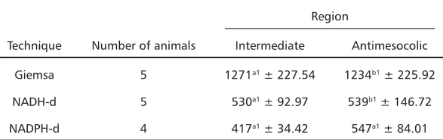
3ABSTRACT - The purpose of this study was to analyze the neuronal density of the myenteric plexus of the intermediate and antimesocolic regions of the descending colon of rats. Whole-mounts were stained with three different techniques of neuronal evidenciation. Through counts of the number of neurons in an area of 6.64 mm2 under light microscopy, we found 1,271 ± 227.54 neurons with Giemsa in the intermediate region and 1,234 ± 225.92 neurons in the antimesocolic region; with the NADH-diaphorase technique we found 530 ± 92.97 neurons in the intermediate region and 539 ± 146.72 neurons in the antimesocolic region; and through the NADPH-diaphorase histochemistry, we found 417 ± 34.42 neurons in the intermediate region and 547 ± 84.01 neurons in the antimesocolic region. We conclude that there is a variation in the density of NADPH-diaphorase positive neurons in the intestinal circumference; that the NADH-diaphorase positive neuronal subpopulation represented 42.7% of that stained with Giemsa; and that the NADPH-diaphorase positive neurons represented 37.8% of the whole myenteric population.
KEY WORDS: myenteric plexus, Giemsa, NADH-diaphorase, NADPH-diaphorase, rat, descending colon.
Diferenças regionais no número e tipo de neurônios mioentéricos do colo descendente de ratos Diferenças regionais no número e tipo de neurônios mioentéricos do colo descendente de ratosDiferenças regionais no número e tipo de neurônios mioentéricos do colo descendente de ratos Diferenças regionais no número e tipo de neurônios mioentéricos do colo descendente de ratos Diferenças regionais no número e tipo de neurônios mioentéricos do colo descendente de ratos
RESUMO - O objetivo deste estudo foi analisar a densidade neuronal do plexo mioentérico das regiões intermediária e antimesocólica do colo descendente de ratos. Preparados de membrana foram corados com três técnicas diferentes de evidenciação neuronal. Através da contagem do número de neurônios, em uma área de 6,64 mm2, sob microscopia de luz, encontramos com a coloração de Giemsa: 1271 ± 227,54 neurônios na região intermediária e 1234 ± 225,92 na região antimesocólica. Utilizando a técnica de marcação da NADH-diaforase, encontramos 530 ± 92,97 neurônios na região intermediária e 539 ± 146,72 na região antimesocólica. Através da histoquímica da NADPH-diaforase, encontramos 417 ± 34,42 neurônios na região intermediária e 547 ± 84,01 na região antimesocólica. Concluímos que há uma variação da densidade de neurônios NADPH-diaforase positivos ao redor da circunferência intestinal; que a subpopulação neuronal NADH-diaforase positivos representou 42,7% da população evidenciada no Giemsa e que os neurônios NADPH-diaforase positivos representam 37,8% do total.
PALAVRAS-CHAVE: plexo mioentérico, Giemsa, NADH-diaforase, NADPH-diaforase, rato, colo descendente.
Este trabalho é parte da dissertação de mestrado apresentada ao Programa de Pós-graduação em Biologia do Instituto de Ciências Biológicas da Universidade Federal de Goiás do primeiro autor 1Professor da Universidade Paranaense (UNIPAR) - Umuarama PR, Brasil; 2Professora associada da Universidade Estadual de Maringá - (UEM) Maringá PR, Brasil; 3 Professor titular da UEM. Apoio financeiro:
UNIPAR, CAPES, UFG, UEM.
Received 24 June 2002, received in final form 14 September 2002. Accepted 17 October 2002.
Dra. Débora de Mello Gonçalves Sant’Ana Instituto de Pesquisa, Estudos e Ambiência Científica (IPEAC), Universidade Paranaense -Praça Mascarenhas de Moraes s/n - 87502-210 Umuarama PR - Brasil.
For most mammals, the large intestine is very ver-satile, once it contributes to the hydroelectrolytic ba-lance, is a potential site of nutrient absorption, con-trols the velocity of formation and elimination of feces, and is the habitat of billions of microorga-nisms1. The large intestine of rats has the capacity
to mix, store and propel feces, as well as absorb fluids2. It is composed of cecum, ascending colon,
descending colon and rectum. In the ascending colon the feces are temporarily stored, favoring the ab-sorption of the excess fluid1-3. The descending colon,
on the other hand, expels the fecal matter and exhi-bits a pattern of intense peristaltic movements, con-trolled trough reflexes triggered by the action of the dehydrated faces2-5, so that it stores them for a short
mainly by the nervous system and modulated by se-cretions such as hormones, bacterial enterotoxins, long-chain fatty acids, and endogenous stimulants1.
The intrinsic nervous control of the colon is made by the Enteric Nervous System, the control of motility being carried out by the myenteric plexus. When we analyze the literature on the myenteric plexus, we verify that many studies approach only the descrip-tive aspects6-9, and a smaller number of authors were
concerned with the quantitative features10-12. It is
scarce the number of authors who observed the neu-ronal density considering the regional differences in the circumference of an organ when carrying out their quantitative analyses13-18. Still smaller is the
number of studies directed to the quantification in the descending colon. In these studies, 359 neurons/ cm2 were found in the descending colon of cats19,
and in the descending colon of guinea pigs it was found that 25% of the enteric neurons are positive to the technique of the NADPH-diaphorase11. Studies
carried out in rats evidenced 2,227 neurons in an unreported area10.
Considering the importance of knowing the en-teric neurons of the descending colon and the paucity of studies on this organ, as well as aiming at providing support for physiological and pharmaco-logical investigations, we propose to analyze, in both qualitative and quantitative terms, the distribution of the population of enteric neurons, in addition to subpopulations stained with NADH-diaphorase and NADPH-diaphorase, in the intermediate and anti-mesocolic regions of the descending colon.
METHOD
We used 14 male Wistar rats (Rattus norvegicus) in age of seven months (body weight 456.03 ± 33.48 g). The animals were handled according to the rules of ethic conduct in animal experimentation20.
The rats were killed through inhalation of ethylic ether. All the experiments were made at the same daily hour and season of the year. The large intestine was removed, measured with millimeter ruler and the descending colon was collected.
The descending colon of five animals was washed in 0.9% saline solution, filled and immersed in fixative solu-tion of acetic formol for 48 hr. After that the segments were dissected and stained with Giemsa (methylene blue) staining solution in Sorensen phosphate buffer (pH 7.0)21. The descending colon of other five animals was filled with Krebs solution (pH 7.3), washed twice (10 min. each) in the same solution and immersed for 5 min. in 0.3% Triton X-100 in Krebs solution. They were again washed twice (10 min. each) in Krebs solution and immersed for 45 min. for evidenciation of the NADH-diaphorase enzyme.
This incubation medium contained in each 100 ml: 25 ml of a 0.5% Nitro Blue Tetrazolium stock solution (NBT: Sig-ma, St. Louis, USA); 25 ml of phosphate buffer 0.1M pH 7.3; 50 ml of distilled water and 50 mg of β-NADH (Sigma, Steinheim, Germany). After incubation the segments were opened at the mesocolic border and immersed in 10% buffered formol solution22.
The descending colon of four animals was washed and filled with phosphate buffer (PB; pH 7.4), fixed in 4% para-formaldehyde in PB 0.1M for 30 min, immersed in 0.3% Triton X-100 (Sigma, St. Louis, USA) in phosphate buffered saline (PBS pH7.4) for 10 min. and washed 10 times (10 min. each) in PBS. Next they were immersed in incubation medium for neuronal evidenciation of the NADPH-diapho-rase enzyme for 2 hours. This medium contained in each 100 ml: 25 mg of NBT; 50 mg de b-NADPH (Sigma, Ste-inheim, Germany), 0.3% Triton X-100 in Tris-HCl buffer (GibcoBRL, New York, USA) 0.1M (pH 6.0). After incubation the segments were opened at the mesocolon and washed three times in PBS (5 min. each) and immersed in 5% para-formaldehyde solution23.
The whole-mounts of the different techniques were prepared under stereomicroscope with trans-illumination through dissection of the mucosa and submucosa. Then they were dehydrated in ascending series of ethylic alcohol, cleared in xylene and mounted between slide and cover glass with synthetic resin Permount (Fischer Chemical, New Jersey, USA).
The quantitative analysis was carried out with all tech-niques in the antimesocolic (120o-240o) and intermediate regions (60o-120o and 240o-300o, with 0o being the inser-tion of the mesocolon) of the descending colon14,17. The neurons were counted in an Olympus BX40 microscope with 40X objective. In each whole-mount, 40 microscopic fields were counted in each region14,17. Half-seen neurons were counted in alternate fields. The area of each micros-copic field was of 0.1735 mm2.
The mean and standard deviation of the obtained data were calculated. To compare the neuronal incidence bet-ween the antimesocolic and intermediate regions in each technique, the unpired test t of Student was employed at the significance level of 5%. To compare the neuronal inci-dence with Giemsa, NADH-diaphorase and NADPH-diapho-rase in each region, the test of Kruskal-Wallis was used at the significance level of 5%.
The photographic documentation was obtained with an Olympus BX50 photomicroscope and PM 10AK photo-graphic equipment.
RESULTS
Fig 1. Whole-mount showing NADPH-diaphorase positive neurons in the myenteric plexus of the descending colon of adult rats. 408X.
Fig 3. Whole-mount showing Giemsa-stained neurons in the myenteric plexus of the descending colon of adult rats. 408X.
are found in ganglia which are often elongated with the major axis oriented longitudinally in relation to the circular layer of the smooth muscle tunica (Figs 1-3).
With the histoenzymelogic technique of the NADPH-diaphorase we verified that in both regions of the intestinal circumference there are thick bun-dles of nerve fibers (primary bunbun-dles) forming a net-work linking the ganglia both transversally and longi-tudinally. Thinner bundles (secondary bundles) con-necting the primary bundles to each other, and se-veral fine bundles and isolated nerve fibers (tertiary bundles) connecting different elements of the myen-teric plexus (Fig 1) were also observed.
The incidence of neurons stained with the diffe-rent techniques is presented in Table 1.
The Giemsa stain revealed a distribution of about 18,314 neurons/cm2 in the intermediate region and
17,780 neurons/cm2 in the antimesocolic region.
With the NADH-diaphorase technique we verified so-me 7,636 neurons/cm2 in the intermediate region
and 7,766 neurons/cm2 in the antimesocolic region.
As for the NADPH-diaphorase positive neurons, we counted about 6,008 neurons/cm2 in the
interme-diate region and 7,636 neurons/cm2 in the
antime-socolic region.
DISCUSSION
The apparently regular distribution of the myen-teric plexus in the intestinal circumference of the des-cending colon of rats differs from that in guinea pigs, where there is a sparse plexus in the region corres-ponding to the antimesocolon and a dense arrange-ment in the regions corresponding to the interme-diate region8. In our study, the analysis of the plexus
was made with the NADPH-diaphorase technique, while in guinea pigs it was made with the NADH-diaphorase, thus revealing different neuronal
sub-populations. It must be stated that the NADPH-dia-phorase clearly stains neuronal cell bodies and nerve fibers and has been used to describe the arrange-ment of the plexus by other authors9.
In all the techniques employed here we observed the location of the cell bodies in ganglia, which are predominantly elongated with the major axis longitu-dinal to the circular smooth muscle. The location of the neurons in ganglia is a common finding in the colon13,24,25,8. As for the ganglion shape, in the guinea
pig colon they were found with irregular sizes13,24,
and polygonal, oval25 and elongated shapes26, the
largest most often in the mesocolic region8. In the
descending colon of mice, the ganglia are also elon-gated27 and in the ascending colon of rats ganglia
of varied shapes were found12,14. As for the
ganglio-nar orientation, a similar distribution was found in other instances: in the jejunum-ileum of mice, guinea pigs and sheeps8; jejunum18 and ileum16 of rats; and
ascending and descending colon of mice27. However,
our results disagree with the perpendicular orienta-tion relative to the circular smooth muscle described in the colon of guinea pigs26.
When comparing the neuronal density around the circumference of the descending colon, we verified with Giemsa that the nerve cells are evenly distribu-ted, similarly to the pattern described in the ascen-ding colon14. On the other hand, in the ileum of rats
there is a greater density of neurons in the mesenteric and intermediate regions than in the antimesenteric16.
The comparison of the neuronal density in the ascending and descending colon with the Giemsa technique shows that there are 30,968 neurons/cm2
in the intermediate region and 29,046 neurons/cm2
in the antimesocolic region of the ascending colon, while there are 18.314 neurons/cm2 and 17,780
neu-Table 1. Incidence of neurons of the myenteric plexus found in 6.64 mm2 of the
intermediate and antimesocolic regions of the descending colon of seven month-old rats, with different techniques of neuronal staining.
Region
Technique Number of animals Intermediate Antimesocolic Giemsa 5 1271a1 ± 227.54 1234b1 ± 225.92
NADH-d 5 530a1 ± 92.97 539b1 ± 146.72
NADPH-d 4 417a1 ± 34.42 547a1 ± 84.01
rons/cm2 in these regions, respectively, in the
des-cending colon. As the Giemsa stain reveals all the neurons14,16, we verified that the populations of
myenteric neurons of the descending colon are 40.9% (intermediate region) and 38.8% (antime-socolic region) smaller than those of the ascending colon. A smaller neuronal density in the final colon in comparison to the ascending colon was also obser-ved in a quantitative analysis in transverse sections of the rat intestine10. In whole-mounts of guinea
pigs13, mice27 and humans28 stained with methylene
blue, a denser plexus was seen in the proximal colon.
The NADH-diaphorase positive neurons are evenly distributed in the descending colon circumference, contrary to early findings in the ascending colon with this technique, in which a larger number of neurons was found in the antimesocolic region14. Differences
in the distribution of the neuronal subpopulation were also observed in the small intestine circumfe-rence. In the rat ileum it was observed a greater den-sity in the intermediate than in the antimesenteric region16. In the jejunum the comparison of the
me-senteric and antimeme-senteric regions showed a greater density on the first18.
When comparing the density of NADH-diaphorase positive neurons between the ascending and des-cending colon, we found on the former first 8,798 neurons/cm2 in the intermediate region and 12,308
neurons/cm2 in the antimesocolic region; and on the
latter 7,636 neurons/cm2 and 7,766 neurons/cm2 in
the intermediate and antimesocolic regions, respec-tively. As with the total neuronal population, this subpopulation is less evident in the descending than in the ascending colon, with a neuronal density 13.2% smaller in the intermediate region and 36.9% smaller in the antimesocolic region.
The NADH-diaphorase positive neurons of the descending colon, when compared to those stained with Giemsa, represent 43.7% of the neuronal po-pulation in the intermediate and 41.7% in the anti-mesocolic region. In the ascending colon, this sub-population represents 42.4% and 28.2% of the to-tal neuronal population of the antimesocolic and intermediate regions, respectively. It was observed that the proportion of myenteric neurons which is NADH-diaphorase positive é greater in the descen-ding than in the ascendescen-ding colon of rats, despite being less numerous. Our data agree with those au-thors which state that the NADH-diaphorase tech-nique does not stain all the neurons14,16, but only
those with greater metabolic activity14,17. The greater
proportion of NADH-diaphorase positive neurons in
the descending colon could be linked to distinct func-tional demands, reflecting the difference of action of these two organs. In spite of the total number of neurons being smaller, there is evidence that the inci-dence of muscle contractions is greater in the descen-ding colon5, which could explain the greater proportion
of metabolically active neurons in this organ. The fact that we did not encounter differences in this subpopulation in the intestinal circumference, as well as in the general neuronal population, allow us to sug-gest that the fecal propulsion in the descending colon is made in a circularly uniform way, as evidenced by fluoroscopy in the descending colon of humans4.
When we compared the density of the NADPH-diaphorase positive neurons in the descending colon circumference, we noticed a greater concentration in the antimesocolic region relative to the intermedia-te region. The opposiintermedia-te was observed in the ileum, that is, a greater density in the intermediate region16.
In the ascending colon the NADPH-diaphorase po-sitive neuronal density was of 8,970 neurons/cm2,
while in the descending colon it was of 6,822 neu-rons/cm2. Similar to what occurred with the total
neuronal population, the incidence of this subpopu-lation was 23.9% smaller in the descending that in the ascending colon. Another investigation, also in rats, showed agreement with our results2.
In proportion to the neurons stained with Giemsa, the NADPH-diaphorase positive neurons represented 29.9% in the ascending and 37.8% in the descending colon. Thus, the proportion of NADPH-diaphorase posi-tive myenteric neurons is greater in the descending colon of rats. A greater proportion of these neurons was also observed in the descending colon of guinea pigs, compared to oral segments of the intestine11.
The studies have been demonstrating that the neurons stained with the NADPH-diaphorase tech-nique are nitric oxide-producers through the nitric oxide synthase29. Nitric oxide is one of the inhibitory
neurotransmitters of the intestinal muscle30, e
the-refore the NADPH-diaphorase positive neurons are inhibitory motor neurons. Despite the greater pro-portion of these neurons in the descending than in the ascending colon, physiological studies comparing these segments have shown that there is a greater propulsive activity of the luminal content in the des-cending colon, once the dehydrated feces are denser and harder to propel2-5. Possibly the greater
are taking place, so as to warrant fecal propulsion. In this way this intestinal segment must have a well-developed musculature, subjected to an efficient neural control to promote intense contractions spa-ced by equally intense relaxations.
The balance of these contractions and relaxations would be related not only to the density of subpo-pulations, but also to the capacity of producing and/ or respond to specific neurotransmitters. In the des-cending colon, although there is a large population of NADPH-diaphorase positive neurons, studies re-vealed that the catalytic activity of the nitric oxide synthase can be smaller in this organ2, and this, to
an extent, reduces the inhibitory ability. On the other hand, studies in guinea pigs showed a greater con-traction response in the descending colon relative to the ascending colon, due to an increase in the cholinergic neuronal response and in the muscle sensitivity to acetilcholine5.
In situations in which the defecation reflex must be inhibited, even when the colon is plenty, the inhi-bitory neurons are possibly put into action and, as this situation may last for relatively long periods, the-re is enough time to produce and stothe-re nitric oxide precursors, making the action of these neurons more efficient. The return of the reflex of propulsive mo-vements would represent the reactivation of the in-trinsic and exin-trinsic cholinergic neuronal responses.
REFERENCES
1. Phillips SF. Functions of the large bowel: an overview. Scand J Gastroenterol 1984;93:1-12.
2. Takahashi T, Owyang C. Regional differences in the nitrergic innervation between the proximal and the distal colon in rats. Gastroenterology 1998;115:1504-1512.
3. Kerlin P, Zinsmeister A, Phillips S. Motor responses to food of the ileum, proximal colon, and distal colon of healthy humans. Gastroenterology 1983;84:762-770.
4. Ritchie JA. Colonic motor activity and bowel function. Gut 1968;9:442-456. 5. Hasler WL, Kurosawa S, Owyang C. Regional cholinergic differences
between distal and proximal colonic myenteric plexus. Am J Physiol 1990;258:G404-G410.
6. Komuro T, Baluk P, Burnstock G. An ultrastructural study of nerves profiles in the myenteric plexus of the rabbit colon. Neuroscience 1982;7:295-305.
7. Christensen J, Stiles MJ, Rick GA, Sutherland J. Comparative anatomy of the myenteric plexus of the distal colon in eight mammals. Gastroenterology 1984;86:706-713.
8. Gabella G. On the plasticity of form and structure of enteric ganglia. J Auton Nerv Syst 1990;30:59-66.
9. Krammer HJ, Zhang M, Kühnel W. Distribution of NADPH-diaphorase-positive neurons in the enteric system of the human colon. Ann Anat 1994; 176:137-141.
10. Alcântara FG, Oliveira JAM. Estudo quantitativo dos neurônios do plexo de Auerbach e sua distribuição no tubo digestivo do rato wistar. Rev Bras Med 1964;21:369-371.
11. Furness JB, Li ZS, Young HM, Förstermann U. Nitric oxide synthase in the enteric nervous system of the guinea-pig: a quantitative description. Cell Tissue Res 1994;277:139-149.
12. Mello EVSL, Stabille SR, Miranda-Neto MH. Effect of maternal protein deprivation on morphological and quantitative aspects of the myenteric plexus neurons of proximal colon in rats. Arq Neuropsiquiatr 1995; 55:106-113.
13. Irwin DA. The anatomy of Auerbach’s plexus. Am J Anat 1931;49:141-165. 14. Sant’ana DMG, Miranda-Neto MH, Souza RR, Molinari SL. Morphological and quantitative study of the myenteric plexus of the ascending colon of rats subjected to proteic desnutrition. Arq Neuropsquiatr 1997;55:687-695.
15. Miranda-Neto MH, Furlan MMDP, Sant’Ana DMG, Molinari SL, Sou-za JA. Evaluation of the areas of neuronal cell bodies and nuclei in the myenteric plexus of the duodenum of adult rats. Arq Neuropsiquiatr 2000;58:246-251.
16. Miranda-Neto MH, Molinari SL, Natali MRM, Sant’Ana DMG. Regio-nal differences in the number and type of myenteric neurons of the ileum of rats. Arq Neuropsiquiatr 2001;59:54-59.
17. Sant’Ana DMG, Molinari, SL, Miranda-Neto, MH. Effects of protein and vitamin B deficiency on blood parameters and myenteric neurons of the colon of rats. Arq Neuropsiquiatr 2001;59:493-498.
18. Zanin SMT, Molinari SL, Sant‘Ana DMG, Miranda-Neto MH. Densi-dade dos neurônios mioentéricos NADH-diaforase positivos do jejuno de ratos (Rattus norvegicus). Arq. Ciênc. Saúde Unipar 2001;5:3-7. 19. Christensen J, Rick GA. Nerve cell density in submucous plexus throughout
the gut of cat and opossum. Gastroenterology 1985;89:1064-1069. 20. Brasil, Lei n. 6.638, de 8 de maio de 1979. Normas para a prática
didá-tico-científica da vivissecção de animais e determinação de outras pro-vidências. Lex 1979;43:416.
21. Barbosa AJA. Técnica histológica para gânglios nervosos intramurais em preparados espessos. Rev Bras Pesq Med Biol 1978;11:95-97. 22. Gabella G. Detection of nerve cells by histochemical technique.
Experientia 1969;25:218-219.
23. Scherer-Singler U, Vincent SR, Kimura H, McGeer EG. Demonstration of a unique population of neurons with NADPH-diaphorase histochemistry. J Neurosci Methods 1983;9:229-234.
24. Matsuo HA. Contribution on the anatomy of Auerbach’s plexus. Jpn J. Med Sci Anat 1934;4:417- 428.
25. Ohkubo K. Studien über das intramural Nervensystem des Verdauungskanals: III. Affe und Mensch. Jpn J Med Sci Anat 1936;6:219-347.
26. Tafuri, WL, Campos FA. Auerbach plexus bei der Maus. Z Naturforsch 1958;13:816-818.
27. Pereira FEL, Presotti M. The myenteric plexus of the large intestine of the mouse: observations with supravital staining with methylene blue. Braz J Morphol Sci 1996;13:231-218.
28. Gomes AO, Souza RR, Liberti EA. A Preliminary investigation of the effects of aging of the nerve cell number in the myenteric ganglia of the human colon.Gerontology 1997;43:210-217.
29. Hope BT, Michael GJ, Knigge KM, Vicent SR. Neuronal NADPH diaphorase is a nitric oxide synthase. Proc Natl Acad Sci USA 1991;88:2811-2814.