Arq Neuropsiquiatr 2008;66(2-A):242-245
242
QUANTITATIVE ANALYSIS OF THE NEURONS FROM
THE MYENTERIC PLEXUS IN THE ILEUM OF RATS
SUBMITTED TO SEVERE PROTEIN DEFICIENCY
Neide Martins Moreira, Catchia Hermes, Carla Simone Leite de Almeida, Evelyne Cruz Santana,
Débora de Mello Gonçales Sant’Ana, Eduardo José de Almeida Araújo
Abstract – The effects of protein malnutrition on the quantitative aspects of the myenteric plexus in the ileum of adult Rattus norvegicus were assessed. Thirty 90-day-old rats were divided into two groups: Control Group (CG, n=15) and Experimental Group (EG, n=15). The CG received 26% protein chow and the EG received 4% protein chow for 90 days. At the end of the experiment, the animals from the CG weighed 369.63±26.33, and the ones from the EG 215.34±56.31. The ileum was submitted to Giemsa, NADH- and NADPH-diaphorase technique in order to evidence nervous cells in the whole-mount preparations. Animals from the EG presented a 41.75% body weight loss in relation to the CG as well as 17.6% length reduction for the ileum-jejunum. Moreover, the organ was 41% lighter for the EG. Giemsa-stained neurons were 17.02% more concentrated in the EG (p>0.05). NADH-diaphorase-stained neurons were 26.6% more concentrated in the EG (p<0.05), while the NADPH-diaphorase were 26.28% more concentrated in this group (p<0.05).
KEy worDs: protein malnutrition, rat ileum, enteric neurons.
Análise quantitativa dos neurônios do plexo mientérico do íleo de ratos submetidos a intensa carência de proteínas
Resumo – Avaliou-se o efeito da desnutrição protéica sobre o número de neurônios mientéricos do íleo de ratos adultos. Foram utilizados 30 animais (90 dias de idade), divididos em dois grupos: controle (GC, n=15) e experimental (GE, n=15), sendo oferecido ao GC ração com teor protéico de 26% e, para o GE, ração com 4% de proteína, durante 90 dias. os animais do grupo controle pesaram 369,63±26,33g e o experimental 215,34±56,31g. Preparados de membrana do íleo foram submetidos à técnica de Giemsa, NADH- e NADPH-diaforase. os animais do GE apresentaram perda de peso de 41,75%, em relação ao GC e redução do comprimento do jejuno-íleo de 17,6%, além disso, o órgão apresentou-se 41% mais leve no GE. os neurônios corados com a técnica de Giemsa apresentaram-se 17,02% mais concentrados no GE (p>0,05). os neurônios NADH-diaforase apresentaram-se 26,60% mais concentrados no GE (p<0,05). E os neurônios NADPH-diaforase apresentaram-se 26,28% mais concentrados neste grupo (p<0,05).
PAlAvrAs-CHAvE: má nutrição protéica, íleo, rato, neurônio entérico.
Paper presented as part of the irst author´s graduation monograph to the UNIPAr Nursing Departament; Experimental Neurogastroenterology labo-ratory – UNIPAr; Umuarama, Pr Brazil. Financial support: UNIPAr.
received 28 september 2007, received in inal form 11 February 2008. Accepted 6 March 2008.
Dr. Eduardo José de Almeida Araújo – Experimental Neurogastroenterology Lab. Universidade Paranaense (UNIPAR) - Praça Mascarenhas de Moraes s/n - 87502-210 Umuarama PR - Brasil.
The digestive tube presents its own nervous system named enteric nervous system (ENs), from the esophagus through the end of the anal canal. The ENs independent-ly and integrativeindependent-ly coordinates all the digestive process-es (nutrient absorption, secretion, and intprocess-estinal motility) even though it differs from the sympathetic and parasym-pathetic nervous systems with respect to its structure1,2.
The ENs is constituted by groups of neurons organized in ganglions and interconnected by nervous iber bundles constituting the intramural plexuses. Among these,
myen-teric and submucous plexuses are the most important for the coordination of the digestive activities. Comprehend-ing the functionComprehend-ing and alterations of the myenteric plex-us is aimed in the scientiic studies1,2. Morphofunctional
alterations in the enteric plexuses may occur due to age and unbalanced diets. Thus, studies inducing nutritional deiciency may aid explaining common clinical signs on the malnourished such as abdominal pain, constipation, fecal incontinence, diarrhea, and malabsorption³.
Arq Neuropsiquiatr 2008;66(2-A)
243
Ileum myenteric plexus: protein deiciency Moreira et al.
may differently occur resulting in either reversible or ir-reversible organic alteration4. All normal metabolic
pro-cesses demand protein participation and all tissues are af-fected by a state of protein malnutrition; however, it does not present the same speed and type of modiication. The former tissues suffering alterations caused by protein de-iciency are those which present high cell renovation rates such as the intestinal mucosa, as the latter are those pre-senting low cell renovation rates such as the nervous sys-tem5. A number of articles demonstrate that the small
in-testine in face of malnutrition situations suffer function-al and morphologicfunction-al function-alterations. Hypoplasia and hypot-rophy of the mucosa in malnourished rats6,7 and the
in-crease of the myenteric neuronal density while reducing the protein level on the chow to 8%6,8-13.
As there is a gap in the literature with respect to the effects of severe malnutrition (4%) on the enteric neurons, this paper analyzed the neuronal density of the neurons from the myenteric plexus in the ileum of malnourished rats subjected to a hypoproteic diet (4%) for 90 days.
METHOD
The experimental protocol was previously approved by the UNIPAr (Universidade Paranaense) Ethics Committee on Ani-mal Experimentation.
Thirty male 90-day-old wistar rats (303.8±29.73g) were di-vided into two groups: Control Group (CG; n=15) and Experimen-tal Group (EG; n=15), housed in individual cages with constant temperature in dark/bright (12/12hr) cycle, receiving water and
chow ad libitum. The CG was maintained on
26%-protein-com-mercial NUvIlAB® (Paraná, Brazil) chow, the GE was maintained on 4%-protein- prepared chow14.
Animals from both groups were weighed and monitored re-garding their water and chow intake weekly.
After 90 days, the animals were anesthetized after a 12-hr fasting according to the following protocol: Acepran (1.26 ml/ Kg) + Ketamine 10% (1.26 ml/Kg) + Xilazine 2% (0.42 ml/Kg) e Atropine 1% (0.22 ml/Kg) intramuscularly administered15.
lapa-rotomy was carried out and the ileum-jejunum from each animal was removed, weighed, and measured with a millimeter ruler.
The terminal ileums from 5 other rats were washed with a 0.9% NaCl solution and immersed in a formal acetic ixation solution for 48 hour, then dissected and stained with Giemsa solution16.
The ileums from other 5 animals from each group were sub-mitted to the NADH-diaphorase histochemistry. segments were irst illed and washed twice (10 min) with Krebs solution (pH 7.3), second immersed in 0.3% Triton X-100 for 5 min in Krebs and washed (2x10 min, each) , then immersed for 45 min in an incu-bation medium containing in every 100 ml: 25 ml of stock solu-tion of the Nitro Blue Tetrazolium (NBT, sigma, st. louis, UsA); 25 ml 0.1M phosphate buffer, pH 7.3; 50 ml of distilled water, and 5 mg of β-NADH (sigma, steinheim, Germany)17.
The ileums of other 5 animals from each group were washed and illed with phosphate buffer (pH 7.4) ixed with 4%
parafor-maldehyde (Merck, Darmstad, Germany) in 0.1M PBs (pH 7.4) for 30 min, immersed in Triton X-100 (sigma, st. louis, UsA) in 0.3% 0.01M PBs (pH 7.4), then washed (10 x 10 min, each) in PBs and submersed for 60 min in an incubation medium containing in ev-ery 200 ml.: 200 ml tris-HCl (GibcoBrl, New york, EUA); 0.05g NBT (sigma, steinheim, Germany); 0.1 g β-NADPH (sigma, stein-heim, Germany), and 0.6 ml Triton X-10018. reactions were
mon-itored in a stereoscopic microscope.
The whole-mount preparations from the different tech-niques were dissected under a transillumination stereomicro-scope by removing the mucosa and the submucosa. Then, they were dehydrated in ascending series of ethanol, diaphanized in xilol and mounted among slides and coverslips with Permount® synthetic resin (Fischer Chemical, New Jersey, UsA).
sampling was carried out in order to quantify the myenter-ic neurons. Forty mmyenter-icroscopmyenter-ic ields were counted on the fol-lowing regions: mesenteric, intermediary, and antimesenteric on all animals and techniques on both groups. A MoTIC B1 micro-scope (40x objective lens) was used for counting. All the neu-rons from each ield were counted considering the half-neu-rons from each alternate ield. The area on each microscopic ield was 0.21 mm2.
All data were irst submitted to the Kolmogorov-smirnov test in order to verify the distribution type. Thus, data from the normal distribution were expressed as mean±standard deviation.
T-test for independent samples was used to compare
informa-tion between the CG and the EG. p values lower than 0.05 were considered statistically signiicant.
RESULTS
The weight from the experimental rats decreased (215.34±56.31g) in comparison with the control group (369.63±26.33g) (p<0.05) in the end of the experiment.
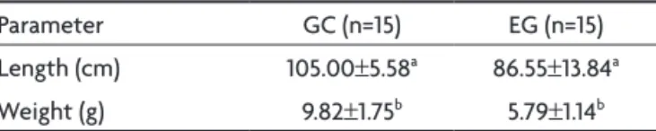
length and body weight of the ileum-jejunum was sig-niicantly low for the EG as described on Table 1.
results from the quantitative analysis of the neurons by three different techniques are described on Table 2.
DISCUSSION
The comparison of the final body weight between the CG and the EG demonstrated a difference of 41.75% (p<0.05). studies with different experimental models of malnutrition carried out with rats with different ages also demonstrated retarded body weight gain for the animals while fed with a 8% hypoprotein diet6,8,9,10,13. Body weight
Arq Neuropsiquiatr 2008;66(2-A)
244
Ileum myenteric plexus: protein deiciency Moreira et al.
Protein level reduction not only commits body devel-opment but also organs and system6. They respond
dif-ferently to malnutrition effects and the digestive tube is usually committed by the amino-acid deiciency5.
The ileum-jejunum was 17.6% smaller and 41% lighter for the EG which demonstrates that the protein level re-duction committed the normal development of the organ (p<0.05). while studying the protein availability reduction to 8% for rats during pregnancy, lactation, and weaning until their 60-day-old, it was observed a 45.21% reduction on the ileum length and wall thickness6. The small size
of the organ may be considered as an adaptive response either to the decreased amount of chow or the reduced metabolic rate19. The reduction of the organs is believed
to be a relex of its own tissue alterations as they may present different degrees of commitment according to their own structural and cellular organization6.
By comparing the total population density of the stained neurons with Giemsa technique, neurons were ex-pected to be 17.60% more concentrated as the organ was 17.60% smaller; however, the concentration was 17.02% - a very close value not statistically signiicant – indicating that there were not any reductions on the total popula-tion of neurons. Previous studies carried out on malnour-ished animals have also showed the preservation of total neuronal population in the ileum of rats6,9,10. other
experi-ments on different segexperi-ments in the small intestine did not evidence any myenteric neuron losses12,20 as well when
rats were submitted to different protein level
malnutri-tion. The protein level was probably suficient to ensure neuron survival in this study reinforcing the hypothesis that the reduction of chow during aging has demonstrated to be a neuronal population protection factor providing a longevity increase21,22.
Positive NADH-diaphorase neurons from the EG were 26.60% more concentrated. Malnutrition does not in-crease the number of total neurons as it is determined since the embryonic life, therefore, the neurons which were not from this class started to do it after malnutri-tion13. subpopulation of positive NADH-d neurons on CG
was 35.85% of the total neuronal population and 40.53% on the EG. As the neurons present the highest metabolic activity23, protein reduction seemed to have activated
neuronal metabolism in this study. rats submitted to 8% protein malnutrition for 120 days have also presented an increase with respect to this neuronal subpopulation in the duodenum12 and 96.7% higher in the jejunum13. on the
other hand, long-term diet restriction demonstrated that positive NADH-diaphorase subpopulation in the ileum de-creased for the 24-month-old animals without reducing total population. The authors associate such reduction to lower enzymatic activity relating to NADH-diaphorase as though it is not associated with neuronal death22.
NADPH-diaphorase neurons were 26.28% more con-centrated than those in the CG. Although NADPH-d his-tochemistry does not evidence neurons the overall neu-ronal population, it has been largely used for evidencing neurons presenting the No synthase responsible for the production of nitric oxide24 – an important mediator of
the intestinal relaxing. The nitrergic subpopulation on the CG was 16.3% and 18.34% on the EG, demonstrating ampliication. As the studied organ was 17.60% smaller, the natural concentration increase of the subpopulation would be 17.60%; however, 26.8% was veriied suggesting a 9.2% neuron increase expressing the Nos. It has been studied that the positive NADPH-d neurons are invulner-able to cellular death in animals under controlled diet22.
studies carried out on young and old rats submitted to chow intake restrictions showed that the NADPH-diapho-rase neurons reduced in average of 50% during the aging period when they were normofed in the CG. Neverthe-less, when they received a 50% normal diet, there were not any neuronal reductions showing that there were not any losses due to aging. In the same study, animals fed with just a 25% normal diet did not present any neuronal losses at 30 months of age as well22.
In this study, we observed that 4% protein malnutri-tion for 90 days of age resulted in the decrease of the body weight, as well as the organ size and weight. when comparing the neuronal density, we observed that mal-nutrition did not cause in any losses with respect to the Table 1. Length and weight of the ileum-jejunum of rats
normal-ly fed (Control Group – CG) and subjected to protein desnutri-tion (Experimental Group – EG).
Parameter GC (n=15) EG (n=15)
length (cm) 105.00±5.58a 86.55±13.84a
weight (g) 9.82±1.75b 5.79±1.14b
Data presented as mean±standard deviation. Means followed by the same letter on the same row showed signiicant difference (ap=0.001; bp<0.0001.
Table 2. Populational density of myenteric neurons of the ileum of rats normally fed (Control Group – CG) and subjected to protein
desnutrition (Experimental Group – EG) in an area of 25.2 mm2.
Technique CG (n=5) EG (n=5)
Giemsa 6,648.6±790.2 8,012.0±1,368.4
NADH-diaphorase 2,383.4±721.84a 3,247.0±337.15a
NADPH-diaphorase 1,083.4±170.37b 1,469.6±231.92b
Arq Neuropsiquiatr 2008;66(2-A)
245
Ileum myenteric plexus: protein deiciency Moreira et al.
overall neuronal population, and resulted in an increase of the positive NADH-diaphorase and NADPH-diaphorase neuron subpopulations possibly relecting the organ func-tional adaptation to protein deiciency.
REFERENCES
1. Brehmer A. Structure of enteric neurons. New york: Springer, 2006:1-5. 2. Furness JB, Costa M. The enteric nervous system.New York: Churchill
Livingstone, 2006:1-28.
3. Wade PR, Cowen T. Neurodegeneration: a key factor in the ageing. Gut Motil 2004;16:19-23.
4. Oliveira FLG. Aspectos clínicos e laboratoriais In Nobrega FJ (Ed). Distúrbios da nutrição: na infância e na adolescência.Rio de Janeiro: Revinter, 2007:195-198.
5. Deo MG. Cell biology of protein-calorie malnutrition. Wld Rev Nutr Diet 1978;32:49-95.
6. Torrejais MM, Natali MRM, Conegero CI, Miranda-Neto MH. Effects of proteic desnutrition after breast-feeding on the morphology of the intestinal wall and enteric neurons of the ileum of rats. Rev UNIMAR 1995;17:315-327.
7. Sarni ROS, Souza FIS. Tratamento da desnutrição energético-protéico
moderado e grave In Nobrega FJ (Ed). Distúrbios da nutrição:na
in-fância e na adolescência. Rio de Janeiro: Revinter, 2007:210. 8. Natali MRM, Miranda-Neto MH. Effects of maternal proteic
undernu-trition on the neurons of the myenteric plexus of duodenum of rats. Arq Neuropsiquiatr 1996;54:273-279.
9. Meillus M, Natali MRM, Miranda-Neto MH. Study of the myenter-ic plexus of the ileum of rats subjected to protemyenter-ic undernutrition. Rev Chil Anat 1998;16:9-14.
10. Fiorini A, Molinari SL, Natali MRM, Miranda-Neto MH. Quantitative morphological analysis of the myenteric neurons of the ileum in rats under experimental desnutrition. Acta Scientiarium 1999;21:404-423. 11. Natali MRM, Miranda-Neto MH, Orsi AM. Ultrastructural features of
myenteric ganglia of adult Wistar rats (Rattus norvegicus). Anat Histol Embryol 2000;29:393-397.
12. Natali MRM, Miranda-Neto MH, Orsi AM. Morphometry and quan-tiication of the myenteric neurons of the duodenum of adult rats fed
with hypoproteic chow. Inter J Morphology2003;21:273-277.
13. Zanin SMT, Molinari SL, Sant’Ana DMG, Miranda-Neto MH. Neurônios NADH-diaforase positivos do jejuno de ratos (Rattus norvegicus)
desnu-tridos. Arq Ciênc Saúde Unipar2003;61:650-653.
14. Araújo EJA, Sant’Ana DMG, Molinari SL, Miranda-Neto MH. Biomet-ric and food consumption parameters of rats subjected to hypoproteic and hypocaloric diet. Arq Ciên �et Zool Unipar 2005;8:133-140.Arq Ciên �et Zool Unipar 2005;8:133-140. 15. Pachaly JR, Sant’Ana DMG, Araújo EJA, Ciffoni EMG, Acco A.
Anes-thesiaof Wistar rats (rattus norvegicus) with allometrically scaled do-ses of ketamine xylazine acepromazine and atropine-prelimanary re-port. Arq Ciênc �et Zool UNIPAR 2003;3:195-197.
16. Barbosa AJA. Técnicahistológica para gânglios nervosos intramurais
em preparados espessos. Rev Bras Pesq Med Biol 1978;11:95-97. 17. Gabella G. Detection of nerve cells by a histochemical technique.
Expe-rientia 1969;23:218-219.
18. Scherer-Singler U, �icent SR, Kimura H, Megeer EG. Demonstration of a unique population of neurons with NADPH-diaphorase histochem-istry. J Neurosci Methods 1983;9:229-234.
19. Araújo AEJ, Sant’Ana DMG, Molinari SL, Miranda-Neto MH. Quantita-tive study of the myenteric plexus of the descending colon of young rats subjected to sintese protein deiciency. Int J Morphol 2006;24:591-597. 20. Natali MRM, Molinari SL, �alentini LC, Miranda-Neto MH. Morpho-quantitative evaluation of the duodenal myenteric neuronal popula-tion in ratos fed with hypoproteic rapopula-tion. Biocell 2004;29:39-46. 21. Johnson RJR, Schemann M, Santer RM, Cowen T. The effects of age on
the overal population and on sub-populations of myenteric neurons in the rat smal intestine. J Anat1998;192:479-488.
22. Cowen T, Johnson RJ, Soubeyre �, Santer RM. Restricted diet rescues rat enteric motor neurones from age related cell death. Gut Online2000; 47:653-660.
23. Sant’Ana DMG, Miranda-Neto MH, Souza RR, Molinari SL. Morplo-logical and quantitative atudy of the myenteric plexus of the ascend-ing colon of rats subjcted to proteic desnutrition. Arq Neuropsiquiatr 1997;55:687-695.