*Correspondence: M. Machinski Jr. Laboratório de Toxicologia, Departamento de Ciências Básicas da Saúde, Universidade Estadual de Maringá. Av. Colombo, 5790. 87020-900 – Maringá - PR, Brasil. E-mail: [email protected]
A
vol. 48, n. 3, jul./sep., 2012
Aflatoxin M
1in the urine of non-carriers and chronic carriers of
hepatitis B virus in Maringa, Brazil
Marcel Padovani Giolo
1, Christiane Minervino de Oliveira
1, Dennis Armando Bertolini
2, Maria
Valdrinez Campana Lonardoni
2, Matheus Sampaio Gouveia
3, Daisy Pontes Netto
4, Suzana Lucy
Nixdorf
3, Miguel Machinski Junior
1,*1Laboratory of Toxicology, Department of Basic Health Sciences, State University of Maringá, 2Laboratory of Immunology,
Department of Clinical Analysis and Biomedicine, State University of Maringá, 3Laboratory of Analytical Chemistry,
Department of Chemistry, State University of Londrina, 4Laboratory of Toxicology, Department of Preventive Veterinary
Medicine, State University of Londrina
Exposure to alatoxins (AFs) in the diet may favour the development of hepatocellular carcinoma (HCC) and the acute exacerbation of hepatitis in chronic hepatitis B virus (HBV) carriers. Measurement of biomarkers such as alatoxin M1 (AFM1), a metabolite of alatoxin B1 (AFB1), in urine allows for the
assessment of populations exposed to alatoxins. The aim of this study was to investigate the occurrence of alatoxin M1 in the urine of HBV carrier and non-carrier patients. One group included 43 randomly
selected HBV carriers treated at two hospitals in the city of Maringa, Brazil, from March to June 2008. Control group consisted of 29 healthy adult volunteers with anti-HBs positive and HBsAg negative test results. Detection of AFM1 was performed by luorescence using high performance liquid chromatography
(HPLC) and post-column derivation with the Kobra Cell®. Of the 72 samples analysed, 05/29 (17.2%)
AFM1 positive samples were from HBV non-carriers, and 16/43 (37.2%) of samples were from chronic
HBV carriers. This study showed AFM1 in the urine of the two surveyed population. However, there is
evidence that the chronic HBV carriers have a higher risk of developing HCC due to additive interaction between AFs and HBV.
Uniterms: Alatoxins. Biomarkers. Hepatitis B. Hepatocellular carcinoma. Urina/análise toxicológica. Epidemiology.
A exposição às alatoxinas (AFs) na dieta é um fator de risco para o desenvolvimento do carcinoma hepatocelular (CHC) e a exacerbação da hepatite aguda em indivíduos portadores do vírus da hepatite B (VHB). O uso de biomarcadores, como a alatoxina M1 (AFM1) na urina, produto de biotransformação
da alatoxina B1 (AFB1), permite avaliar se a população está exposta às AFs. O objetivo do presente
estudo foi investigar ocorrência de AFM1 na urina de portadores e não portadores crônicos do VHB.
Foi selecionado um grupo, de forma aleatória, representado por 43 portadores do VHB atendidos em dois hospitais da cidade de Maringá, Brasil, no período de Março a Junho/2008. O grupo controle foi composto por 29 voluntários adultos saudáveis anti-HBs positivo e HBsAg negativo. A determinação de AFM1 foi realizada por meio de detecção por luorescência em sistema de cromatograia a líquido
de alta eiciência com derivação pós-coluna utilizando Kobra Cell®. Das 72 amostras analisadas, 05/29
(17,2%) foram positivas para AFM1 em indivíduos não portadores do VHB, e 16/43 (37,2%) de pacientes
portadores do VHB. Este estudo demonstrou a ocorrência de AFM1 na urina dos dois grupos estudados.
Entretanto, há evidências de que os portadores do VHB possuam alto risco no desenvolvimento do CHC devido ao efeito aditivo pela interação entre alatoxinas e VHB.
Unitermos: Alatoxinas. Biomarcadores. Hepatite B. Carcinoma hepatocelular. Análise toxicológica.
INTRODUCTION
Aflatoxins (AFs) are a group of mycotoxins of major importance in foods such as peanuts and corn. The most important toxigenic fungi producing alatoxins are
Aspergillus flavus, A. parasiticus and A. nomius. The
four alatoxins naturally found in foods are B1 (AFB1), B2 (AFB2), G1 (AFG1) and G2 (AFG2). Alatoxin M1 (AFM1) is a hydroxylated product of AFB1 that can be secreted through body luids (Creppy, 2002). The action of these toxins can cause alterations in the growth of children and
adolescents, neurological and immunological disorders
and hepatocellular carcinoma (Eaton, Gropman, 1994;
Zhang et al., 2012).
Human exposure to alatoxins is a global concern, as studies have reported an association between exposure to alatoxins in the diet and infection with hepatitis B virus (HBV) that may favour the development of hepatocellular carcinoma (HCC) and the acute exacerbation of hepatitis in chronic HBV carriers (Peers et al., 1987; Kensler et al.,
2003). In areas with high incidence of HBV and alatoxin contaminated foods, such as sub-Saharan Africa, Southeast
Asia and China, the risk of HCC is high. Of the
550,000-600,000 new HCC cases worldwide each year, about 25,200-155,000 may be attributable to alatoxin exposure (Liu, Wu, 2010).
One method for estimating exposure to alatoxins in the diet is to determine the concentration and rate of consumption in contaminated foods, but several fac
-tors complicate this assessment. Thus, the detection of mycotoxins or their metabolites in body fluids such as milk, blood and urine allows a quantitative assessment of exposure (Groopman, Wogan, 1994; Bando et al., 2007).
AFB1, an indicator of recent exposure and AFB-albumin adducts, which is indicator of long-term exposure, can be detected in blood samples (Wang et al., 2001; Bando et al.,
2007). Alatoxin M1 (AFM1), alatoxin P1 (AFP1), alatoxin
Q1 (AFQ1) and AFB1-N7-guanine adducts are biotransfor -mation products that can be detected in urine (Wang et al.,
2001). The determination of AFM1 in urine is correlated with the amount of AFB1 ingested, and as such is a good biomarker of exposure (Groopman et al., 1992). The aim
of this study was to evaluate the occurrence of AFM1 in the urine of non-carriers and chronic carriers of HBV in the population of Maringa, southern Brazil.
MATERIAL AND METHODS
Study population
The study population consisted of 72 randomly se
-lected individuals divided into two groups: non-carriers and chronic HBV carriers. The group of non-carriers included 29 healthy adults with anti-HBs positive and HBsAg (surface antigen) negative results. All had been
vaccinated against HBV. Age, race and schooling level similar to the group exposed. The group of chronic HBV
carriers was composed of 43 patients adults diagnosed at two hospitals in the city of Maringa (Brazil) from March to June 2008, with HBsAg positive by more than six months. The voluntary participants each completed individual questionnaires. All participants signed a free and informed consent form. This study was approved by the Research Ethics Committee Involving Human Sub
-jects (COPEP), State University of Maringa (CAAE N. 0007.0.093.000-08).
Data collection
The self-administered questionnaire for chronic HBV carriers covered personal data, alcohol and medica
-tion use, dura-tion of HBV infec-tion and laboratory data. Personal data including age and gender was collected from HBV non-carriers. Participants were provided with instructions regarding completion of the questionnaire and the collection of biological materials.
Blood samples were collected from HBV non-carriers for detection of anti-HBs and HBsAg serologic markers. For both study groups, samples from irst morn
-ing urine were collected in a 300 mL polyethylene bottle, identiied and stored at -20 °C until analysis for AFM1.
Serological markers of HBV
Analysis of serologic markers for HBV antibody against surface antigen “S” (anti-HBs) and surface antigen
S (HBsAg) was performed using the AxSym® and Che
-miluminescence System methods, respectively, according to the manufacturer’s instructions.
Urine AFM1 analyses
AFM1 and β-glucuronidase were purchased from Sigma Chemicals Co. (St. Louis, MO, USA). The solvents and chemical reagents used were analytical grade aceto
-nitrile (Mallinckrodt Baker, Xalostoc, Mexico), HPLC grade methanol (Mallinckrodt Baker, Phillipsburg, NJ, USA), potassium bromide (F. Maia Indústria e Comércio Ltda, Cotia, Brazil), acetone and anhydrous sodium sul
(CAQ, Diadema, Brazil) and hydrochloric acid (Merck, Darmstadt, Germany). Nitrogen was purchased from AGA (Sundyberg, Switzerland) and Alaprep®immunoafinity columns were obtained from R-Biopharm Rhône Ltd. (Glasgow, Scotland).
AFM1 standard was diluted in acetonitrile to cre -ate a stock solution of 7.61 µg/mL. Concentration of the prepared solution was performed according to the meth
-odology 971.22 described by the Association of Oficial Analytical Chemists (AOAC, 2005). Working solutions for
the calibration curve, prepared by diluting in acetonitrile-water (30:70 v/v), were 0.01, 0.03, 0.1, 0.2 and 0.3 ng/mL. Urine samples were thawed at room temperature, homogenised by inversion and iltered through standard ilter paper. Ten mL of 0.1 M sodium acetate buffer pH 5.0 containing 0.4 mg/mL (500 U/mL) β-glucuronidase was added to 10 mL of iltered urine and incubated in a EV 015T water bath (Evlab, Londrina, Brazil) at 37 oC for 17 hours (Kussak, Andersson, 1995).
After hydrolysis, AFM1 was extracted from urine samples on immunoaffinity columns. The column was conditioned with 10 mL of Milli-Q Plus ultrapure wa
-ter (Millipore, Bedford, MA, USA). Twenty mL of the sample was added, the column was washed with 10 mL water, dried by air low, and AFM1 was eluted with 2 mL of acetonitrile in previously silanised tubes under a low of less than 2 mL/min. The eluate was concentrated under nitrogen low in a concentrator Tech Vap TE 0194 (Tecnal, Piracicaba, Brazil). The residue was frozen at -20 oC until
chromatographic determination.
The eluate was resuspended in 400 µL acetonitrile-water (30:70 v/v) and agitated in a KMC-1300V vortex (Vision, Korea). It was then filtered through a Sun-Sri (PTFE Titan 2 HPLC filter Yellow 17 mm, 0.45 µm) membrane. The standards and the iltered samples were analysed in an Alliance® HPLC system (Waters, Dublin, Ireland) attached to a quaternary pump and an automatic sample injection system at a volume of 100 µL. The luo
-rescence detector used was a 2475 model (Waters, Dublin, Ireland) with an excitation wavelength of 365 nm and emission at 440 nm. A phenyl C-18 column 250 x 4.6 mm with 5 µm particles was used (XTerra, Dublin, Ireland). The isocratic system included an acetonitrile-water (30:70 v/v) mobile phase containing 1 mM potassium bromide and 1 mM nitric acid at a low rate of 1.5 mL/min. Post-column derivation was performed with Kobra Cell® (R-Biopharm Rhône Ltd., Glasgow, Scotland) at 20A con
-nected to a 500 x 0.55 mm PTFE tube.
Quantiication of AFM1 was performed using the calibration curve obtained from working solutions (0.01 to 0.3 ng/mLor 0.8 to 24 pg/mL, depending on the dilu
-tion factor of the sample). Each solu-tion was injected three times and the average of the replicates was used to construct the calibration curve. Linearity of the calibration curve was 0.9949. The detection limit was 0.13 pg/mLand
the limit of quantiication for the method was 0.40 pg/mL. The AFM1 retention time was 5.0 minutes. Precision and accuracy (recovery) were 5.9% and 91.7% in a concentra
-tion of 10 pg/mL (n=6).
Statistical analysis
For the statistical evaluation between the means of AFM1 detected in the urine of individuals in each group, the “t test” with a 95% conidence interval was performed using BioEstat 5.0 and Sigma Statistic 2.3.
RESULTS
The HBV non-carriers included 29 subjects with a mean age of 33 years (20 to 57 years) who presented anti-HBs positive and anti-HBsAg antigen negative results. There were 5 males and 24 females in this group. There were 43 chronic HBV carriers with a mean age of 46 years (22 to 60 years). Of these, 26 were males and 17 were females. Questionnaires that were answered appropriately provided the following data: 86.5% of patients were Caucasian, 2.7% were Asian and 10.8% were African descent. The average time from diagnosis of HBV until the present study was six years. Laboratory tests previously carried out included: HBsAg (94.7%), HBs (57.9%), anti-HBe (52.6%), anti-HBc (47.4%), anti-HBeAg (36.8%) and anti-HBc IgM (31.6%). Only 9.5% of patients reported having undergone viral load testing for hepatitis B.
Regarding the consumption of alcoholic beverages, 73.7% of individuals reported that they do not drink and 26.3% reported that they consume alcohol ‘frequently’. Of the 25% of patients that underwent treatment, lamivudine (14.3%) and interferon (3.6%) were the most common medications used. Other unspecified medications ac
-counted for 7.1% of the HBV carrier population.
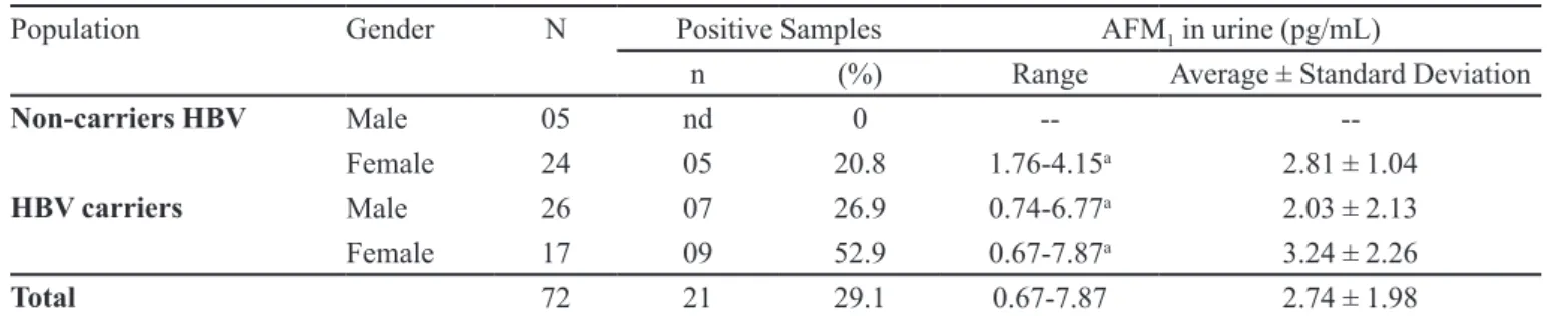
Of 72 urine samples obtained from non-carriers and chronic HBV carriers, 21 (29.2%) presented quantiiable levels of AFM1 in urine, ranging from 0.67 to 7.87 pg/mL (Table I).
positive for AFM1 in chronic HBV carriers. Nine (56.3%) were from female patients and 7 (43.8%) were from males, ranging between 0.6 to 8.0 pg/mL. The number of samples with concentrations ranging from 0.6 to 3.0, 3.01 to 5.50 and 5.51 to 8.00 pg/mL were 5, 3 and 1 for females, and 6, 0 and 1 for males, respectively.
DISCUSSION
The levels found of AFM1 in urine, 0.67 to
7.87 pg/mL (29.2%), were lower than the value reported by
Romero et al. (2010), who showed a prevalence of 65%,
with levels ranging from 1.8 to 39.9 pg/mLin a Brazilian population. The difference between the data may have oc
-curred, since in our study only one sample was analyzed for each subject, and the seasonal variation and dietary data were not investigated.
These results showed a lower incidence and con
-centration of AFM1 in urine than other studies. Jolly et al.
(2006) conducted a cross-sectional study in Ghana and detected AFM1 in the urine of 83 (91.2%) of the 91 partici -pants studied, ranging between 10 and 11,562.36 pg/mg creatinine. In the Czech Republic, the occurrence of AFM1 in urine was 57.6% of samples, ranging between 0.019 and 19.219 pg/mg creatinine (Malir et al., 2006). Cheng et al. (1997) analysed 32 urine samples from patients in
Taiwan and 138 from China, and AFM1 was found in 66% and 64% of the samples, respectively.
In China, Mykkanen et al.(2005) demonstrated
the presence of AFM1 in the urine of 47.3% of young individuals (18-24 years) with an average level of 40 pg/mL. Concentrations ranged between 10 and 330 pg/mL. The presence of this biotransformation product in urine was not statistically different between subjects presenting HBsAg positive and HBsAg negative results. Our results were similar to this study in that there was no signiicant statistical difference between the average level of AFM1 in the urine of individuals from the non-carrier group FIGURE 1 - Distribution of AFM1 levels in the urine of 29 HBV non-carriers (control) and 43 chronic HBV carriers in Maringá,
Brazil. (nd = not detected)
TABLE I - AFM1 in 72 urine samples from non-carriers and chronic HBV carriers from Maringa, Brazil
Population Gender N Positive Samples AFM1 in urine (pg/mL)
n (%) Range Average ± Standard Deviation
Non-carriers HBV Male 05 nd 0 --
--Female 24 05 20.8 1.76-4.15a 2.81 ± 1.04
HBV carriers Male 26 07 26.9 0.74-6.77a 2.03 ± 2.13
Female 17 09 52.9 0.67-7.87a 3.24 ± 2.26
Total 72 21 29.1 0.67-7.87 2.74 ± 1.98
and from the chronic HBV carrier group (p=0.925). There was no signiicant statistical difference when levels of AFM1 in the urine of females and males were compared (p=0.257). In our study, the group of chronic HBV carriers showed AFM1 in 16 (37.2%) of 43 samples analysed. This result showed that this population was exposed to AFB1, and is therefore at risk of developing HCC, as sev -eral studies have shown high morbidity from HCC due to exposure to alatoxins and a high rate of chronic HBV infection (Qian et al., 1994; Sun et al., 1999; Wang et al., 2001). Sun et al. (1999) demonstrated that detectable
urinary AFM1 levels above 3.6 ng/L were associated with increased 3.3-fold risk of HCC in male HBsAg carriers with chronic hepatitis. This fact is due to mutations in the third base of codon 249 of the p53 gene, since the studies showed a positive correlation between alatoxin exposure and HCC with mutation 249ser (Sun et al., 1999; Bando et
al., 2007). Anwar et al. (2008) concluded that have strate
-gies to reduce HCC with the use of markers of HBV, HCV (hepatitis C virus) and AFB1 exposure.
AFM1 is the major metabolite of AFB1 in quantita -tive terms and excretion represents exposure within a period of 24 hours of sampling (Eaton, Gropman. 1994). Thus, the levels of AFM1 in urine were used as a biomarker for short-term exposure to AFB1 (Gan et al., 1988). This study showed AFM1 in the urine of 29.2% of the surveyed population, although differences were not statistically sig
-niicant between the non-carrier group and chronic HBV carriers regarding recent exposure to AFB1. However, there is evidence that the chronic HBV carriers have a higher risk of developing HCC due to additive interaction between AFB1 and HBV (Wu et al., 2009). Further studies using long-term exposure biomarkers, such as AFB1-N7 -guanine and AF-albumin adducts, are required to assess the actual levels of long-term exposure to alatoxins in the Brazilian population.
ACKNOWLEDGEMENTS
We want to thank the Unidade Gestora do Fundo Paraná, Secretaria de Ciência, Tecnologia e Ensino Su-perior do Estado do Paraná - SETI-PR, TC N. 2807 for
inancial support. We also thank the volunteers and chronic HBV patients from the two hospitals in Maringa, Brazil.
REFERENCES
ANWAR, W.A.; KHALED, H.M.; AMRA, H.A.; EL-NEZAMI, H.; LOFFREDO, C.A. Changing pattern of hepatocellular carcinoma (HCC) and its risk factors in Egypt: possibilities for prevention. Mutat. Res., v.659, p.176-184, 2008.
ASSOCIATION OF OFFICIAL ANALYTICAL CHEMISTS
(AOAC). Oficial Methods of Analysis of the Association
of Oficial Analytical Chemists. Arlington: AOAC, 2005. chap.49, p.41-42.
BANDO, E.; GONÇALVES, L.N.; TAMURA, N.K.; MACHINSKI JR, M. Biomarkers for assessment of human exposure to mycotoxins. J. Bras. Patol. Med. Lab., v.43, p.175-180, 2007.
CHENG, Z.; ROOT, M.; PAN, W.; CHEN, J.; CAMPBELL, C.T.
Use of improved method for analysis of urinary alatoxin M1 in a survey of Mailand China e Taiwan. Cancer Epidemiol. Biomark. Prev., v.6, p.523-529, 1997.
CREPPY, E.E. Update of survey, regulation and toxic effects of mycotoxins in Europe. Toxicol. Lett., v.127, p.19-28, 2002.
EATON, D.L.; GROPMAN, J.D. The toxicology of alatoxins:
human health, veterinary, and agricultural signiicance. San Diego: Academic Press, 1994. 544 p.
GAN, L.S.; SKIPPER, P.L.; PENG, X.; GROOPMAN, J.D.;
CHEN, J.S.; WOGAN, G.N.; TANNENBAUM, S.R.
Serum albumin adducts in the molecular epidemiology of alatoxin carcinogenesis: correlation with alatoxin B1 intake and urinary excretion of alatoxin M1. Carcinogenesis, v.9, p.1323-1325, 1988.
GROOPMAN, J.D.; WOGAN, G.N. Molecular biomarkers for aflatoxinas and their application to human cancer
prevention. Cancer Res., v.54, p.1907-1911, 1994.
GROOPMAN, J.D.; ZHU, J.Q.; DONAHUE, P.R.; PIKUL, A.; LISHENG, Z.; JUN-SHI, C.; WOGAN, G.N. Molecular dosimetry of urinary aflatoxin-DNA adducts in people living in Guangxi Autonomous region, People’s Republic of China. Cancer Res., v.1, p.45-52, 1992.
JOLLY, P.; JIANG, Y.; ELLIS, W.; AWUAH, R.; NNEDU, O.; PHILLIPS, T.; WANG, J.S.; AFRIYIE-GYAWU, E.; TANG,
L.; PERSON, S.; WILLIAMS, J.; JOLLY, C. Determinants
of alatoxin levels in Ghanaians: sociodemographic factors knowledge of alatoxin and food handling and consumption
practices. Int. J. Hyg. Environ. Health, v.209, p.345-358,
2006.
KENSLER, T.W.; QIAN, G.S.; CHEN, J.G.; GROOPMAN,
KUSSAK, A.; ANDERSSON, K. Immunoaffinity column clean-up for the high-performace liquid chromatographic of alatoxinas B1, B2, G1, G2, M1 and Q1 in urine. J. Chromatogr.
B, v.672, p.253-259, 1995.
LIU, Y.; WU, F. Global burden of aflatoxin-induced
hepatocellular carcinoma: a risk assessment. Environ. Health Perspect., v.118, p.818-824, 2010.
MALIR, F.; OSTRY, V.; GROSSE, Y.; ROUBAL, T.; SKARKOVA, J.; RUPRICH, J. Monitoring the mycotoxin in food and their biomarkers in the Czech Republic. Mol. Nutr. Food Res., v.50, p.513-518, 2006.
MYKKANEN, H.; ZHU, H.; SALMINEN, E.; JUVONEN, R.O.; LING, W.; MA, J.; POLYCHRONAKI, N.;
KEMILAINEN, H.; MYKKANEN, O.; SALMINEN, S.
Fecal and urinary excretion of aflatoxin B1 metabolites
(AFQ1, AFM1 and AFB-N
7-guanine) in young Chinese
males. Int. J. Cancer, v.115, p.879-884, 2005.
PEERS, F.; BOSCH, X.; KALDOR, J.; LINDSELL, A.; PLUIJMEN, M. Aflatoxin exposure, hepatitis B virus infection and liver cancer in Swaziland. Int. J. Cancer, v.39,
p.545-553, 1987.
QIAN, G.S.; ROSS, R.K.; YU, M.C.; YUAN, J.M.; GAO, Y.T.; HENDERSON, B.E.; WOGAN, G.N.; GROOPMAN, J.D. A follow-up study of urinary markers of alatoxin exposure and liver cancer risk in Shanghai, People’s Republic of
China. Cancer Epidemiol. Biomark. Prev., v.3, p.3-10, 1994.
ROMERO, A.C.; FERREIRA, T.R.B.; DIAS, C.T.S.;
CALORI-DOMINGUES, M.A.; GLORIA, E.M. Occurrence of AFM1
in urine samples of a Brazilian population and association
with food consumption. Food Control, v.21, p.554-558,
2010.
SUN, Z.; LU, P.; GAIL, M.H.; PEE, D.; ZHANG, Q.; MING,
L.; WANG, J.; WU, Y.; LIU, G.; WU, Y.; ZHU, Y. Increase
risk of hepatocellular carcinoma in male hepatitis B surface antigen carriers with chronic hepatitis who have detectable urinary alatoxin metabolite M1. Hepatology, v.30, p.379-83, 1999.
WANG, J.S.; ABUBAKER, S.; HE, X.; SUN, G.; STRICKLAND, P.T.; GROOPMAN, J.D. Development of alatoxin B1-lysine adduct monoclonal antibody for human
exposure studies. Appl. Environ. Microbiol., v.67, p.2712-2717, 2001.
WU, H.C.; WANG, Q.; YANG, H.I.; AHSAN, H.; TSAI, W.Y.; WANG, L.Y.; CHEN, S.Y.; CHEN, C.J.; SANTELLA,
R.M. Alatoxin B1 exposure, hepatitis B virus infection, and hepatocellular carcinoma in Taiwan. Cancer Epidemiol. Biomark. Prev., v.18, p.846-853, 2009.
ZHANG, Y.J.; WU, H.C.; YAZICI, H.; YU, M.W.; LEE, P.H.; SANTELLA, R.M. Global hypomethylation in hepatocellular carcinoma and its relationship to alatoxin
B
1 exposure. World J. Hepatol., v.4, p.169-175, 2012.