356
Fernandes RCL et al. TCS findings in Parkinson’s disease and essential tremor
Radiol Bras. 2012 Nov/Dez;45(6):356–358
Transcranial sonography findings in Parkinson’s disease
and essential tremor: cases report
*
Achados de ultrassonografia transcraniana na doença de Parkinson e no tremor essencial: relato de casos
Rita de Cassia Leite Fernandes1, Ana Lucia Zuma de Rosso2, Maurice Borges Vincent3, Paulo Roberto Valle Bahia4, Celia Maria Coelho Resende5, Nordeval Cavalcante Araujo6
Over the last years, transcranial sonography has been investigated as a diagnostic tool in neurology. It allows a good visualization of midline brain structures, a frequent site of involvement in movement disorders. The authors discuss cases of Parkinson’s disease and essential tremor where transcranial sonography could suggest the diagnosis of the condition.
Keywords: Transcranial sonography; Parkinson’s disease; Essential tremor.
A ultrassonografia transcraniana tem sido objeto de investigação como ferramenta diagnóstica em neurologia nos últimos anos. Ela permite boa visualização de estruturas cerebrais situadas na linha média, sítio frequente de anormalidades nas doenças do movimento. Relatamos os casos de pacientes com a doença de Parkinson e o tremor essencial em que a ultrassonografia transcraniana foi capaz de sugerir o diagnóstico.
Unitermos: Ultrassonografia transcraniana; Doença de Parkinson; Tremor essencial.
Abstract
Resumo
* Study conducted at Hospital Universitário Clementino Fraga Filho – Universidade Federal do Rio de Janeiro (HUCFF-UFRJ), Rio de Janeiro, RJ, Brazil.
1. Master, Ministry of Health Physician, Fellow PhD degree, Post-graduation Program in Medical Practice (Concentration area: Neurology), Faculdade de Medicina da Universidade Federal do Rio de Janeiro (UFRJ), Rio de Janeiro, RJ, Brazil.
2. PhD, Head of Movement Disorders Clinic, Hospital Univer-sitário Clementino Fraga Filho – Universidade Federal do Rio de Janeiro (HUCFF-UFRJ), Rio de Janeiro, RJ, Brazil.
3. PhD, Assistant Professor, Universidade Federal do Rio de Janeiro (UFRJ), Head of the Neurologic Clinic, Hospital Universi-tário Clementino Fraga Filho – Universidade Federal do Rio de Janeiro (HUCFF-UFRJ), Rio de Janeiro, RJ, Brazil.
4. PhD, Assistant Professor, Universidade Federal do Rio de Janeiro (UFRJ), Head of the Radiodiagnosis Unit, Hospital Uni-versitário Clementino Fraga Filho – Universidade Federal do Rio de Janeiro (HUCFF-UFRJ), Rio de Janeiro, RJ, Brazil.
5. PhD, Head of the Unit of Ultrasonography, Hospital Univer-sitário Clementino Fraga Filho – Universidade Federal do Rio de Janeiro (HUCFF-UFRJ), Rio de Janeiro, RJ, Brazil.
6. PhD, Associate Professor of Post-graduation in Medical Sciences, Universidade do Estado do Rio de Janeiro (UERJ), Rio de Janeiro, RJ, Brazil.
Mailing Address: Dra. Rita C. L. Fernandes. Rua Marques de Abrantes, 171/502, Flamengo. Rio de Janeiro, RJ, Brazil, 22230-060. E-mail: [email protected]
Received May 30, 2012. Accepted after revision August 8, 2012.
Fernandes RCL, Rosso ALZ, Vincent MB, Bahia PRV, Resende CMC, Araujo NC. Transcranial sonography findings in Parkinson’s dis-ease and essential tremor: cases report. Radiol Bras. 2012 Nov/Dez;45(6):356–358.
0100-3984 © Colégio Brasileiro de Radiologia e Diagnóstico por Imagem CASE REPORT
eases coursing with parkinsonian symp-toms, particularly PD and ET(4). TCS is
harmless, low-cost, performed with widely available equipment and does not require patient sedation. The greatest limitation of this technique is dependence on the tempo-ral acoustic window to allow the ultrasound waves to overcome the bone barrier and generate two-dimensional images of intrac-ranial structures. This accounts for 10% to 20% of failure of the method to obtain use-ful diagnostic images(1).
Many authors reported key TCS finding in PD patients – expanded area of SN echogenicity > 0.20 cm –, in up to 90% of cases(5). In the Hospital Universitário Clementino Fraga Filho da Universidade Federal do Rio de Janeiro (HUCFF-UFRJ), a study is currently going on about the use-fulness of the method for the diagnosis of PD in the Brazilian population. In a pilot study, 88% of the PD patients presented expanded area of SN echogenicity, cor-roborating the good sensitivity of the method for detecting the disease(6).
How-ever, the finding of the marker in individu-als within the control group (findividu-alse-positive results), reduced the technique specificity to approximately 82%. Recent prospective ditions present distinctive clinical courses
– PD is more severe –, as well as distinc-tive therapeutic approaches. For that rea-son, neuroimaging studies such as brain computed tomography and magnetic reso-nance imaging are requested, however without providing a sure diagnosis of nei-ther of the two entities, restricting the role of those imaging methods to that of ruling out causes of secondary parkinsonism, such as hydrocephalus, cerebrovascular disease, etc.(2). Functional neuroimaging,
such as positron emission tomography (PET) or single-photon emission tomogra-phy (SPECT), might play a relevant role in the differential diagnosis because of their capability of identifying reduced striatal dopaminergic activity; but their high cost and limited availability outside of the large Brazilian research centers make their rou-tine utilization unfeasible(1,2).
Research on transcranial sonography (TCS) started in 1995 when investigators observed increased echogenicity at the mesencephalic substantia nigra (SN) site in patients with PD(3). The technique has been studied in several countries, with a high number of publications validating its use-fulness in the differential diagnosis of dis-INTRODUCTION
con-357
Fernandes RCL et al. TCS findings in Parkinson’s disease and essential tremor
Radiol Bras. 2012 Nov/Dez;45(6):356–358 multicentric studies suggest that healthy individuals with expanded SN area mea-sured at TCS present increased risk for developing PD(7), therefore making such
echographic change a candidate for a risk marker of the disease(1).
With the aim of presenting to the radi-ologists an example of the application of TCS, the authors present two cases of PD patients and ET patients paired by gender and age, where TCS could provide addi-tional diagnostic information regarding one or the other disease.
The present study was approved by the Committee for Ethics in Research of HUCFF-UFRJ. The patients were selected in the Movement Disorders Clinic of the HUCFF as follows: two PD patients diag-nosed according to the United Kingdom Brain Bank criteria(8); and two ET patients,
diagnosed according to the Movement Dis-orders Society criteria(9). The scans were
performed with an AcusonX300 equipment (Siemens; Erlangen, Germany) with a 1.5– 2.5 MHz phased-array transducer, as the previous described method(4,6). Axial
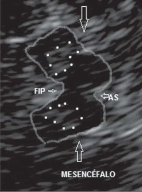
sec-tions were obtained bilaterally by insonat-ing the preauricular region. A hypoechoic, butterfly-shaped image of the mesencepha-lon (outlined on the figures), surrounded by the hyperechogenic basal cisterns was vi-sualized on the plane parallel to the orbitomeatal line. The hyperechogenic area of the SN visualized on the cerebral pe-duncle ipsilateral to the insonation region was measured. Values > 0.20 cm2 were
considered as being increased, according to the international consensus(4).
CASES REPORT
Case 1 – A 70-year-old woman with PD for ten years, at Hoehn and Yahr stage II. USTC demonstrated bilaterally increased echogenicity of the mesencephalic SN area (Figure1). The SN area at right measured 0.32 cm2 and, at left, 0.27 cm2.
Case 2 – A 51-year-old man with PD for 9 years, at Hoehn and Yahr stage II. The pa-tient presented important unilateral in-crease of the SN at ultrasonography: right SN echogenic area with 0.27 cm2 and the
left with 0.17 cm2 (Figure 2).
Case 3 – A 70-year-old woman with ET for 40 years. SN echogenicity area at right
measuring 0.12 cm2 and at left, 0.16 cm2
(Figure 3).
Case 4 – A 48-year-old man with ET since his youth. The right-sided SN echogenic area measured 0.11 cm2 and the
left-sided, 0.10 cm2 (Figure 4).
DISCUSSION
The images of the encephalon visual-ized at TCS provide important morphomet-ric data for the diagnosis of some neuro-logical disorders(4,5). Because of the
physi-Figure 4. Transcranial sonography – case 4. Left preauricular insonation in a patient with essential tremor. Outlined mesencephalon. The echogenic area of the left substantia nigra is outlined by dot-ted lines.
Figure 3. Transcranial sonography – case 3. Right preauricular insonation in a patient with essential tremor. Outlined mesencephalon. The echogenic area of the right substantia nigra is outlined on the cerebral peduncle. (SN, substantia nigra).
Figure 2. Transcranial sonography – case 2. Right preauricular insonation in a patient with Parkinson’s disease. Outlined mesencephalon. The echogenic area of the right substantia nigra is measured (A1 = 0.27 cm2). (SND, right substantia nigra). Figure 1. Transcranial sonography – case 1. Left
358
Fernandes RCL et al. TCS findings in Parkinson’s disease and essential tremor
Radiol Bras. 2012 Nov/Dez;45(6):356–358 cal characteristics of the method and the
need to overcome the bone barrier, the most appropriate image demonstrates structures located on the midline of the brain stem and basal ganglia(4). Such characteristics make
the method suitable for the study of move-ment disorders whose origin is the dysfunc-tion of such structures.
An echogenic SN area > 0.20 cm2, either
unilateral or bilaterally, is strongly corre-lated with PD diagnosis, and is considered as a stable marker since it does not undergo changes with the disease progression(3–5).
Unilateral SN enlargement is associated with the side of the symptoms(4). The four
described cases correspond to individuals with clinically confirmed diagnosis of both long-lasting PD and ET. The measurement of the echogenic area at the SN site could differentiate the two diseases. Such area was > 0.20 cm2 in the two PD cases (bilaterally
in case 1 and unilaterally in case 2), and was normal in the two ET cases (cases 3 and 4). Enlargement of the SN echogenic area may also be found in healthy individuals, in those with atypical parkinsonism (progres-sive supranuclear palsy, multisystems atro-phy, etc.), and even in ET patients, which would contribute to the reduction of the method specificity, besides(5,10).
The cause of increased SN echogenicity still remains subject of debate.
Consider-ing that such characteristic is present since the onset of the condition and does not change with the disease progression, it must not be caused by neuronal degenera-tion(4,7). There are evidences that the
in-creased iron content in the SN site might be responsible for the finding(11).
CONCLUSION
Considering that TCS is an innocuous and low-cost method, providing relevant information to the neurologist, TCS de-serves to be further investigated for the diagnosis of movement disorders. The limi-tations of this method include dependence on the temporal acoustic bone window and low specificity, which would place it in the category of screening methods(10). Some
Brazilian studies have already confirmed the validity of TCS findings for the diag-nosis of movement disorders among us(6,12).
REFERENCES
1. Stern MB, Lang A, Poewe W. Toward a redefini-tion of Parkinson’s disease. Mov Disord. 2012; 27:54–60.
2. Doepp F, Plotkin M, Siegel L, et al. Brain paren-chyma sonography and (123)I-FP-CIT SPECT in
Parkinson’s disease and essential tremor. Mov Disord. 2008;23:405–10.
3. Becker G, Seufert J, Bogdahn U, et al. Degenera-tion of substantia nigra in chronic Parkinson’s disease visualized by transcranial color-coded real-time sonography. Neurology. 1995;45:182–4.
4. Walter U, Behnke S, Eyding J, et al. Transcranial brain parenchyma sonography in movement dis-orders: state of the art. Ultrasound Med Biol. 2007;33:15–25.
5. Vlaar AMM, Bouwmans A, Mess WH, et al. Transcranial duplex in the differential diagnosis of parkinsonian syndromes: a systematic review. J Neurol. 2009;256:530–8.
6. Fernandes RCL, Rosso ALZ, Vincent MB, et al. Transcranial sonography as a diagnostic tool for Parkinson’s disease: a pilot study in the city of Rio de Janeiro, Brazil. Arq Neuropsiquiatr. 2011;69: 892–5.
7. Berg D, Seppi K, Behnke S, et al. Enlarged sub-stantia nigra hyperechogenicity and risk for Parkinson disease: a 37-month 3-center study of 1847 older persons. Arch Neurol. 2011;68:932– 7.
8. Hughes AJ, Daniel SE, Kilford L, et al. Accuracy of clinical diagnosis of idiopathic Parkinson’s disease: a clinico-pathological study of 100 cases. J Neurol Neurosurg Psychiatry. 1992;55:181–4.
9. Deuschl G, Bain P, Brin M. Consensus statement of the Movement Disorders Society on Tremor. Ad Hoc Scientific Committee. Mov Disord. 1998; 13 Suppl 3:2–23.
10. Lau…kait• K, Rastenyt• D, Òurkien• D, et al. Specificity of transcranial sonography in parkin-son spectrum disorders in compariparkin-son to degen-erative cognitive syndromes. BMC Neurol. 2012; 12:12.
11. Berg D, Roggendorf W, Schröder U, et al. Echo-genicity of the substantia nigra: association with increased iron content and marker for suscepti-bility to nigroestriatal injury. Arch Neurol. 2002; 59:999–1005.