353
Niemeyer B et al. Lethal midline granuloma syndrome
Radiol Bras. 2012 Nov/Dez;45(6):353–355
Lethal midline granuloma syndrome: a diagnostic dilemma
*
Síndrome do granuloma letal da linha média:um dilema diagnóstico
Bruno Niemeyer de Freitas Ribeiro1, Paulo Roberto Valle Bahia2, Ana Luiza Vianna Sobral de Magalhães Oliveira3, João Luiz Marchon Júnior4
The rare lethal midline granuloma syndrome is difficult to diagnose because of the wide array of related diseases and lack of knowledge by the majority of physicians. In the present report, the authors describe the case of a patient with this disease, caused by squamous cell carcinoma, drawing attention to differential diagnoses and to clinical and radiological findings that may be useful to define the diagnosis.
Keywords: Lethal midline granuloma; Non-Hodgkin’s lymphoma; Wegener’s granulomatosis; Squamous cell carci-noma.
A rara síndrome do granuloma letal da linha média apresenta difícil diagnóstico, em razão da grande variedade de doenças que podem causá-la e um desconhecimento pela maioria da classe médica. No presente artigo relatamos caso de paciente com esta doença, provocada por carcinoma epidermoide, chamando a atenção para os diagnósticos diferenciais e aspectos clínico-radiológicos que podem auxiliar no diagnóstico.
Unitermos: Granuloma letal da linha média; Linfoma não Hodgkin; Granulomatose de Wegener; Carcinoma epider-moide.
Abstract
Resumo
* Study developed at Hospital Universitário Clementino Fraga Filho – Universidade Federal do Rio de Janeiro (HUCFF-UFRJ), Rio de Janeiro, RJ, Brazil.
1. MD, Resident of Radiology, Hospital Universitário Clemen-tino Fraga Filho – Universidade Federal do Rio de Janeiro (HU-CFF-UFRJ), Rio de Janeiro, RJ, Brazil.
2. Assistant Professor, Department of Radiology, Universidade Federal do Rio de Janeiro (UFRJ), Head of the Unit of Radiology and Imaging Diagnosis, Hospital Universitário Clementino Fraga Filho – Universidade Federal do Rio de Janeiro (HUCFF-UFRJ), Rio de Janeiro, RJ, Brazil.
3. MD, Resident of Medical Practice, Hospital Federal da La-goa, Rio de Janeiro, RJ, Brazil.
4. MD, Radiologist, Head of the Unit of Computed Tomography, Hospital Federal da Lagoa, Rio de Janeiro, RJ, Brazil.
Mailing Address: Dr. Bruno Niemeyer de Freitas Ribeiro. Es-trada do Capenha, 1431, ap. 202, bloco 01, Freguesia, Jaca-repaguá. Rio de Janeiro, RJ, Brazil, 22743-041. E-mail: [email protected]
Received August 4, 2012. Accepted after revision Septem-ber 11, 2012.
Niemeyer B, Bahia PRV, Oliveira ALVSM, Marchon Júnior JL. Lethal midline granuloma syndrome: a diagnostic dilemma. Radiol Bras. 2012 Nov/Dez;45(6):353–355.
0100-3984 © Colégio Brasileiro de Radiologia e Diagnóstico por Imagem
CASE REPORT
gressing with fast growth and a predomi-nantly vegetative behavior.
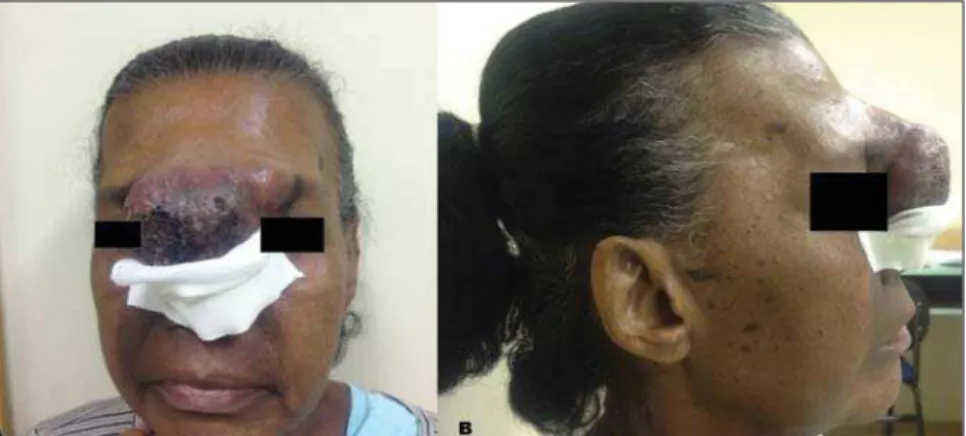
Three months after the first evaluation, the patient was admitted for investigation of an already large-sized lesion (Figure 1). Laboratory tests did not demonstrate any relevant findings, and fine-needle aspira-tion biopsy was inconclusive. Skull com-puted tomography (CT) (Figure 2) demon-strated the presence of a vegetative, het-erogeneous lesion in association with ex-tensive bone destruction predominantly af-fecting the nasal septum and the frontal sinus.
The majority of cases involve natural killer/T cell non-Hodgkin’s lymphoma and Wegener’s granulomatosis, and epidermoid carcinoma should be considered as differ-ential diagnosis since it is the most preva-lent malignant neoplasm of the head, cor-responding to 5% of all tumors occurring in the world population(2,3).
CASE REPORT
A female, black, 58-year-old patient presented a small ulcerative lesion in the glabellar region in February/2012,
pro-INTRODUCTION
Lethal midline granuloma syndrome (LMG) comprises a condition whose diag-nosis is difficult to be made because of the wide array of related diseases and nonspe-cific symptoms. Midline destructive le-sions of the face were first described in 1897, and later a variety of terms were coined to describe them. A factor that is common to all of such lesions is the devel-opment of an ulcerative/vegetative process culminating with destruction of the nasal region, resulting in functional and cosmetic deformity(1).
354
Niemeyer B et al. Lethal midline granuloma syndrome
Radiol Bras. 2012 Nov/Dez;45(6):353–355
Complementary magnetic resonance imaging (MRI) (Figure 3) performed 13 days after the CT scan demonstrated the presence of heterogeneous isointensity at T1-weighted, and mild hyperintensity at T2-weighted and FLAIR sequences, with subtle gadolinium enhancement. A syn-chronous lesion similar to the previously mentioned one was also observed, but without contiguity and with close contact with the maxillary palatine process.
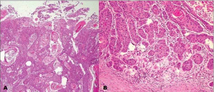
At follow-up, biopsy was performed (Figure 4), including the synchronous
le-sions, with anatomopathological results compatible with epidermoid carcinoma.
DISCUSSION
LMG is rarely found and hardly diag-nosed because of nonspecific symptoms, many times requiring several biopsies for a correct diagnosis(4).
Main diseases implied in this clinical condition include natural killer/T-cell non-Hodgkin’s lymphoma and Wegener’s granulomatosis, but with a wide range of
differential diagnoses, particularly epider-moid carcinoma, like in the present case.
Epidermoid carcinoma originates from suprabasal keratinocytes, predominantly affecting men, with incidence peak in the age range between 50 and 70 years. Risk factors are related to the disease location, thus smoking and alcohol consumption represent main risk factors for mucosal le-sions, while ultraviolet radiation exposure, chronic ulcers and fistulas, for skin lesions. Amongst head and neck neoplasms, epider-moid carcinoma is the most common ma-Figure 2. Contrast-enhanced skull computed
to-mography. Axial section demonstrate heteroge-neous vegetative lesion with soft tissue density, extensive bone destruction and subtle contrast-en-hancement, particularly in the periphery of the lesion.
Figure 3. A: Axial, FLAIR sequence demonstrating the presence of heterogeneous, mildly hyperintense lesion with significant midline destruction. B: Axial, T2-weighted STIR image demonstrating a small syn-chronous lesion (arrow) with subtle hypersignal intensity, adjacent to the lateral wall of the left nasal ostium, in close contact with the maxillary palatine process. Biopsy revealed the presence of epidermoid carci-noma.
355
Niemeyer B et al. Lethal midline granuloma syndrome
Radiol Bras. 2012 Nov/Dez;45(6):353–355
lignant neoplasia, corresponding to 5% of cancer cases.
The symptoms are generally insidious and are related to the lesion’s site of origin. Metastases usually involve lymph nodes, and the treatment of choice is surgery in association with radiotherapy and chemo-therapy in selected cases.
Both at CT and MRI, epidermoid car-cinomas are not distinctive from other le-sions, typically appearing with irregular margins, bone destruction and heteroge-neous contrast enhancement. In a study developed by Groell et al.(5) with 27 pa-tients with epidermoid carcinoma, it was suggested that delayed CT images acquired at 180 seconds following contrast injection would allow a better delimitation of the lesion.
It is interesting to note that 8%(2) of epi-dermoid carcinomas present as synchro-nous lesions, like in the present case, con-stituting an indication for positron emission tomography. Recent studies highlight the utilization of diffusion weighted MRI in the evaluation of LMG, demonstrating that apparent diffusion coefficient values < 1.22 × 10⁻³ mm2/s(6,7) are suggestive of the pres-ence of malignant lesions, and values < 0.84 × 10⁻³ mm2/s(6,7) are more compatible with lymphomas. In the present case, with the same protocol utilized in those studies, the lesion presented an apparent diffusion
coefficient of 0.91 × 10⁻³ mm2/s,
corrobo-rating the previously described findings. Differential diagnoses include princi-pally, among others, natural-killer/T-cell non-Hodgkin’s lymphoma, with highest prevalence in Asian individuals aged above 50 and in association with Epstein-Barr vi-rus. The differentiation of the disease by means of imaging methods remains diffi-cult. Lymph node enlargement represents an uncommon finding, bone involvement is less severe than in cases of epidermoid carcinoma, and involvement of the anterior wall of the maxillary sinus is rarely ob-served(8).
Wegener’s granulomatosis is another possible diagnosis to be considered, pref-erentially affecting men aged between 40 and 50 years, and classically presenting with airways lesion, glomerulonephritis and disseminated vasculitis. In such cases, c-ANCA testing plays a relevant role in the diagnosis.
Other differential diagnoses include polymorphic reticulosis, tuberculosis, leishmaniasis, cocaine abuse, giant cell granuloma, cholesterol granuloma and lobular capillary hemangioma, these last three conditions being associated with trauma.
Finally, the diagnosis of epidermoid car-cinoma should be considered in LMG, since it is the main head and neck
neo-plasm. Additionally, the presence of syn-chronous involvement should be investi-gated.
REFERENCES
1. Parker NP, Pearlman AN, Conley DB, et al. The dilemma of midline destructive lesions: a case se-ries and diagnostic review. Am J Otolaryngol. 2010;31:104–9.
2. Dammann F, Horger M, Mueller-Berg M, et al. Rational diagnosis of squamous cell carcinoma of the head and neck region: comparative evaluation of CT, MRI, and 18FDG PET. AJR Am J Roent-genol. 2005;184:1326–31.
3. Mendonça VF, Carvalho ACP, Freitas E, et al. Tu-mores malignos da cavidade nasal: avaliação por tomografia computadorizada. Radiol Bras. 2005; 38:175–80.
4. Lessa MM, Goto EY, Voegels RL, et al. Granuloma de linha media: revisão de 17 casos. Arq Int Otorri-nolaringol. 2001;5(1). [acessado em 29 de maio de 2012]. Disponível em: http://www.arquivosdeorl. org.br/conteudo/acervo_port.asp?id=146 5. Groell R, Doerfler O, Schaffler GJ, et al.
Contrast-enhanced helical CT of the head and neck: im-proved conspicuity of squamous cell carcinoma on delayed scans. AJR Am J Roentgenol. 2001;176: 1571–5.
6. Wang J, Takashima S, Takayama F, et al. Head and neck lesions: characterization with diffusion-weighted echo-planar MR imaging. Radiology. 2001;220:621–30.
7. Gonçalves FG, Ovalle JP, Grieb DFJ, et al. Diffu-sion in the head and neck: an assessment beyond the anatomy. Radiol Bras. 2011;44:308–14. 8. Ooi GC, Chim CS, Liang R, et al. Nasal