Sao Paulo Med J. 2015; 133(1):51-4 51
SHORT COMMUNICATION
DOI: 10.1590/1516-3180.2013.1912814Frequency of Epstein-Barr virus DNA sequences in
human gliomas
Frequência de sequências de DNA do vírus Epstein-Barr em gliomas humanos
Renata Fragelli Fonseca
I, Siane Lopes Bittencourt Rosas
II, José Antônio Oliveira
III, Anselmo Teixeira
III, Gilda Alves
IV,
Maria da Glória Costa Carvalho
VMolecular Pathology Laboratory, Pathology Department, Clementino Fraga Filho University Hospital, Universidade Federal do
Rio de Janeiro (UFRJ), and Applied Genetics Laboratory, Instituto Nacional de Câncer (INCA), Rio de Janeiro, Brazil
ABSTRACT
CONTEXT AND OBJECTIVE: The Epstein-Barr virus (EBV) is the most common cause of infectious mononucleosis and is also associated with several human tumors, including Burkitt’s lymphoma, Hodgkin’s lymphoma, some cases of gastric carcinoma and nasopharyngeal carcinoma, among other neoplasms. The aim of this study was to screen 75 primary gliomas for the presence of speciic EBV DNA sequences by means of the polymerase chain reaction (PCR), with conirmation by direct sequencing.
DESIGN AND SETTING: Prevalence study on EBV molecular genetics at a molecular pathology laboratory in a university hospital and at an applied genetics laboratory in a national institution.
METHODS: A total of 75 primary glioma biopsies and 6 others from other tumors from the central nervous system were obtained. The tissues were immediately frozen for subsequent DNA extraction by means of traditional methods using proteinase K digestion and extraction with a phenol-chloroform-isoamyl alcohol mixture. DNA was precipitated with ethanol, resuspended in bufer and stored. The PCRs were carried out using primers for ampliication of the EBV BamM region. Positive and negative controls were added to each reaction. The PCR products were used for direct sequencing for conirmation.
RESULTS: The viral sequences were positive in 11/75 (14.7%) of our samples.
CONCLUSION: The prevalence of EBV DNA was 11/75 (14.7%) in our glioma collection. Further molecular and epidemiological studies are needed to establish the possible role played by EBV in the tumorigenesis of gliomas.
RESUMO
CONTEXTO E OBJETIVO: O vírus Epstein-Barr (EBV) é a causa mais comum de mononucleose infecciosa e também está associado com vários tumores humanos, tais como: linfoma de Burkitt, linfoma de Hodgkin, alguns casos de carcinoma gástrico e carcinoma nasofaríngeo, entre outras neoplasias. O objetivo deste estudo foi rastrear a presença de sequências de DNA-especíicas do vírus EBV em biópsias de 75 gliomas primários usando reação em cadeia da polimerase (PCR), com conirmação por sequenciamento direto. TIPO DE ESTUDO E LOCAL: Estudo de prevalência sobre genética molecular de EBV em laboratório de patologia molecular de hospital universitário, e em laboratório de genética aplicada de instituição nacional. MÉTODOS: Foram obtidas 75 biópsias de gliomas primários e 6 de outros tumores do sistema nervoso central. Os tecidos foram imediatamente congelados para a extração posterior de DNA, através do método tradicional, usando a digestão por proteinase K e a extração pela mistura defenol-clorofórmio-álcool isolamílico. O DNA foi precipitado com etanol, ressuspendido em tampão e armazenado. As PCRs foram realizadas com iniciadores para ampliicar a região BamM do EBV. Controles positivos e negativos foram adicionados a cada reação. Os produtos de PCR foram usados para sequenciamento direto para conirmação.
RESULTADOS: As sequências virais foram positivas em 11/75 (14,7%) de nossas amostras.
CONCLUSÃO: A prevalência de DNA de EBV foi de 11/75 (14.7%) na nossa coleção de gliomas. Mais estudos moleculares e epidemiológicos são necessários para esclarecer o possível papel do EBV na tumorigênese dos gliomas.
IBSc, PhD. Postdoctoral Researcher, Congenital
Malformations Laboratory, Department of Genetics, Universidade Federal do Rio de Janeiro (UFRJ), Rio de Janeiro, Brazil.
IIBSc, PhD. Postdoctoral Researcher, Molecular
Oncology Laboratory, Clementino Fraga Filho University Hospital, Universidade Federal do Rio de Janeiro (UFRJ), Rio de Janeiro, Brazil.
IIIMD. Neurosurgeon, Neurosurgery Service,
Instituto Nacional de Câncer (INCA), Rio de Janeiro, Brazil.
IVPhD. Biologist, Instituto Nacional de Câncer
(INCA), Rio de Janeiro, Brazil.
VMD, PhD. Professor, Molecular Pathology
Laboratory, Pathology Department, Clementino Fraga Filho University Hospital, Universidade Federal do Rio de Janeiro (UFRJ), Rio de Janeiro, Brazil.
KEY WORDS: Astrocytoma. Glioma. Brain neoplasms. Polymerase chain reaction. Herpesvirus 4, human.
PALAVRAS-CHAVE: Astrocitoma. Glioma.
SHORT COMMUNICATION | Fonseca RF, Rosas SLB, Oliveira JA, Teixeira A, Alves G, Carvalho MGC
52 Sao Paulo Med J. 2015; 133(1):51-4
INTRODUCTION
Little is known about the etiology of gliomas, although they are the most common histological type of tumor in the central nervous system (CNS). Recently, it was suggested that some virus families could be important contributors towards glioma development. Viruses may contribute towards human tumor development by inducing immunosuppression, modifying host cells through inducing oncoproteins, or altering the expression of host cell proteins at viral integration sites.
Herpes viruses can infect humans easily. he timing of infec-tion is related to living condiinfec-tions, and several members of the herpes family possess known transformational properties, nota-bly the Epstein-Barr virus (EBV). About 90% of the world’s pop-ulation is estimated to be infected by EBV. Primary EBV infec-tion is spread mainly through saliva transfer between individuals. Following primary infection, which may be either symptomatic or silent, this virus has two distinct life cycles in the human host: a lytic cycle, during which the production of new virions occurs; and a latent form, which remains in the host.1
EBV is associated with several human malignancies includ-ing Burkitt’s lymphoma, Hodgkin’s disease, nasopharyngeal car-cinoma (NPC), peripheral T-cell lymphoma, thymoma and gas-tric cancer.2-5
EBV has been intensely studied, not only because of its ability to cause lifelong persistent infection but also because it is causally associated with a number of diseases in the CNS (infectious mono-nucleosis, demyelinating disease, acute encephalitis, acute cerebel-lar ataxia, myelitis or meningitis).6 EBV is thought to be
respon-sible for a number of neurological syndromes, such as difuse or focal encephalitis, aseptic meningitis, Guillain-Barré syndrome and peripheral neuropathy, among others.7,8 It has been
demon-strated that astrocyte cell lines and human fetal astrocytes are the only brain cells that express complement receptor type 2 (CR2), the major cellular receptor for EBV.9 EBV has been found to be able
to infect astrocyte cell lines.10 hese indings together support the
idea that EBV could act as an etiological agent in brain diseases.
OBJECTIVE
he aim of this study was to screen 75 primary astrocytomas for the presence of speciic EBV DNA sequences, by means of the polymerase chain reaction (PCR), with conirmation using direct sequencing.
METHODS
Patients and tumor samples
Between 1997 and 2001, a total of 75 primary glioma biopsies and six others from other tumors from the CNS were obtained during surgery performed by the neurosurgery service at a cancer treat-ment institution in the city of Rio de Janeiro, Brazil. his work was
approved by the institution’s ethics board, under registration number 35/02. he patients’ participation was voluntary and their data was conidential. To conirm the participation, the volunteer signed a clear statement of informed consent, ater having been informed of all aspects of the study. When the patient was underage, his/her parents were asked to sign the informed consent. he number of patients recruited was based on the number of new registrations in the Neurosurgery Service between the years 1997 and 2001.
he tissues were immediately snap-frozen in liquid nitro-gen, and subsequently stored at - 70 °C until deoxyribonucleic acid (DNA) extraction. Histological diagnoses were irst made on the specimens during surgery and were later on conirmed by the pathology service of the same institution. hese patients had no clinical evidence demonstrating that the tumors generated metastasis. We followed the World Health Organization (WHO) brain tumor classiication.
DNA extraction
he samples were subjected to proteinase K digestion (100 mg/ml) in the presence of 0.5% SDS at 37 °C overnight. his was followed by phenol-chloroform-isoamyl alcohol (25:24:1) extraction and etha-nol precipitation. he DNA was resuspended in bufer and stored at -20 °C until molecular analysis was performed.11
Polymerase chain reaction
he PCRs were carried out in a inal volume of 25 ml contain-ing 100 ng of DNA, 1 mM of each primer, 10 mM of Tris-HCl (pH 8.3), 50 mM of KCl, 1.5 mM of MgCl2, 200 µM of each nucleotide and 0.125 U of Taq polymerase. he primers were selected so as to amplify a 288 bp DNA product from the EBV BamM region: CAGGCTTCCCTGCAATTTTACAAGCGG and CCCAGAAGTATACGTGGTGACGTAGA.12 A negative DNA–
free control and a positive control from the EBV-positive Raji cell lineage were included in each assay.
hermal cycling was performed using the following condi-tions: initial denaturation at 95 °C for ive minutes; 40 cycles of ramping to 94 °C for one minute; cooling to 55 °C for two min-utes; heating to 72 °C for one minute; and a inal extension step at 72 °C for seven minutes. PCR fragments were separated by means of electrophoresis on 8% PAGE gel in 1X TBE bufer, co-migrat-ing with a 100 bp ladder marker, and this was followed by sil-ver staining as previously described.13 As an internal control for
DNA quality, we used primers to amplify exon 5 of the tumor sup-pressor gene TP53: GCAACCAGCCCTGTCGTGTCTCCA and GAATTCTGTTCACTTGTGCCCTGACTTTCAAC.14 All the samples
analyzed for the presence of EBV ampliied the suppressor gene TP53.
Sequencing reaction
Frequency of Epstein-Barr virus DNA sequences in human gliomas | SHORT COMMUNICATION
Sao Paulo Med J. 2015; 133(1):51-4 53 UK), in accordance with the manufacturer’s instructions, and
this was used for direct sequencing in an ABI 3130 device (Life Technologies, CA, USA). he alignments were obtained through the GenBank online Blast-N sotware, which is available from the National Center for Biotechnology Information (http://www. ncbi.nlm.nih.gov).
RESULTS
he glioma specimens were classiied as astrocytoma grade I (7/75), astrocytoma grade II (25/75), astrocytoma grade III (16/75), glioblastoma multiforme (18/75), ependymoma (5/75), oligodendroglioma (2/75) and oligoastrocytoma (2/75). hese tumors were from 40 males and 35 females; 25 patients were chil-dren (under 21 years of age) and 50 were adults. he location of the tumors in the brain was variable: temporal lobe, fourth ven-tricle, thalamus, cerebellar vermis, brain stem, cerebral venven-tricle, supra tentorial, occipital lobe and parietal lobe.
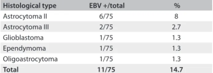
he DNA extracted from the gliomas was screened for the presence of EBV DNA. he result was positive in 11/75 (14.7%) of the tumor specimens. he tumors positive for EBV DNA were: astrocytoma grade II (6/11); astrocytoma grade III (2/11); oli-goastrocytoma (1/11); ependymoma (1/11); and glioblastoma multiforme (1/11) (Table 1).
Figure 1 shows a gel containing positive PCR reactions.
Additionally, the presence of EBV DNA was also screened for in other CNS tumors (one neurilemma, two non-Hodgkin lympho-mas, two pituitary adenomas and one cortical dysplasia) and they were all negative for EBV DNA.
he homology between the EBV sequences found in the astrocytoma patients was compared with the published EBV sequence, by examining their similarity. he identicalness rate observed (95.5%) conirmed that the virus found was EBV.
DISCUSSION
Over the past decade, several groups have examined the evidence for the presence and expression of viruses in CNS tumors and have analyzed the quantity of viral material. Reports on SV40 (simian virus 40), JCV (John Cunningham virus) and HCMV (human cytomegalovirus) have been made, with diverse indings.15-24 More recently, JCV and HCMV have been accepted
as being associated with CNS tumors.25 Discordant indings may
occur because of the sensitivity of the PCR/in situ techniques used, diiculty in working with parain-embedded tissues and also small numbers of samples. In the present study, it is important to note that we used 75 fresh frozen tissue samples and conventional PCR, which has the advantage of amplifying small target sequences. In this study, in order to search for EBV DNA in diferent histological types/grades (Table 1), we used the simple method of conventional PCR, which had been used in previous work.12 he prevalence of EBV DNA found was 11/75 (14.7%).
here is evidence showing that EBV emerges from latency in immunosuppressed individuals. Many cancer patients pres-ent immunosuppression, which may be caused by aging, by the disease itself or even by medications, and EBV and other silent viruses may be activated in this manner.26,27 he EBV genome is
complex and comprises at least 12 known genes; however, the most famous EBV transforming protein is LMP1 (latent-infec-tion membrane protein 1).28 In spite of all of this knowledge, the
role of EBV in glioma tumorigenesis is still not understood. An EBV vaccine has been tested (in a phase I clinical trial) on Chinese nasopharyngeal carcinoma patients to determine the safe and immunogenic dose.29 In that study, it was concluded
that the vaccine is both safe and immunogenic, thus allowing the highest dose to be moved forward to phase II studies. From our data, it is possible that in the future, in the same way as seen among those nasopharyngeal carcinoma patients, tests using an EBV vaccine may be found to beneit glioma patients.
CONCLUSION
he prevalence of EBV DNA was 11/75 (14.7%) in our glioma samples. Further molecular and epidemiological studies are needed in order to establish the possible role played by EBV in the tumorigenesis of gliomas.
REFERENCES
1. Pattle SB, Farrell PJ. The role of Epstein-Barr virus in cancer. Expert
Opin Biol Ther. 2006;6(11):1193-205.
2. Weiss LM, Strickler JG, Warnke RA, Purtilo DT, Sklar J. Epstein-Barr viral
DNA in tissues of Hodgkin’s disease. Am J Pathol. 1987;129(1):86-91.
Histological type EBV +/total %
Astrocytoma II 6/75 8
Astrocytoma III 2/75 2.7
Glioblastoma 1/75 1.3
Ependymoma 1/75 1.3
Oligoastrocytoma 1/75 1.3
Total 11/75 14.7
Table 1. Histological subtype and number of samples positive for Epstein-Barr virus (EBV) deoxyribonucleic acid (DNA)
Figure 1. Silver-stained 8% PAGE gel showing examples of polymerase chain reaction (PCR) products from the Epstein-Barr virus (EBV), from glioma biopsies.
SHORT COMMUNICATION | Fonseca RF, Rosas SLB, Oliveira JA, Teixeira A, Alves G, Carvalho MGC
54 Sao Paulo Med J. 2015; 133(1):51-4
3. Musacchio JG, Carvalho Mda G, Morais JC, et al. Detection of
free circulating Epstein-Barr virus DNA in plasma of patients with
Hodgkin’s disease. Sao Paulo Med J. 2006;124(3):154-7.
4. Lima MAP, Rabenhorst SHB. Associação do vírus Epstein-Barr (EBV)
com tumores sólidos [Association of Epstein-Barr virus (EBV) with
solid tumors]. Rev Bras Cancerol. 2006;52(1):87-96.
5. de Aquino PF, Carvalho PC, da Gama Fischer JS, et al. Epstein-Barr
virus DNA associated with gastric adenocarcinoma and adjacent
non-cancerous mucosa in patients from Manaus, Brazil. Genet Mol
Res. 2012;11(4):4442-6.
6. Fujimoto H, Asaoka K, Imaizumi T, et al. Epstein-Barr virus infections of
the central nervous system. Intern Med. 2003;42(1):33-40.
7. Cleary TG, Henle W, Pickering LK. Acute cerebellar ataxia associated
with Epstein-Barr virus infection. JAMA. 1980;243(2):148-9.
8. Gilbert JW, Culebras A. Cerebellitis in infectious mononucleosis.
JAMA. 1992;220(5):727.
9. Gasque P, Chan P, Mauger C, et al. Identiication and characterization
of complement C3 receptors on human astrocytes. J Immunol.
1996;156(6):2247-55.
10. Menet A, Speth C, Larcher C, et al. Epstein-Barr virus infection of
human astrocyte cell lines. J Virol. 1999;73(9):7722-33.
11. Martins GA, Gattas CR, da Costa Carvalho MG. Free DNA induces
modiication on the protein synthesis proile of human peripheral
blood mononuclear cells of healthy donors. Int J Mol Med.
2000;5(5):511-3.
12. Saito I, Servenius B, Compton T, Fox RI. Detection of Epstein-Barr virus
DNA by polymerase chain reaction in blood and tissue biopsies from
patients with Sjogren’s syndrome. J Exp Med. 1989;169(6):2191-8.
13. Rosenbaum V, Riesner D. Temperature-gradient gel electrophoresis.
Thermodynamic analysis of nucleic acids and proteins in puriied
form and in cellular extracts. Biophys Chem. 1987;26(2-3):235-46.
14. Pestaner JP, Bibbo M, Bobroski L, Seshamma T, Bagasra O. Potential
of the in situ polymerase chain reaction in diagnostic cytology. Acta
Cytol. 1994;38(5):676-80.
15. Kouhata T, Fukuyama K, Hagihara N, Tabushi K. Detection of simian
virus 40 DNA sequence in human primary glioblastomas multiforme.
J Neurosurg. 2001;95(1):96-101.
16. Poltermann S, Schlehofer B, Steindorf K, et al. Lack of association of
herpesviruses with brain tumors. J Neurovirol. 2006;12(2):90-9.
17. Neves AM, Thompson G, Carvalheira J, et al. Detection and
quantitative analysis of human herpesvirus in pilocytic astrocytoma.
Brain Res. 2008;1221:108-14.
18. Bhattacharjee B, Renzette N, Kowalik TF. Genetic analysis of
cytomegalovirus in malignant gliomas. J Virol. 2012;86(12):6815-24.
19. Cobbs CS, Harkins L, Samanta M, et al. Human cytomegalovirus
infection and expression in human malignant glioma. Cancer Res.
2002;62(12):3347-50.
20. Sabatier J, Uro-Coste E, Pommepuy I, et al. Detection of human
cytomegalovirus genome and gene products in central nervous
system tumours. Br J Cancer. 2005;92(4):747-50.
21. Lau SK, Chen YY, Chen WG, et al. Lack of association of cytomegalovirus
with human brain tumors. Mod Pathol. 2005;18(6):838-43.
22. Mitchell DA, Xie W, Schmittling R, et al. Sensitive detection of
human cytomegalovirus in tumors and peripheral blood of patients
diagnosed with glioblastoma. Neuro Oncol. 2008;10(1):10-8.
23. Lucas KG, Bao L, Bruggeman R, Dunham K, Specht C. The detection
of CMV pp65 and IE1 in glioblastoma multiforme. J Neurooncol.
2011;103(2):231-8.
24. Fonseca RF, Kawamura MT, Oliveira JA, et al. The prevalence of human
cytomegalovirus DNA in gliomas of Brazilian patients. Mem Inst
Oswaldo Cruz. 2012;107(7):953-4.
25. Alibek K, Kakpenova A, Baiken Y. Role of infectious agents in the
carcinogenesis of brain and head and neck cancers. Infect Agent
Cancer. 2013;8(1):7.
26. Martelius T, Lappalainen M, Palomäki M, Anttila VJ. Clinical
characteristics of patients with Epstein Barr virus in cerebrospinal
luid. BMC Infect Dis. 2011;11:281.
27. Kleinschmidt-DeMasters BK, Damek DM, Lillehei KO, Dogan A,
Giannini C. Epstein Barr virus-associated primary CNS lymphomas in
elderly patients on immunosuppressive medications. J Neuropathol
Exp Neurol. 2008;67(11):1103-11.
28. Kaye KM, Izumi KM, Kief E. Epstein-Barr virus latent membrane
protein 1 is essential for B-lymphocyte growth transformation. Proc
Natl Acad Sci U S A. 1993;90(19):9150-4
29. Hui EP, Taylor GS, Jia H, et al. Phase I trial of recombinant modiied
vaccinia ankara encoding Epstein-Barr viral tumor antigens in
nasopharyngeal carcinoma patients. Cancer Res. 2013;73(6):1676-88.
Acknowledgements: We thank Marcelo Soares Mota for technical assistance and all members of the Neurosurgery Service (INCA)
Sources of funding: This work was supported by Conselho Nacional de Desenvolvimento Cientíico e Tecnológico (CNPq) (Brazil), number
302924/2001-6; and Programa de Oncobiologia (Rio de Janeiro, Brazil)
Conlict of interest: None
Date of irst submission: December 19, 2013 Last received: September 13, 2014 Accepted: September 23, 2014
Address for correspondence: Gilda Alves Brown
Laboratório de Genética Aplicada
Serviço de Hematologia
Instituto Nacional de Câncer
Praça da Cruz Vermelha 23, 6o andar
Rio de Janeiro (RJ) — Brasil
CEP 20230-130
Tel. (+55 21) 3207-1805