ABSTRACT
Effect of leaching residual methyl methacrylate
concentrations on
in vitro
cytotoxicity of heat
polymerized denture base acrylic resin processed
with different polymerization cycles
Canan BURAL1 2, Günnur DENIZ34, Gülsen BAYRAKTAR5
1- DDS, PhD, Assistant Professor, Department Prosthodontics, Istanbul University, Faculty of Dentistry, Istanbul, Turkey.
2- MsC, PhD, Assistant Professor, Department of Immunology, Institute of Experimental Medicine, Istanbul University, Istanbul, Turkey. 3- MsC, PhD, Professor, Department of Immunology, Institute of Experimental Medicine, Istanbul University, Istanbul, Turkey. 4- DMD, Professor, Department of Biochemistry, Istanbul Medical Faculty, Istanbul University, Istanbul, Turkey.
5- DDS, PhD, Professor, Department of Removable Prosthodontics Istanbul University, Faculty of Dentistry, Istanbul, Turkey.
Corresponding address: Dr. Canan Bural - Istanbul University, Faculty of Dentistry - Department of Removable Prosthodontics - Capa, 34390 - Istanbul - Turkey - Phone: +90212 414 20 20 (ext: 30256) - Fax: +90212 525 35 85 - e-mail: [email protected]
!!"#$%&'(%$')*+!!#-($'7!!
O
bjectives: Residual methyl methacrylate (MMA) may leach from the acrylic resin denture bases and have adverse effects on the oral mucosa. This in vitro study evaluated andcorrelated the effect of the leaching residual MMA concentrations ([MMA]r) on in vitro
! acrylic resin specimens were fabricated using 4 different polymerization cycles: (1) at 74ºC for 9 h, (2) at 74ºC for 9 h and terminal boiling (at 100ºC) for 30 min, (3) at 74ºC for 9 h and terminal boiling for 3 h, (4) at 74ºC for 30 min and terminal boiling for 30 min. Specimens were eluted in a complete cell culture medium at 37ºC for 1, 2, 5 and 7 days. [MMA]r in eluates was measured using high-performance liquid chromatography. In vitro
"#$" using a tetrazolium salt XTT (sodium 3´-[1-phenyl-aminocarbonyl)-3,4-tetrazolium]bis(4-methoxy-6-nitro)benzenesulphonic acid) assay. Differences in [MMA]r of eluates and cell proliferation values between polymerization cycles were statistically analyzed by Kruskal-Wallis, Friedman and Dunn’s multiple comparison tests. The correlation between [MMA]r of eluates and cell proliferation was analyzed by Pearson’s correlation test (p<0.05). Results:
[MMA]r#%&'***+%" !#
without terminal boiling after elution of 1 and 2 days. Cell proliferation values for all cycles #%&/**+#" 8 between [MMA]r and cell proliferation values was negative after all elution periods, showing %&/**;+ " " resin throughout 7 days and leaching concentrations markedly reduced after elution of 1 and 2 days. Conclusion: Due to reduction of leaching residual MMA concentrations, use of terminal boiling in the polymerization process for at least 30 min and water storage of the heat-polymerized denture bases for at least 1 to 2 days before denture delivery is clinically recommended for minimizing the residual MMA and possible cytotoxic effects.
Key words: Acrylic resins. Denture bases. Methylmethacrylate. Cytotoxicity. Cell
polymerization cycles
INTRODUCTION
Heat-polymerization is the most widely used method of polymerization for acrylic resin denture base fabrication and usually is accomplished in a
heated water-bath10,12,26.
The nature and duration of the conditions to which the molded acrylic resin subjected is
described as the polymerization cycle14,26. The
conventional method of polymerization cycle is a long, slow-temperature water-bath polymerization where the heat-polymerization of acrylic resin is
processed for 9 h at 74ºC26. There are also
post-polymerization cycles such as terminal boiling at 100ºC for durations of 30 min (short-term) or
longer than 1 h (long-term)10,12,14,32. Furthermore,
it has been reported that total polymerization time shorter than 2 h is widely preferred than the long
polymerization cycles3.
Although there has been several reported variations of polymerization temperature and time, the conversion of monomer is not complete and this might result in unreacted, residual monomer
in the denture base acrylic resin2-5,10,12,14,25. Methyl
methacrylate (MMA) was the predominant residual
monomer in the acrylic resin32. In addition, it has
been stated that the residual MMA content might
change due to polymerization method4,5,31 and
cycle10,12,14,25,32.
Denture base is in continuous contact with the great part of the oral mucosa. It is important to evaluate the effect of the residual monomer, which
has been shown to leach into water21,31,32,saliva2,4,30
$20, on the oral mucosa that is
adjacent to the denture base. Leaching residual monomer have been suggested as potentially high enough to cause irritation of oral mucosa, irritation or even an allergic reaction. This might especially # =>
lacerated mucosa11,23.
In vitro cytotoxic effects of denture base
acrylic resins have also been attributed to the
leaching components4,8,15,17-19,22-24,30. Although there
are a number of reports of in vitro cytotoxicity
of denture base acrylic resins, which were processed with various polymerization methods and cycles8,11,16-19,22-24,30, there is no available study
that correlated in vitro cytotoxicity and the leaching
concentrations of residual MMA ([MMA]r) of
heat-polymerized denture base acrylic resin, processed with different polymerization cycles. The aims of
the present study were to evaluate 1) [MMA]r, 2)
in vitro cytotoxicity, 3) the correlation between the
leaching [MMA]r and in vitro cytotoxicity of
heat-polymerized specimens, heat-polymerized with different polymerization cycles after elution in a cell culture medium for 1, 2, 5 and 7 days. The hypothesis was that terminal boiling would reduce the leaching
[MMA]r and in vitro cytotoxicity of heat-polymerized
denture base acrylic resin.
MATERIAL AND METHODS
Specimen preparation
Stainless steel discs (1 mm thick x 10 mm
diameter)22-24 were conventionally molded in Type
II dental stone (Moldano, Heraus Kulzer, Germany) with a powder/liquid ratio of 100 g/30 mL under aseptic conditions. Flasks were kept under hydraulic pressure (Kavo Elektrotechnisches Werk, GmBH, Allgäu, Germany) of 2 atm for 45 min.
Heat-polymerizing, PMMA based denture base acrylic resin (Meliodent Heat-cure Denture JNQ"U"!VJQYZ=Q"= Germany) without cadmium was tested in the present study. Acrylic resin was mixed in accordance with manufacturers’ recommendations, with a powder/liquid ratio of 23.4 g/10 mL for 60 s at room temperature (23±2ºC). After 5 min of doughing time, unpolymerized resin was packed in molds and >\#\""" 2 atm for 45 min.
H e a t- p o l y m e r i z a t i o n wa s p e r f o r m e d i n thermostatically controlled water bath (Kavo EWL Typ 5506; Kavo Elektronisches Werk) with 4 different polymerization cycles (Figure 1). After completion !=>\# at room temperature (23±2ºC) for 2 h. Specimens were transferred into sterile centrifuge tubes (TPP Centrifuge Tubes, Switzerland), containing 50 mL of distilled water at room temperature (23±2ºC) and then ultrasonically cleaned (Metu Elektromekanik;
Ultrasonic Cleaner, Istanbul, Turkey) for 5 min16-19.
Thirty six (n=36) acrylic resins specimens were fabricated for each polymerization cycle with a total of 144 specimens.
Eluate preparation
Complete cell culture medium without serum11
was used as elution medium. Eluates of specimens were prepared by placing 3 disks into a sterile vial
with 9 mL11,17 {"|%"
(DMEM)/ F-12 (Biological Industries, Haemek, Israel) supplemented with 1% antibiotics solution (100 IU/mL penicillin, 100 μg/mL streptomycin, 25 μg/mL Amfoterisin-B; Biological Industries), 1% vitamin solution (MEM-Vitamins Solution 100X; Biological Industries), 2% non-essential amino acid solution (MEM-Eagle non-essential amino acid solution 100X; Biological Industries), and 1% L-Glutamine (L-Glutamine Solution; 200 mM, Biological Industries). The DMEM was maintained at pH 7.3 by adding 25 mM HEPES (HEPES BUFFER;
Biological Industries)7. The ratio of surface area
of the discs to the volume of culture medium was
a recommended polymerization cycle by the manufacturer
Code Polymerization cycle
H1 at 74ºC, for 9 h (Conventional)
H2 at 74ºC, for 9 h + at 100ºC, for 30 min (Short-term terminal boil)
H3 at 74ºC, for 9 h + at 100ºC, for 3 h (Long-term terminal boil)
H4a at 74ºC, for 30 min + at 100ºC, for 30 min (Short-term polymerization)
Figure 1- Polymerization cycles used
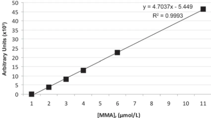
Figure 2- Standard calibration curve for methyl methacrylate (MMA)
cm2/mL as recommended by the International
Standards Organization (ISO) 10993-513.
Specimens were eluted with complete cell culture medium at 37ºC for 1, 2, 5 and 7 days in a
" ;Z2, 95% air. After
each elution period, the eluates were removed and the specimens were transferred into new vials and fresh cell culture medium. Cell culture media without acrylic resin specimens were also incubated
to serve as negative controls11,16-19.Eluates were
!* " (FCS Heat-inactivated; Biological Industries) were added. Eluates were stored at -20ºC until the determination of the concentration of leaching
residual MMA and in vitro cytotoxicity tests.
Determination of leaching residual MMA concentration ([MMA]r)
[MMA]r in eluates was determined using High
Performance Liquid Chromatography (HPLC) with the HPLC pump (Waters 600 E, Millford, MA, USA) equipped with a gradient controller (Waters Model 600), autosampler (Waters 717 plus), tunable UV-Vis detector (Waters 486) and a reversed phase C18 with stainless steel analytical column (μ Bondapak 3.9x300 mm, 10 μ particle size, 125 Aº).
The analysis was performed at room temperature (23±2ºC) under the following conditions: chromatographic grade methanol (Merck, KGaA, Darmstadt, Germany)/distilled water (1:1) mobile N*>#* nm.
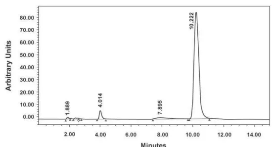
Known serial concentrations of 1, 2, 3, 5 and 10 μmol/L (standards) of MMA dissolved in methanol was analyzed and a calibration curve (Figure 2) was obtained using chromatographic MMA peak at retention time of 10.22 min (Figure 3).
Eluates were diluted with methanol (1:5 v/v) and injected into column with 10 μL volume. Peak area of each eluate was put into equation obtained
from the calibration curve (Figure 2) and [MMA]r in
each eluate was expressed as μmol/L.
Twenty-four chromatographic analyses for each polymerization cycle and 6 for each elution period with a total of 96 analyses were performed.
Cell culture
L-929 murine fibroblasts (American Type Z"" Z= ZZ = Z8Z 929) were used in the study. Cells were cultured
in 75 cm2 culture flasks (TPP, Tissue Culture
Dish, Switzerland) with the complete cell culture medium described above and incubated at 37ºC
" ;Z2, 95% air.
Cell proliferation
Figure 3- High performance liquid cromatography chromatogram of methyl methacrylate (MMA) and characteristic peak at approximately 10.22 min of retention time.
polymerization cycles
and an activation solution. 5x10³ cells were plated
in each well of 96 well-plates and incubated at
Z" ;Z2, 95%
air for 24 h. 100 μL of eluates were added to each
well and further incubated for 24 h7,19. 5 mL of XTT
reagent were mixed with 0.1 mL activation solution in accordance with manufacturer’s instructions to obtain a solution which will react with cells. 50 μL of reaction solution were added to each well and " Z" ;
CO2, 95% air for 2 h. Twenty-four cell proliferation
measurements for each polymerization cycle and six for each elution period with a total of 96 measurements were performed.
After incubation, colorimetric absorbance was measured at 450 nm (reference wavelength at 670 nm) using a microtiter plate reader (Universal Microplate Reader ELX 800; Bio-Tek Instruments Inc., Winooski, VT, USA). Cell proliferation was expressed as a percentage of negative controls7,11,16-19.
Data were analyzed statistically using GraphPad Prisma Version 3 (San Diego, California, USA)
[MMA]r and cell proliferation values between the
polymerization cycles were analyzed with
Kruskal-Wallis test and the change in [MMA]r and cell
proliferation values within the cycles were analyzed
with Friedman tests. Post-hoc comparisons were
performed by Dunn’s multiple-comparisons test.
The correlation between [MMA]r in eluates and cell
proliferation values were analyzed by Pearson’s %$ **;
RESULTS
Leaching [MMA]r
Mean and standard deviation values of [MMA]r
of each polymerization cycle after elution of 1, 2, 5
and 7 days are presented in Table 1. For elution of
1 and 2 days, leached [MMA]r of long-term terminal
%&Q+#%&'***+# than polymerization cycles with no terminal boiling (H1) and short-term polymerization with short-term terminal boiling (H4). For elution of 5 and 7 days,
leached [MMA]r of short-term terminal boiling cycle
&Q+ # % # # % &Q+&'**;+% %&Q+&'**;+=$
Leached [MMA]r"%&/**+
polymerization cycle with no terminal boiling (H1) between elution of 1 and 2 days and increased %&/**;+%% cycle (H3) between elution of 2 and 7 days.
Cell proliferation
Mean and standard deviation of cell proliferation values of each polymerization cycle after elution of specimens for 1, 2, 5 and 7 days are shown in Table 2. For elution of 1 and 2 days, cell proliferation values of long-term terminal boiling cycle (H3) were significantly higher than cycles with no terminal boiling (H1) (p<0.01) and short-term polymerization with short-term terminal boiling (H4) (p<0.05). Cell proliferation values changed % &/**;+ % boiling cycle (H3) with an increase between elution of 2 and 7 days.
The correlation between leached [MMA]r and cell
proliferation values was negative after elution of 1, 2, 5 and 7 days. The correlation was statistically
%" &r=-0.573, p<0.01)
and 2 days (r=-0.491= '**;+ 8
#%$#%
at elution of 5 days (r=-0.116, p>0.05) and 7 days
Cycle Elution
Day 1 Day 2 Day 5 Day 7
H1 67.03±3.18a 74.26±8.7 72.26±6.13 71.88±10.39
H2 73.67±7.99 84.56±5.71 82.88±10.36 72.73±9.96
H3 84.67±9.17a 87.31±15.3b 75.2±10.63 66.84±6.56b
H4 72.99±4.23a 75.04±1.96 70.77±4.04 71.29±2.84
Table 2- Mean cell proliferation (%) at the end of days 1, 2, 5 and 7 of elution. The same letters indicate the statistically VLJQL¿FDQWGLIIHUHQFHEHWZHHQF\FOHVSd0.05)
Cycle Elution (μmol/L)
Day 1 Day 2 Day 5 Day 7
H1 6.45±2.27a,e 3.23±1.16b 3.41±1.04c 4.04±1.19
H2 2.29±0.89 1.69±0.55 2.02±3.66c 2.38±2.02d
H3 0.92±0.40a 0.71±0.39b,f 2.66±3.62 5.87±2.56d,f
H4 4.39±1.66a 2.12±0.51b 3.54±0.96 4.70±1.13
Table 1- Mean residual ± standard deviation methyl methacrylate [MMA]r values at the end of days 1, 2, 5 and 7 of elution. 7KHVDPHOHWWHUVLQGLFDWHWKHVWDWLVWLFDOO\VLJQL¿FDQWGLIIHUHQFHEHWZHHQF\FOHVSd0.05)
DISCUSSION
As far as the existing scientific data on in
vitro cytotoxicity of denture base materials are
=$" "%" 8"#" that residual MMA leached into eluates. It was
observed in this study that in vitro cytotoxicity
changed depending on the leached [MMA]r. In
other words, increased [MMA]r in the eluates
produced reduced cell proliferation, thus increasing
in vitro cytotoxicity. The hypothesis of this study
was accepted, since terminal boiling reduced the
leaching of [MMA]r, which in turn decreased the in
vitro cytotoxic effects of heat-polymerized denture
base resin.
Salivary concentrations of substances might
diffuse from denture base acrylic resin2,4,20,21,30,32
and show cytotoxic effects22,depending on the time
and the refreshing saliva. At the end of each elution period, eluates were collected and the tubes were
# """11,22.This in vitro
experimental design was preferred to simulate the
in vivo removal of saliva into gastrointestinal tract
by swallowing and salivary refreshment.
It has been shown that leached residual MMA reduced when the polymerization temperature
and time were increased10,12,32,depending on
the decreased residual MMA content21,29.In the
present study, the use of terminal boiling produced
marked reductions in [MMA]r of eluates. The
results of previous studies10,12,14,25,32 that reported
the reduction in residual monomer content with increased terminal boiling time supports the present
% r between long-term
(H3) and short-term (H2 and H4) terminal boiling % % #
leached [MMA]r of the short polymerization cycle
together with short-term terminal boiling (H4) were lower than the polymerization cycle with no terminal boiling (H1), observed after 1 and 2 days "%""
previous studies3,5,12,14,25,32 that indicate the use
of a terminal boiling stage at least for 30 min in the heat-polymerization to minimize the leaching residual MMA.
In the present study, XTT assay was used for the cell proliferation measurements. The reasons for use of XTT assay are higher sensitivity, production #" and provides faster determination than other methods. The use of soluble formazans, such as XTT, has been suggested to eliminate the error-prone solubilization step which is required for the microculture tetrazolium assays which employ MTT6,9,27.
According to the ISO13 (1999) 10993-5 standard,
% cytotoxic when cell proliferation is more than 75%, slightly cytotoxic when 50 to 75%, moderately cytotoxic when 25 to 50% and highly cytotoxic when less than 25%. Use of a terminal boiling stage has been previously attributed to produce improved cytotoxicity due to reduced residual monomer
levels15,18. The finding of negative correlation
between leaching [MMA]r and cell proliferation
values indicates that leaching residual MMA content
affects in vitro cytotoxicity of heat-polymerized
polymerization cycles
tested, the non-cytotoxic effect (the highest cell proliferation values) was determined in the cycles with short- (H2) and long-term (H3) terminal boiling. However, this trend was observed at some elution periods and the degree of cytotoxicity produced by all the polymerization cycles tested was "%
elution of 7 days. A previous study1 has reported
higher cell survival rates of 92%, 82%, 83% 91% and 92% for heat-polymerized specimens after elution of 1 h, 1, 3, 5 and 7 days, respectively. The differences in cytotoxicity levels might be due to differences in the experimental designs, such as elution conditions or cell proliferation assay.
In vitro cytotoxicity of denture base acrylic resins
were previously described mostly after 1 to 2 days of elution7,8,16-19,22,23.There are also few studies1,11,24,28,30
that investigated the in vitro cytotoxicity of denture
base materials eluted for longer periods than 2 days of elution. In the present study, the shortest experimental period was 1 day for elution. The main
reason for choosing this period was that the ISO13
(1999) 10993-5 standard recommends a minimum of 24 h for elution (extraction) process. There is
only one report1 of elution for 1 h of denture base
acrylic resins. However measurement of earlier % $ about leaching mechanism of residual components and in vitro cytotoxic effects of denture base acrylic
resins.
%
changes in [MMA]r (except H3) between elution
of 5 and 7 days might be explained by a possible delayed or resistant leaching behavior of residual MMA from heat-polymerizing denture base acrylic
resin2. After elution of 1 and 2 days, leached [MMA]
r
of this polymerization cycle was also the lowest. In addition, the decreasing trend in cell proliferation values throughout days 5 and 7 might be due to the
increasing trend in [MMA]r on the same days. For
elution of 5 and 7 days, the lowest cytotoxic effect was observed in long-term polymerization cycle with short-term terminal boiling (H2). Although no
%%r andcell proliferation
values of this polymerization cycle was observed between the elution periods, this cycle has produced
the lowest leaching [MMA]r values and cytotoxic
effect. The possibility of presenting [MMA]r values
that clinically induce a toxic effect on oral mucosa or gastrointestinal tract seems to be low.
It has previously been stated that water storage of acrylic resin denture bases can lead to reduction
of residual MMA by diffusion into water5,18,29,31.
Based on the present findings of reduction of
leaching [MMA]r into liquid cell culture media and
slightly cytotoxic effects, water storage of at least 1 to 2 days can be recommended to minimize the risk potential of toxic or adverse effects of
heat-polymerized prosthetic appliances.
The results from in vitro cytotoxicity tests cannot
be directly applied to in vivo conditions. However,
in vitro measures play an important role in the
analysis of denture base acrylic resins. Testing of dental materials by cell culture methods is relatively simple to perform, reproducible, controllable and
cost effective12. In vitro tests may provide vital
information about the biological behavior of dental ! the effect of confounding variables. The results of cytotoxicity tests have limitations with regard to their applicability to their clinical use. The materials used in dentures are subjected to changes in the
moist environments of the oral cavity18. Therefore,
findings of in vitro or in vivo tests cannot be
extrapolated to the clinical setting15.
""" " of the leaching components or their derivatives in the moist environment. The correlation between leaching components and their effects on different cellular mechanisms may be interesting topics of future investigations.
CONCLUSION
Under the experimental protocol and within the
limitations of this in vitro study, it can be concluded
for heat-polymerized denture base acrylic resin: """ ! " affect the leaching concentrations of residual MMA.
"""%" days of elution periods and leaching concentrations markedly reduced after elution of 1 and 2 days
""""
in vitro cytotoxicity.
8 !# least 30 min of terminal boiling may minimize the
leached residual MMA and in vitro cytotoxicity.
ACKNOWLEDGEMENTS
This study was supported by The Research Support Unit of Istanbul University as the project no T-412/08032004.
REFERENCES
1- Ata SO, Yavuzyilmaz H. In vitro comparison of the cytotoxicity
of acetal resin, heat-polymerized resin, and auto-polymerized resin as denture base materials. J Biomed Mater Res B Appl Biomater. 2009;91:905-9.
2- Austin AT, Basker RM. The level of residual monomer in acrylic " # " method of analysis. Br Dent J. 1980;149:281-6.
4- Baker S, Brooks SC, Walker DM. The release of residual monomeric methyl methacrylate from acrylic appliances in the human mouth: an assay for monomer in saliva. J Dent Res. 1988;67:1295-9.
; J\ V= V"$ J= J" Z= >" polymerization method, curing process, and length of time of storage in water on the residual methyl methacrylate content in dental acrylic resins. J Biomed Mater Res B Appl Biomater. 2006;76:340-5.
6- Bean TA, Zhuang WC, Tong PY, Eick JD, Chappelow CC, Yourtee DM. Comparison of tetrazolium colorimetric and 51Cr release assays for cytotoxicity determination of dental biomaterials. Dent Mater. 1995;11:327-31.
7- Bouillaguet S, Shaw L, Gonzalez L, Wataha JC, Krejci I. Long-term cytotoxicity of resin-based dental restorative materials. J Oral Rehabil. 2002;29:7-13.
8- Cimpan MR, Cressey LI, Skaug N, Halstensen A, Lie SA, Gjertsen BT, et al. Patterns of cell death induced by eluates from denture base acrylic resins in U-937 human monoblastoid cells. Eur J Oral Sci. 2000;108:59-69.
9- Goodwin CJ, Holt SJ, Downes S, Marshall NJ. Microculture tetrazolium assays: a comparison between two new tetrazolium salts, XTT and MTS. J Immunol Methods. 1995;13;179:95-103. 10- Honorez P, Catalan A, Angnes U, Grimonster J. The effect of three processing cycles on some physical and chemical properties of a heat-cured acrylic resin. J Prosthet Dent. 1989;61:510-7. 11- Huang FM, Tai KW, Hu CC, Chang YC. Cytotoxic effects of denture base materials on a permanent human oral epithelial
" in vitro. Int J
Prosthodont. 2001;14:439-43.
12- Huggett R, Brooks SC, Bates JF. The effect of different curing cycles on levels of residual monomer in acrylic resin denture base materials. Quintessence Dent Technol. 1984;8:365-71.
13- International Organization for Standardization. ISO 10993-5: biological evaluation of medical devices - Part 5: Tests for in vitro
cytotoxicity. Geneva: ISO; 1999.
14- Jagger RG. Effect of the curing cycle on some properties of a polymethylmethacrylate denture base material. J Oral Rehabil. 1978;5:151-7.
15- Jorge JH, Giampaolo ET, Machado AL, Vergani CE. Cytotoxicity of denture base acrylic resins: a literature review. J Prosthet Dent. 2003;90:190-3.
16- Jorge JH, Giampaolo ET, Vergani CE, Machado AL, Pavarina AC, Carlos IZ. Biocompatibility of denture base acrylic resins evaluated in culture of L929 cells. Effect of polymerisation cycle and post-polymerisation treatments. Gerodontology. 2007;24:52-7. 17- Jorge JH, Giampaolo ET, Vergani CE, Machado AL, Pavarina AC, Carlos IZ. Cytotoxicity of denture base resins: effect of water bath and microwave postpolymerization heat treatments. Int J Prosthodont. 2004;17:340-4.
18- Jorge JH, Giampaolo ET, Vergani CE, Machado AL, Pavarina AC, Carlos IZ. Effect of post-polymerization heat treatments on the cytotoxicity of two denture base acrylic resins. J Appl Oral Sci. 2006;14:203-7.
19- Jorge JH, Giampaolo ET, Vergani CE, Pavarina AC, Machado AL, Carlos IZ. Effect of microwave postpolymerization treatment and of storage time in water on the cytotoxicity of denture base and reline acrylic resins. Quintessence Int. 2009;40:e93-100. 20- Koda T, Tsuchiya H, Yamauchi M, Hoshino Y, Takagi N, Kawano J. High-performance liquid chromatographic estimation of eluates from denture base polymers. J Dent. 1989;17:84-9.
21- Koda T, Tsuchiya H, Yamauchi M, Ohtani S, Takagi N, Kawano " $ Dent Mater. 1990;6:13-6.
22- Lefebvre CA, Knoernschild KL, Schuster GS. Cytotoxicity of eluates from light-polymerized denture base resins. J Prosthet Dent. 1994;72:644-50.
23- Lefebvre CA, Schuster GS. Biocompatibility of visible light-cured resin systems in prosthodontics. J Prosthet Dent. 1994;71:178-85.
24- Lefebvre CA, Schuster GS, Marr JC, Knoernschild KL. The effect of pH on the cytotoxicity of eluates from denture base resins. Int J Prosthodont. 1995;8:122-8.
25- Lung CY, Darvell BW. Minimization of the inevitable residual monomer in denture base acrylic. Dent Mater. 2005;21:1119-28. 26- Phoenix RD. Denture base resins. In: Anusavice KJ, ed. Phillips’ science of dental materials. 11th ed. China: Saunders Elsevier;
2003. p:721-57.
27- Roehm NW, Rodgers GH, Hatfield SM, Glasebrook AL. An improved colorimetric assay for cell proliferation and viability utilizing the tetrazolium salt XTT. J Immunol Methods. 1991;142:257-65.
28- Sheridan PJ, Koka S, Ewoldsen NO, Lefebvre CA, Lavin MT. Cytotoxicity of denture base resins. Int J Prosthodont. 1997;10:73-7.
29- Stafford GD, Brooks SC. The loss of residual monomer from acrylic orthodontic resins. Dent Mater. 1985;1:135-48.
30- Tsuchiya H, Hoshino Y, Tajima K, Takagi N. Leaching and cytotoxicity of formaldehyde and methyl methacrylate from acrylic resin denture base materials. J Prosthet Dent. 1994;71:618-24. 31- Vallittu PK, Miettinen V, Alakuijala P. Residual monomer content and its release into water from denture base materials. Dent Mater. 1995;11:338-42.
![Table 1- Mean residual ± standard deviation methyl methacrylate [MMA] r values at the end of days 1, 2, 5 and 7 of elution](https://thumb-eu.123doks.com/thumbv2/123dok_br/14981820.511008/5.892.114.766.380.520/table-residual-standard-deviation-methyl-methacrylate-values-elution.webp)
