Article
ISSN 0102-695X
http://dx.doi.org/10.1590/S0102-695X2011005000138 Received 29 Jul 2010 Accepted 15 Jan 2011 Available online 12 Aug 2011
braziliensis
infected hamsters
José C. C. Freitas,
*,1Diana C. S. Nunes-Pinheiro,
1Adriana W.
P. Pessoa,
2Luis C. R. Silva,
1Virgínia C. C. Girão,
3Belarmino E.
Lopes-Neto,
2Michelle S. Agostinho,
2Cyntia R. A. Abreu
11Programa de Pós-graduação em Ciências Veterinárias, Universidade Estadual do
Ceará, Brazil,
2Faculdade de Veterinária, Universidade Estadual do Ceará, Brazil,
3Curso de Medicina, Centro de Ciências da Saúde, Universidade de Fortaleza, Brazil.
Abstract: The objective of this study was to evaluate the treatment with ethyl acetate extract (EAE) from husk fiber water of Cocos nucifera L., Arecaceae, in L. braziliensis (Lb) infected hamsters. Twelve male hamsters were randomly allocated in three groups (n=4): G1 received only EAE; G2 was infected with Lb only and G3 received EAE after Lb infection. The infection was carried 28 days prior to the treatment with EAE, which was administrated (0.2 mL, 300 mg.kg-1) for 21
consecutive days. Infection was evaluated through skin lesions and infected footpad edema. Haematological evaluation was done on -28th, 0 and 21st days. Imprint
footpad and lymph node weight were evaluated on 21st day. Lb infection significantly
inhibited the peripheral leukocytes blood. However, neutrophils and lymphocytes values did not have significant alterations. G3 presented eosinophilia in relation to G2. The treatment with EAE did not reduce edema of infected footpad neither weight of drainage lymph node. Infected footpad imprints revealed amastigotes forms and cellular infiltration. Animals from G3 presented skin lesions on 7th day,
shown a reduction of these lesions in day 14. Therefore, the treatment with EAE did not alter the etiological agent elimination in these conditions. However, EAE presents a healing activity in this experimental model.
Keywords:
Cocos nucifera
hematological parameters
in vivo infection immune response
Leishmania braziliensis
Introduction
Leishmaniasis is a group of infectious diseases caused by different species of protozoan parasites of the genus Leishmania, which affect about two million people a year around the world. This infection has a broad clinical spectrum, ranging from subclinical disease to cutaneous and/or visceral infections, causing high morbidity and mortality. Leishmania braziliensis
is one of the main agents of cutaneous leishmaniasis, which is usually unresponsive to treatment known (Mendonça-Filho et al., 2004). Many mammals, such as canines (Gramiccia & Gradoni, 2005) and felines (Simões-Mattos et al., 2005), may serve as reservoir and may develop the disease caused by L. braziliensis.
Leishmaniasis has been considered as an important zoonosis due to its ability to circulate among humans and animals that are present in domiciliary and peridomiciliar areas (Pereira & Alves, 2008). There is no treatment for animals and the seropositive dogs are being sacrificed as a way to control this disease. However, there is no study that has conclusively shown
that this kind of control is effective (Ashford et al., 1998; Ribeiro, 2007).
Cocos nucifera L., Arecaceae, is widely distributed in northeastern of Brazil. The products from its fruit have been used in popular medicine in this region as treatment for various diseases (Esquenazi et al., 2002). The liquid obtained from the husk fiber has antiproliferative (Kirszberg et al., 2003), antioxidant and analgesic activities (Alviano et al., 2004). Fractions of this liquid obtained with ethyl acetate solvent showed antibacterial, antiviral (Esquenazi et al., 2002) and leishmanicidal activities (Mendonça-Filho et al., 2004).
It has been reported by veterinarians in Ceará state the use of aqueous extract of the husk fiber of C. nucifera for the treatment of leishmaniasis infect dogs. However, there are no scientific data to validate this use.
Therefore, the aim of this work was to evaluate the ethyl acetate extract from C. nucifera treatment in
Materials and Methods
Extraction and fraction isolation
Husk fibers of Cocos nucifera L., Arecaceae, were dried in the sun, finely ground and the powder soaked for 3 h in 6 L of boiling distilled water. The extract obtained was filtered and lyophilized and partitioned with ethyl acetate. This fraction was named EAE (Mendonça-Filho et al., 2004). For use EAE was dissolved in saline with DMSO 5%.
Phytochemical study of EAE
EAE was yield to phytochemical study to evaluate the presence of cyanogenic glycosides, phenols, tannins, anthocyanidins, anthocyanins, flavonoids, catechins, flavanons, flavonols, flavanonols, xantons, steroids, triterpenoids, saponins, alkaloids compounds according to Matos (1997). It was used a HPLC column to evaluate the purification of EAE fraction. Spots were visualized by spraying vanillin-sulfuric acid reagent (Mendonça-Filho et al., 2004).
Animals
Twelve male hamsters (Mesocricetus auratus), 3-4 months old, weighing 120-140 g, obtained commercially, were used for the experiment. After acclimatization period, they were individually housed in clean polyethylene cages under standard experimental conditions of humidity (40-45%), temperature (23-25 ºC), and 12 h light and dark cycle. The animals had free access to normal pellet diet and acidified water. All efforts were made to minimize the number of animals used and their suffering. The Ethics Committee for Use of Animals of State University of Ceará, approved (protocol number 07465010-6) the procedure in accordance to Brazilian laws and ethical principles.
Parasites
The L. braziliensis strain MHOM/BR/94/H-3227 was used after brief passages in culture medium. It was isolated from cutaneous ulcer of a leishmaniasis patient from Ceará state. The isolate was identified as L. braziliensis by monoclonal antibodies and PCR technique. Promastigotes were grown in Schneider medium (Sigma, St Louis, MO) at 25 ºC supplemented with 10 % heat-inactivated fetal calf serum, 100 U.mL-1
of penicillin and 100 µg.mL-1 streptomycin (all from
Gibco). Stationary phase promastigotes were used for infection. Before use, promastigotes were harvested from culture, washed in sterile saline and resuspended to get the right concentration. The parasites were given
by Federal University of Ceará Parasitology Laboratory. The animals were infected with 2x10⁷ of promastigotes
forms.
Infection and experimental protocol
The animals were randomly allocated in three groups (n=4): G1 received only EAE (negative control); G2 was infected (positive control) and G3 was infected and treated with EAE. Promastigotes forms of
L. braziliensis (2x10⁷) were inoculated in the right hind
footpad of hamsters from G2 and G3, in the day -28. After 28 days of infection (Day 0 of the experiment), G1 and G3 received EAE 300 mg.kg-1 (Silva et al., 2009)
of Cocos nucifera, by gavage, during 21 consecutive days. The effect of EAE treatment was evaluated on 21st day by performing edema of footpad, skin lesions
and leukocyte parameters. At the end of the experiment, drainage popliteal lymph nodes (PLN) were weighed and imprints of footpad were examined.
Measurement of footpad edema
Infection evolution was monitored weekly through measurement of infected footpad edema. Footpad thickness was measured with a dial gauge calliper (Mitutoyo, 0.01 mm sensitivity). Edema was represented by the difference in the thickness (mm) between the infected footpad and contralateral uninfected footpad. These measurements were initiated after inoculation of promastigotes (day-28) and followed weekly on days -21, -14, -7, 0 and 21.
Weight popliteal lymph node
At 21st day, all animals were sacrificed and
the left and right popliteal lymph nodes (PLN) were collected and weighed (mg). The results were expressed as the difference between right and left PLN and compared among the groups.
Leukocyte counts
Peripheral blood samples of all animals were collected, in days -28, 0 and 21 of the experiment, by retro-orbital plexus, with a Pasteur pipette previously heparinised to prevent the coagulation. All samples were processed in a hematological analyzer to determine total and differential leukocytes counts.
Skin lesions evaluation
or absent. The exam was realised in double blind.
Imprint of footpad
At the 21st day following the treatment
with EAE, all animals were sacrificed and imprint of subcutaneous of the left and right footpad was obtained. These imprints were submitted to a routine of hematoxilin and eosin staining. Slides were analyzed at optical microscopy (400x) to detect the presence or absence of amastigotes forms of L. braziliensis and cellular infiltrate. The microscopic evaluation was realised in double blind.
Statistical analysis
Footpad edema (mm), weight of PLN (mg) and the leukocyte counts were expressed as the mean±SD. The data were submitted to analysis of variance (ANOVA) followed by the Tukey’s test (p<0.05).
Results
The phytochemical study of EAE revealed the presence of condensed tannins, flavanonols, flavanons and flavonols. HPLC analysis indicated high concentrations of polyphenolic compounds such as catechins and epicatechins. Our results are according to others authors (Esquenazi et al., 2002; Mendonça-Filho et al., 2004).
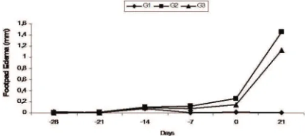
The evolution of the footpad infection is demonstrated in Figure 1. There was a gradual increase in the thickness of infected footpad in animals of G2 and G3 when compared to G1, although there was no difference between them (p<0.05). Regarding the lymph node drainage (Figure 2), it was observed increase in weight of the infected PLN in G2 and G3 (p<0.01) when compared to G1. G2 differ significantly from G3 (p<0.05).
Figure 1. Effect of EAE of Cocos nucifera on the hind footpad edema of L. braziliensis infected hamsters. G1 negative control group, G2 positive control group and G3 infected and treated group.
The results of the leukocyte parameters are presented at Table 1. Leishmania infection (G2)
inhibited significantly the peripheral leukocytes blood, when compared to the -28th and zero days. However,
neutrophils and lymphocytes values did not have significant alterations. The infected and treated group (G3) presented eosinophilia in relation to the infected and no treated group (G2).
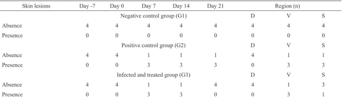
On clinical evaluation, it was verified the presence of body lesions in the infected animals with
L. brasiliensis (Table 2). In G2 and G3 were verified alopecia areas (7th day) in dorsal and ventral regions and
ulcers (7th and 14th day) in dorsal regions of the animals.
However, the lesions remained in G2 until the end of the experiment, while in G3 the lesions disappeared on 21st day. Nails, eyes and ears were normal. The snout
lesions were observed in the animals from G2 (n=3) and G3 (n=1). G1 did not present skin lesions during the experiment.
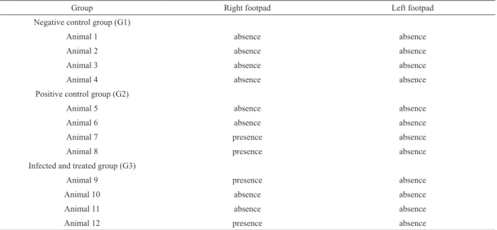
Table 3 presents the effect of EAE on subcutaneous imprints of L. braziliensis infected footpads
that presented amastigotes forms and cellular iniltration. The inlammatory process was characterized by the
presence of macrophages, lymphocytes and giant cells. Amastigotes forms of L. braziliensis were not found on opposite footpads. In most infected animals (G2 and G3) the amastigotes forms of Leishmania were not visualized in the imprints.
Regarding the lymph node drainage, it was observed a significant increase in weight of the infected PLN in G2 and G3 (p<0.01) when compared to G1. G2 differ from G3 (p<0.05).
Figure 2. Effect of EAE of Cocos nucifera on PLN weight in
L. braziliensis infected hamsters. The results were expressed as the difference in weight between the right and left PLN. G1 negative control group, G2 positive control group and G3 infected and treated group. *p<0.01 compared to G1; #p<0.05 compared to G3.
Discussion
Table 1. Effect of EAE of Cocos nucifera on leukocyte counts from L. braziliensis infected hamsters. G1 negative control group, G2 positive control group and G3 infected and treated group.
Day -28
Parameter G1 G2 G3
Leukocytes (x103/mm3) 13.1±0.6 12.9±0.8 12.3±1.3
Neutrophils (%) 29.9±1.5 26.7±1.2 27.1±1.1
Lymphocytes (%) 73.5±2.3 74.2±3.1 73.1±2.6
Eosinophils (%) 1.1±0.8 1.4±0.6 1.2±0.7
Monocytes (%) 2.5±0.2 2.6±0.4 2.4±0.3
Basophils (%) 1.1±0.1 1.3±0.3 1.4±0.3
Day 0
Leukocytes (x103/mm3) 8.4±2.3 4.9±1.3 5.3±1.8
Neutrophils (%) 25.6±4.3 21.3±8.8 14.9±9.3
Lymphocytes (%) 64.3±8.6 73.8±12.5 72.9±14.1
Eosinophils (%) 5.6±3.2 2.0±2.4 4.8±3.7
Monocytes (%) 2.4±1.2 1.6±1.4 5.2±1.7
Basophils (%) 1.9±1.1 1.2±1.8 2.2±2.1
Day 21
Leukocytes (x103/mm3) 8.5±1.3 5.0±1.6 5.1±0.9
Neutrophils (%) 29.8±15.2 33.8±21.3 22.0±21.4
Lymphocytes (%) 63.2±20.2 54.9±23.5 58.7±21.5
Eosinophils (%) 3.8±6.1 0.6±0.7 9.1±12.1
Monocytes (%) 2.8±1.6 10.4±10.9 9.5±12.9
Basophils (%) 0.3±0.4 0.2±0.3 0.6±0.7
Table 2. Presence of lesions from L. braziliensis on different skin regions of hamsters.
Skin lesions Day -7 Day 0 Day 7 Day 14 Day 21 Region (n)
Negative control group (G1) D V S
Absence 4 4 4 4 4 4 4 4
Presence 0 0 0 0 0 0 0 0
Positive control group (G2) D V S
Absence 4 4 1 1 1 4 1 1
Presence 0 0 3 3 3 0 3 3
Infected and treated group (G3) D V S
Absence 4 4 1 1 4 4 1 3
Presence 0 0 3 3 0 0 3 1
Legend: D: dorsal; V: ventral; S: snout.
control of some microorganisms, including bacteria, fungi and protozoa (Esquenazi et al., 2002).
Estevez et al. (2007) studying the leishmanicidal activity in vitro of 27 plant extracts from the Amazon region, obtained results in only two species against the amastigote form of L. amazonensis, and in only one specie against the promastigote form. Ahua et al. (2007) studying 64 extracts of 21 plants used in traditional medicine in Africa, obtained significant activity in only seven extracts of six different plants, against both amastigotes and promastigotes of L. major. Lamidi et al. (2005) studying the leishmanicidal activity in
vitro of 67 crude extracts obtained from fifteen plants used in traditional medicine in Africa, observed that plants of the genus Corynanthe, which is rich in alkaloids, and fractions of Polyalthia suaveolens
both have activity against promastigote forms of L. major.
infected with L. braziliensis and showed inhibitory activity in the two development stages, promastigotes and amastigotes forms, of L. amazonensis. These authors suggested an immunomodulatory effect on infected macrophages. In our study, EAE did not modulate the immune response in L. braziliensis infected animals. This fact was verified by the presence of amastigotes forms of L. braziliensis and the cellular infiltrate in footpad, and an increase of drainage lymph nodes. On the other hand, the oral treatments with Kalanchoe pinnata
(Da Silva et al., 1999), Chenopodium ambrosioides
(Patrício et al., 2008), Tinospora sinensis (Singh et al., 2008) were able to control the dissemination of
Leishmania sp. infection.
In this study, the infected and treated animals (G3) presented eosinophilia, monocytosis and accentuated lymphopenia, but the total leukocytes peripheral blood values and neutrophils remained unaltered when compared to G1 and to G2. It has been reported that in naturally infected patients with
Leishmania species, it is possible to detect alterations in the haematological parameters. These alterations varied from leukopenia with neutropenia to a moderate lymphocytosis and monocytosis. It is also common to detect anemia in these patients (Herwaldt, 1999).
Our results suggest that the treatment with EAE of Cocos nucifera L. did not alter the parameters studied in L. braziliensis infected animals in these conditions. Therefore, the popular use has no scientific support, because it does not provide adequate stimulation of the immune system to eliminate the pathogen. But, the EAE seems to present a healing activity in this experimental model. However, the mechanism whereby EAE of
Cocos nucifera L. modulated the healing response should be investigated further.
saponins (Delorenzi et al., 2001) and limonene (Arruda et al., 2009). Our results show that EAE of Cocos nucifera L. presents high concentrations of catechins and epicatechins. These compounds are known to have immunomodulatory (Mendonça-Filho et al., 2004), anti-inflammatory and antioxidant (Silva et al., 2009) activities.
In the present study, hamsters were used as experimental model because these animals present great susceptibility to L. braziliensis infection. This information was confirmed by Neal & Hale (1983), that compared BALB/C, CBA/H, CDI mouse to hamster infections.
The infection with L. braziliensis promastigotes in hamsters footpad induced a progressive increase in the lesion size in all animals (G2 and G3). The treatment with EAE of Cocos nucifera, for 21 consecutive days, did not reduce neither the footpad edema nor the PLN weight. However, the animals from G3 (infected and treated animals) that presented skin lesions on 7th day, showed a reduction of these lesions on the 14th day. This fact suggests that the EAE may stimulate a healing activity in this infectious process. It has been reported that L. braziliensis infected animals present lesions like papules, nodules and ulcer, which can progress to a secondary bacterial infection (Herwaldt, 1999). The effects observed in our experiment may be mistaken as the heal of infected animals that present skin lesions after treatment with C. nucifera extract. However, the animals were not able to eliminate the parasite with the treatment.
C. nucifera L. has been largely used in popular medicine (Duke, 1992). Mendonça-Filho et al. (2004) demonstrated in vitro activity of EAE on macrophages
Table 3. Evaluation of the presence of L. braziliensis in footpad subcutaneous imprints.
Group Right footpad Left footpad
Negative control group (G1)
Animal 1 absence absence
Animal 2 absence absence
Animal 3 absence absence
Animal 4 absence absence
Positive control group (G2)
Animal 5 absence absence
Animal 6 absence absence
Animal 7 presence absence
Animal 8 presence absence
Infected and treated group (G3)
Animal 9 presence absence
Animal 10 absence absence
Animal 11 absence absence
References
Ahua KM, Ioset JR, Ioset KN, Diallo D, Mauel J, Hostettmann K 2007. Antileishmanial activities associated with plants used in Malian traditional medicine. J Ethnopharmacol 110: 99-104.
Alviano DS, Rodrigues KF, Leitao SG, Rodrigues ML, Matheus ME, Fernandes PD, Antoniolli AR, Alviano CS 2004. Antinociceptive and free radical scavenging activities of Cocos nucifera L. (Palmae) husk fiber aqueous extract. J Ethnopharmacol 92: 269-273. Arruda DC, Miguel DC, Yokoyama-Yasunaka JKU, Katzin
AM, Uliana SRB 2009. Inhibitory activity of limonene against Leishmania parasites in vitro and in vivo. Biomed Pharmacother 63: 643-649.
Ashford DA, David JR, Freire M, David R, Sherlock I, Eulálio MC, Sampaio DP, Badaro R 1998. Studies on control of visceral leishmaniasis: impact of dog control on canine and human visceral leishmaniasis in Jacobina, Bahia, Brazil. Am J Trop Med Hyg 59: 53-57.
Da-Silva SAG, Costa SS, Rossi-Bergmann B 1999. The anti-leishmanial effect of Kalanchoe is mediated by nitric oxide intermediates. Parasitology 118: 575-582. Delorenzi JC, Attias M, Gattass CR, Andrade M, Rezende
C, Cunha-Pinto A, Henriques AT, Bou-Habib DC, Saraiva EMB 2001. Antileishmanial activity of an indole alkaloid from Pesquiera australis. Antimicrob Agents Chemoter 45: 1349-1354.
Duke JA 1992. Handbook of phytochemical constituents of Grãs Herbs and other economic plants. CRC Press: Boca Raton.
Esquenazi D, Wigg MD, Miranda MMFS, Rodrigues HM, Tostes JBF, Rozental S, Da Silva, AJR, Alviano CS 2002. Antimicrobial and antiviral activities of polyphenolics from Cocos nucifera Linn. (Palmae) husk fiber extract. Res Microbiol 153: 647-652. Estevez Y, Castillo D, Pisango MT, Arevalo J, Rojas R, Alban
J, Deharo E, Bourdy G, Sauvain M 2007. Evaluation of the leishmanicidal activity of plants used by Peruvian Chayahuita ethnic group. J Ethnopharmacol 114: 254-259.
Gramiccia M, Gradoni L 2005. The current status of zoonotic leishmaniasis and approaches to disease control. Int J Parasitol 35: 1169-1180.
Herwaldt BL 1999. Leishmaniasis. Lancet 354: 1191-1199. Kirszberg C, Esquenazi D, Alviano CS, Rumjanek VM 2003.
The effect of a catechin-rich extract of Cocos nucifera
on lymphocytes proliferation. Phytother Res 17: 1054-1058.
Lamidi M, Digiorgio C, Delmas F, Favel A, Eyele Mve-Mba C, Rondi ML, Ollivier E, Nze-Ekekang L, Balansard G 2005. In vitro cytotoxic, antileishmanial and antifungal activities of ethnopharmacologically
selected Gabonese plants. J Ethnopharmacol 102: 185-190.
Matos FJA 1997. Introdução à fitoquímica experimental. 2ª ed. Fortaleza: Imprensa Universitária.
Mendonça-Filho RR, Rodrigues IA, Alviano DS, Santos ALS, Soares RMA, Alviano CS 2004. Leishmanicidal activity of polyphenolic-rich extract from husk fiber of Cocos nucifera Linn. (Palmae). Res Microbiol 155: 136-143.
Neal RA, Hale C 1983. A comparative study of susceptibility of inbred and outbred mouse strains compared with hamsters to infection with New World cutaneous leishmaniasis. Parasitology 87: 7-13.
Patrício FJ, Costa GC, Pereira PVS, Aragão-Filho WC, Sousa SM, Frazão JB, Pereira WS, Maciel MCG, Silva LA, Amaral FMM, Rebelo JMM, Guerra RNM, Ribeiro MNS, Nascimento FRF 2008. Efficacy of the intralesional treatment with Chenopodium ambrosioides in the murine infection by Leishmania amazonensis.J Ethnopharmacol 115: 313-319. Pereira BA, Alves CR 2008. Immunological characteristics
of experimental murine infection with Leishmania (Leishmania) amazonensis. Vet Parasitol 158: 239-255.
Ribeiro VM 2007. Leishmaniose visceral Canina: aspectos de tratamento e controle. Clín. Vet. 71: 66-76.
Silva LCR, Nunes-Pinheiro DCS, Morais SM, Lopes-Neto BE, Santos GJL, Campello CC 2009. Avaliação toxicológica e efeito do extrato acetato de etila da fibra de Cocos nucifera L. (Palmae) sobre a resposta inflamatória in vivo. Rev Bras Plantas Med 11: 429-434.
Simões-Mattos L, Mattos MRF, Teixeira MJ, Oliveira-Lima JW, Bevilaqua CML, Prata-Júnior RC, Holanda CM, Rondon FCM, Bastos KMS, Coêlho ZCB, Coêlho ICB, Barral A, Pompeu MML 2005. The susceptibility of domestic cats (Felis catus) to experimental infection with Leishmania braziliensis. Vet Parasitol 127: 199-208.
Singh N, Kumar A, Gupta P, Chand K, Samant M, Maurya R, Dube A 2008. Evaluation of antileishmanial potential of Tinospora sinensis against experimental visceral leishmaniasis. Parasitol Res 102: 561-565.
*Correspondence
José C. C. Freitas
Programa de Pós-graduação em Ciências Veterinárias, Universidade Estadual do Ceará
Avenida Paranjana, 1700, Campus do Itaperi, 60740-002, Fortaleza-CE, Brazil