Resveratrol Enhances Neuroplastic Changes,
Including Hippocampal Neurogenesis, and
Memory in Balb/C Mice at Six Months of Age
Mario Torres-Pérez1, Ruth Ivonne Tellez-Ballesteros1, Leonardo Ortiz-López1,
Muhammad Ichwan2,3, Nelly Maritza Vega-Rivera4, Mario Castro-García4, Ariadna Gómez-Sánchez1, Gerd Kempermann2,5*, Gerardo Bernabe Ramirez-Rodriguez1*
1Laboratory of Neurogenesis, Division of Clinical Investigations, National Institute of Psychiatry“Ramón de la Fuente Muñiz”, Calz. México-Xochimilco 101, 14370, México, D.F., México,2CRTD - Center for Regenerative Therapies Dresden, Tatzberg 47–79, 01307, Dresden, Germany,3Department of Pharmacology and Therapeutic, Faculty of Medicine, Universitas Sumatera Utara, Jalan Dr. Mansur 5, Medan, Indonesia,4Laboratory of Neuropsychopharmacology, Division of Neurosciences, National Institute of Psychiatry“Ramón de la Fuente Muñiz”, Calz. México-Xochimilco 101, 14370, México, D.F., México,
5DZNE, German Center for Neurodegenerative Diseases, Dresden, Tatzberg 47–49, 01307, Dresden, Germany
*[email protected](GK);[email protected](GBRR)
Abstract
Resveratrol (RVTL) is a flavonoid found in red wine and has been publicized heavily as an anti-aging compound. Indeed, basic research confirms that although there is much hype in the promotion of RVTL, flavonoids such as RVTL have a wide range of biological effects. We here investigated the effects of RVTL treatment on hippocampal plasticity and memory performance in female Balb/C mice, a strain with low baseline levels of adult neurogenesis. Two weeks of treatment with RVTL (40 mg/kg) induced the production of new neurons in vivo by increasing cell survival and possibly precursor cell proliferation. In addition, RVTL decreased the number of apoptotic cells. The number of doublecortin (DCX)-expressing intermediate cells was increased. RVTL stimulated neuronal differentiation in vitro without effects on proliferation. In the dentate gyrus, RVTL promoted the formation and maturation of spines on granule cell dendrites. RVTL also improved performance in the step down pas-sive avoidance test. The RVTL-treated mice showed increase in the levels of two key sig-naling proteins, phospho-Akt and phospho-PKC, suggesting the involvement of these signaling pathways. Our results support the vision that flavonoids such as resveratrol deserve further examination as plasticity-inducing compounds in the context of successful cognitive aging.
Introduction
In the context of healthy successful aging, there is hope that natural compounds would pro-mote or at least help to stabilize neural plasticity and thus contribute to maintaining cognitive
a11111
OPEN ACCESS
Citation:Torres-Pérez M, Tellez-Ballesteros RI, Ortiz-López L, Ichwan M, Vega-Rivera NM, Castro-García M, et al. (2015) Resveratrol Enhances Neuroplastic Changes, Including Hippocampal Neurogenesis, and Memory in Balb/C Mice at Six Months of Age. PLoS ONE 10(12): e0145687. doi:10.1371/journal.pone.0145687
Editor:Robert N. Pechnick, Case Western Reserve University School of Dental Medicine, UNITED STATES
Received:June 25, 2015
Accepted:December 6, 2015
Published:December 22, 2015
Copyright:© 2015 Torres-Pérez et al. This is an open access article distributed under the terms of the Creative Commons Attribution License, which permits unrestricted use, distribution, and reproduction in any medium, provided the original author and source are credited.
Data Availability Statement:All relevant data are within the paper.
function in old age. It has been proposed and popularized that certain natural compounds have direct positive effects on neuronal plasticity [1,2,3].
Flavonoids have received prominent attention in this context [4–6] and among these RVTL has been much publicized, presumably because it is found in red grape skin and hence red wine, thus (at least at face value) becoming a plausible single-factor mediator of the positive effects of the mediterranean diet on aging [7,8]. However, several clinical studies with RVTL could not confirm relevant beneficial effects. Nevertheless, RVTL remains neurobiologically interesting, for example because of its anti-aging effects through the deacetylase sirtuin-1 (Sirt1) [9].
The clinical evidence is also not terminally conclusive, because many important questions are open. Among these are the relevant biological targets of the intervention and the related question of how a suitable group of subjects would have to be stratified to observe an effect of RVTL [10]. After all, the acute treatment within the study context is different from the poten-tially essenpoten-tially lifelong intake in the context of the mediterranean diet.
Resveratrol is a phytoalexin found in grapes, raspberries, mulberries and peanuts [9] and exists in bothcis- ortrans-forms.Trans-RVTL has anti-cancer and anti-inflammatory effects [11–13]. RVTL crosses the blood brain barrier [14], where RVTL reverted deficits in spatial memory after stroke [15] and acted as a neuroprotective agent [16]. Finally, treatment with RVTL extended the life span [9] and in addition to its interaction with Sirt-1, which suggests a mechanism involving epigenetic changes, the effects of RVTL also involved signaling pathways such as the AMP-activated protein kinase (AMPK) [17,18].
Our general hypothesis is that adult hippocampal neurogenesis provides“neurogenic reserve”that allows for the maintenance of cognitive flexibility in older age [19]. Given the pre-sumed profile of bioactivity of RVTL an effect on neural precursor cells and adult hippocampal neurogenesis seems likely. However, previous studies indicated that RVTL would decrease adult hippocampal neurogenesis and spatial memory in young C57Bl/6 mice [20], and reduce proliferation of neural precursor cells from the adult hippocampus [21]. In contrast, positive effects of RVTL on neurogenesis were seen in prenatally stressed rats [22] and in a mouse model of chronic fatigue [23] as well as in a knockout for the calcitonin gene related peptide (CGRP) [24]. Long-term treatment with RVTL prevented age-related cognitive decline and was associated with maintained greater level of adult neurogenesis [25]. In that study, the sur-prising aspect was that only four weeks of treatment in late mid-life was reported to have very long-term effects into old age.
In the present study we investigated six month old rather than younger mice, using a strain of mice (Balb/C) as a genetically defined cohort different from previous studies and with low baseline levels of adult neurogenesis [26], and applied RVTL for 14 days. We chose a protocol, in which RVTL was applied after labeling of hippocampal precursor cells with BrdU, so that the effect of RVTL on the survival of newborn cells rather than the proliferation of the precur-sor cells was in the center of interest. At the age of 6 months physiological levels of adult neuro-genesis have declined to very low baseline levels [27,28].
We thus hypothesized that RVTL might be effective in influencing structural hippocampal plasticity by regulating adult hippocampal neurogenesis and dendritic spines in a 14 days treat-ment model in female Balb/C mice at six months of age and improved behavioral performance. Balb/C mice were chosen because they show medium to low levels of baseline adult hippocam-pal neurogenesis but high relative numbers of surviving newborn cells in comparison to CD1 mice [26] and at least in one study was observed to have a very large sensitivity to activity-induced regulation [29]. Our previous work on the effects of melatonin on adult hippocampal neurogenesis had found strong effects on the survival of newborn neurons in this strain of mice [30].
and GK were supported by a CONACYT-BMBF cooperation project for exchange researchers (GRR: 147377; GK:01DN12086). The funders had no role in study design, data collection and analysis, decision to publish, or preparation of the manuscript.
Materials and Methods
Animals and ethics statement
One hundred two month-old female Balb/C mice were obtained from Harlan (México, D.F. México). Animals were housed in standard laboratory cages (12-hour light/12-hour dark cycles at a temperature of 23 ± 1°C) following the institutional and legal regulations regarding to ani-mal ethics and handling. Mice had access to food and waterad libitumand were left to acclima-tize in their environment until they reached the age of 6 months. The light/dark cycle
corresponded to the timing of lights on (Zeitgeber time 0; ZT0) at 0700 hours (h) and to the timing of lights off (Zeitgeber time 12; ZT12) at 1900 h. All institutional and legal regulations regarding animal ethics and handling were followed for thein vivoexperiments (Ethical Com-mittee for Research of the National Institute of Psychiatry approved the present study with the number IACUC SIC092025). In our study we decided to use female instead of male mice based on reports of increased aggression in rodent males, while this was not observed in females [31].
Experimental design, RVTL treatment and BrdU injections
Once mice had reached 6 months of age, they received two injections of BrdU (50 mg/kg; Sigma, St. Louis, MO, USA) 2 h apart (Fig 1A). Then, the animals were treated with RVTL (Sigma, St. Louis, MO, USA) in concentrations (0.0044, 0.022, 0.044, 0.088, 0.176 mg/ml) to yield differ-ent doses (1, 5, 10, 20 or 40 mg/kg of body weight) per day for 14 days. The estimated daily RVTL intake for each mouse was based on an average daily water consumption rate of 5 ml per day. Res-veratrol was dissolved in minimum absolute ethanol and then added to drinking water at a final ethanol concentration of 0.3%. Fresh RVTL solution was prepared every third day and provided in feeding bottles that were protected from light. Mice in the control groups received water con-taining 0.3% of ethanol. The water consumption was similar for all experimental groups. Control-and RVTL-treated mice were housed under normal conditions in groups of 4 animals.
Brain dissection and tissue processing for immunohistochemistry
One day after the end of the treatment, mice were sacrificed by an overdose of ketamine and perfused transcardially with 4%p-formaldehyde in 0.1 M phosphate buffer (pH 7.4). Brains were removed and post-fixed for 24 h before they were stabilized in 30% sucrose in phosphate buffer [32]. Brains were cut into 40μm coronal sections on a tabletop sliding microtome (Leica, Buffalo Grove, IL, USA). The sections were stored at 4°C in cryoprotectant solution and stained following the free floating immunohistochemistry method and pretreated for BrdU-immunodetection by incubation in 2 N HCl for 30 minutes at 37°C followed by 2 washes in 0.1 M borate buffer (pH 8.5) for 10 minutes each [30,32].Immunohistochemistry and cell quantification
To analyze cell proliferation, survival and the immature neurons in the subgranular or in the granular cell layer of the dentate gyrus, we identified specific markers of each step with the per-oxidase method, as described previously [32]. In addition, we analyzed the apoptotic cells in the granular cell layer.
Grove, PA, USA). Sections were clarified and mounted in Neomount medium (Merck, White-house Station, NJ, USA).
The number of Ki67-, BrdU-, DCX- or caspase-3 (active)-labeled cells was determined in series of every 6thsection from all animals. Positive cells were counted using a 40x objective throughout the rostro-caudal extent of the granule cell layer on a light microscope coupled to a camera (Leica, Buffalo Grove, IL, USA). Counting was done as described previously, with a modified version of the optical fractionator. The cells appearing in the uppermost focal plane were not counted in order to avoid oversampling [32]. The estimated total number of labeled cells per granular cell layer was calculated from estimated resulting numbers multiplied by 6.
Phenotypic analysis
Newly mature formed neurons in the dentate gyrus of adult mice were analyzed in one-in-twelve series of sections from animals of control- or RVTL (40 mg/kg)-groups by immunofluo-rescent double staining [32]. From the percentages of every phenotype and to the total number of BrdU-labeled cells the total number per phenotype was calculated. The newborn neurons were identified by the BrdU/NeuN co-labeling. Analysis was performed by confocal micros-copy (Zeiss LSM 510 Meta; Thornwood, NY, USA) in sequential scanning mode to avoid cross-bleeding between channels. Double-labeling was confirmed by three-dimensional recon-structions of z-series covering the entire nucleus (or cell) in question. The primary antibodies were: monoclonal mouse NeuN (1:100; Chemicon, Temecula, CA, USA) and rat BrdU antibody (1:250; Accurate, Westbury, NY, USA). Fluorophore-coupled secondary anti-bodies were: anti-rat TRITC or anti-mouse FITC. All secondary antianti-bodies were raised in don-key and diluted 1:250 (Jackson Immunoresearch, West Grove, PA, USA). Sections were mounted in polyvinyl alcohol with diazabicyclo-octane as anti-fading agent (PVA-DABCO).
Hippocampal neurosphere cultures
Precursor cells were isolated from the adult dentate gyrus and collected into neurosphere medium consisting of mouse DMEM/F12 basal medium (Gibco, Life Technologies, Karlsruhe, Germany) plus mouse NeuroCult NSC Proliferation Supplements (Stem Cell Technologies, Grenoble, France) with 2% bovine serum albumin (Gibco, Life Technologies, Karlsruhe, Ger-many), 2μg/ml heparin (Sigma-Aldrich, Munich, Germany). Culture medium was comple-mented with 20 ng/ml purified mouse receptor-grade epidermal growth factor (EGF; BD Biosciences, Heidelberg, Germany) and 10 ng/ml recombinant bovine FGF-2 (Roche, Mann-heim, Germany). Cells were incubated in humidified 5% CO2for 12 d to permit neurosphere
formation in the presence of RVTL (0, 0.001, 0.01, 0.1, 1 or 10μM).
Adherent adult hippocampal precursor cell cultures
For adherent adult hippocampal neural precursor cell cultures, cells were isolated as previously reported [33,34]. After the brains were removed from the skull, the dentate gyrus was dissected and dissociated by enzymatic digestion. The resulting cell suspension was separated by centri-fugation using a Percoll gradient. Precursor cells were plated on laminin pre-coated coverslips
cells show significant increase in cell survival in mice treated with 10, 20 and 40 mg/kg of RVTL (**p= 0.005,*p= 0.017, respectively).F)Representative micrograph of cells expressing caspase 3 (active) is shown. (G)Apoptotic cells show significant decrease in mice treated with 40 mg/kg of RVTL
(*p= 0.043). However, lower doses of RVTL (1, 5, 10 and 20 mg/kg) did not significantly decrease the absolute number of caspase 3 (active)-labeled cells (p= 1,p= 0.94,p= 0.17,p= 0.14, respectively). Data are expressed as the mean±S.E.M. of the absolute number of cells. Bonferroni’s post hoc test was performed after one-way ANOVA for cell proliferation, survival and apoptotic cells. Micrographs also show the granule cell layer (GCL), molecular layer (ML), subgranular zone (SGZ) and the hilus (H). Scale bar = 160μm. Scale bar in amplification of panels B, D and F = 30μm.n= 4 mice per group.
or 96 multi-well plates and cultured with 20 ng/ml of human Epidermal Growth Factor (EGF) and 20 ng/ml of human Fibroblast Growth Factor-2 (FGF2; both from PeproTech, México, D. F. México) in Neurobasal medium supplemented with B27 (Gibco, Life Technologies, Grand Island, NY, USA) for 1 day followed by 2 additional days in the presence of growth factors plus RVTL (0, 0.001, 0.01, 0.1, 1 or 10μM). Cell proliferation was determined by manual quantifica-tion of BrdU immunocytochemistry in an epifluorescence microscope Nikon Eclipse Ti U (Nikon, Melville, NY, USA).
For differentiation studies, adherent precursor cells were cultured in medium containing EGF and FGF2 for 1 day followed by 2 additional days in the presence of growth factors plus RVTL (0, 0.001, 0.01, 0.1, 1 or 10μM) before cells were switched to a medium containing only RVTL (0, 0.001, 0.01, 0.1, 1 or 10μM) for 5 days. Culture media was replaced every third day. Cell survival and the phenotype of differentiated precursor cells were assessed by quantitative immunocytochemistry. Positive cells were identified with specific primary antibodies: rabbit anti-GFAP (1:2000; Dako, Via Real, CA, USA); mouse anti-βIII-tubulin (1:2000; Promega, Madison, WI, USA) or monoclonal anti-MAP2 (1:500; Sigma, St. Louis, MO, USA).
Golgi-Cox impregnation
For metal impregnation, brains were removed from the skull and immersed in a Golgi-Cox solu-tion during 21 days. Every third day, the Golgi-Cox solusolu-tion was replaced with fresh solusolu-tion. Brains were transferred to a 30% sucrose solution and sliced on a table-top sliding microtome (Leica). Coronal brain sections (200μm) were collected in 3% sucrose solution and mounted onto pre-gelatinized glass slides. Golgi-Cox impregnation was revealed with 18.7% ammonia solution and fixed in Kodak rapid fix (Kodak, Rochester, NY, USA). Brain slices were dehydrated and clarified in Neoclear before they were coverslipped with Neomount medium (Merck, White-house Station, NJ, USA) [35,36]. Twenty Golgi-stained neurons in the granule cell layer (GCL) of the dentate gyrus were analyzed to determine dendritic spine density in every 10μm of sec-ondary dendrites of rodents exposed to either control- or RVTL (40 mg/kg). Control- and RVTL-treated mice were housed under normal conditions in groups of 4–5 animals.
Behavioral experiments
Step-down passive avoidance test (SDPA). Sixteen mice were used in the SDPA test (8 mice per group). This test has two phases. During the training phase the animal is punished for making the instinctive response of moving from a brightly lid chamber into a darkened one [37]. After a specified delay interval, the rodent is tested under the same conditions to deter-mine the length of time, after which the animal moves into the dark side of the chamber (test phase). The latency to repeat the punished response is considered as an index of memory. Here, we used an automated testing apparatus (Ugo Basile; Comerio, VA, Italy) located in a well-illuminated room. One day after the end of the treatment, the training in the SDPA was performed (day 15). Mice were subjected to a foot-shock (0.4 mA, 5 s) as soon as they completely descended onto the grid floor of the dark side of the apparatus. Retention tests without shocks were performed 1.5 (day 15) and 24 h (day 16). A researcher blind to the exper-imental groups analyzed the parameters.
and sessions were continued until the mouse had collected all cereal-chocolate pellets, at the latest after 10 minutes. After each trial, the 8-RAM was carefully cleaned with antiseptic solu-tion containing ethanol (70%). All trials were video recorded with a Canon Power Shot SX50 HS (Canon, Japan) and analyzed by a researcher blind to the experimental condition. The parameters analyzed were: 1) working memory errors (entries into baited arms that had already been visited during the same trial), 2) reference memory errors (entries into unbaited arms), and 3) the locomotor activity (total number of entries).
Western blot for key signaling protein levels in the hippocampus
Hippocampi from control- or RVTL-treated mice were dissected and lysed as reported previ-ously [35,36]. Control- and RVTL-treated mice were housed under normal conditions in groups of 4–5 animals. The lysate was obtained in RIPA buffer (150 mM NaCl, 10% glycerol, 0.5 mM EDTA, 0.5% triton X-100, 1 mM PMSF, 25μg/ml leupeptin, 25μg/ml aprotinin and 1 mM sodium ortho-vanadate in 50 mM Tris-HCl, pH 7.6), homogenized with an ultrasonic homogenizer for 30 seconds and centrifuged at 3300 xg for 15 minutes. Total protein content was quantified using Bradford reagent (Bio-Rad, Philadelphia, PA, USA). Protein separation was performed by the Laemmli method [38] and transferred to PVDF or nitrocellulose paper. Membranes were blocked with 5% skim milk in 0.05% Tween 20-TBS (TTBS) and incubated with rabbit-anti-phospho Akt or rabbit anti-Akt (Cell Signaling, Danvers, MA, USA); mouse monoclonal anti-phospho-p44/42 MAP kinase or rabbit anti-p44/42 MAP kinase (Cell Signal-ing); rabbit CREB or rabbit anti-CREB (Cell-SignalSignal-ing); rabbit anti-phospho-PKC (Cell Signaling). The mouse anti-GAPDH antibody (Chemicon, Temecula, CA, USA) was used for the loading control. Blots were washed 3 times with TTBS and incubated for 1 hour in a 1:3000 dilution of the appropriate secondary antibody: phosphatase-conjugated don-key anti-mouse; dondon-key anti-goat, or dondon-key anti-rabbit. Proteins were visualized with the enhanced chemiluminescence detection system (Merck-Millipore). Images were scanned and densitometry analysis was performed with the ImageJ software (NIH, Bethesda, MA, USA).
Statistical Analysis
Analysis was performed using SigmaStat 3.1 software. The results are presented as
mean ± standard error of the mean SEM. Forin vivocell proliferation, survival and apoptosis mean differences between groups were analyzed using one-way ANOVA followed by Bonferro-ni’s post-hoc test, where appropriate. Doublecortin-labeled cells were analyzed using a Krus-kal-Wallis-ANOVA on ranks followed by Student-Newman-Keuls comparison because the equal variance test failed. Mature new neurons, dendritic spines number, proportions of den-dritic spines and relative protein levels were analyzed by unpaired t-test. The results of the hip-pocampal dependent behavior (SDPA-test) and the results of 8-RAM test were analyzed with a repeated measures two-way ANOVA, both followed by the appropriate post-hoc test. Two-tailed unpaired t-tests were used for comparison between two groups. The results derived from precursor cells were analyzed using one-way ANOVA followed by Tukey post-hoc test. Differ-ences were considered statistically significant atp0.05.
Results
Resveratrol affects cell survival and doublecortin-cells without affecting
cell proliferation in vivo
gyrus of mice at six months of age as indicated by an increase in the number of BrdU-labeled cells compared with the control group (Fig 1E). The number of surviving cells in the dentate gyrus of mice treated with the highest doses of RVTL (10, 20 or 40 mg/kg) reflected an increase of 31%, 28%, 31%, respectively (control: 182 ± 5.3; RVTL 10 mg/kg: 240 ± 8.83; RVTL 20 mg/ kg: 234 ± 12; RVTL 40 mg/kg: 240 ± 13; F5,23= 12.28;p= 0.005, 0.017, 0.005, respectively).
However, the lowest dose of RVTL caused a decrease in cell survival without reaching statistical significance (RVTL 1 mg/kg: 160 ± 7.2; F5,23= 12.28;p= 0.79). To rule out that the pro-survival
effect of RVTL might be only a consequence of an increased cell proliferation, we also analyzed Ki67-expression as an intrinsic proliferation marker (Fig 1B and 1C). Quantification at 14 days after treatment revealed that the dose of 1 mg/kg decreased the absolute number of prolifer-ative cells in the dentate gyrus without reaching statistical significance (control: 174 ± 22.04 versus RVTL 1 mg/kg: 133 ± 14.77; F5,23= 6.12;p= 0.17;Fig 1B and 1C). In contrast, the
high-est doses of RVTL (20 and 40 mg/kg) showed an increase of cell proliferation, however none of these differences were statistically significant (RVTL 20 mg/kg: 199 ± 6.6; RVTL 40 mg/kg: 220 ± 4.5; F5,23= 6.12;p= 0.83, 0.086, respectively;Fig 1B and 1C) in comparison to control
treated mice. However, the analysis of apoptotic cells revealed that RVTL (40 mg/kg) decreased the number of caspase 3 (active)-labeled cells (40%) in comparison to the control group (con-trol: 187 ± 9.66 versus RVTL 40 mg/kg: 111 ± 4.41; F5,23= 3.22;p= 0.027;Fig 1F and 1G).
Thus, RVTL shows a pro-survival effect on the newborn cells in the dentate gyrus of female Balb/C mice at six months of age, which in general cannot be explained by an effect on precur-sor cell proliferation.
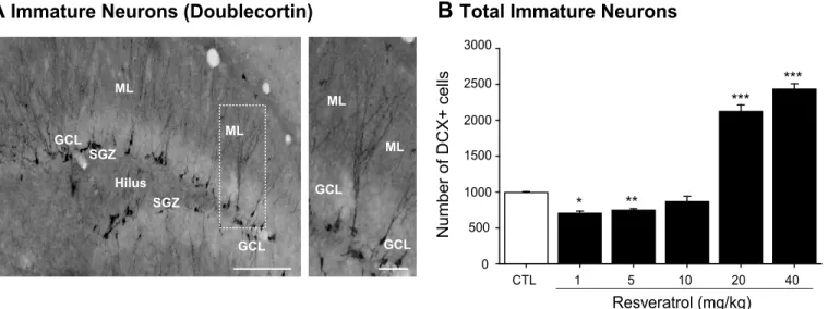
Additionally, we next evaluated the population of DCX-expressing cells, corresponding to the intermediate progenitor cells (type-2b and -3) and early post-mitotic stages of development (Fig 2A and 2B). Quantification at 14 days after treatment with RVTL revealed that the doses of 1 and 5 mg/kg significantly decreased (29% and 24%, respectively) the absolute number of DCX-cells in comparison to control mice (H = 25.10; control: 993 ± 12.1; RVTL 1 mg/kg: 704 ± 30.15; RVTL 5 mg/kg: 747 ± 20.40; q = 4.3, q = 5, respectively;p0.001). However, the doses of 20 and 40 mg/kg of RVTL significantly increased (113% and 144%, respectively) the number of DCX-labeled cells (RVTL 20 mg/kg: 2122 ± 90.90; RVTL 40 mg/kg: 2433 ± 75.33; q = 4.58, q = 5.2, respectively;p0.001) compared to the control mice. These results suggest that the lower doses of RVTL (1 and 5 mg/kg) affected DCX-cells, while higher doses of RVTL (20 and 40 mg/kg) favor the DCX-expressing intermediate population of cells.
Resveratrol increases net hippocampal neurogenesis
Considering that the highest dose of RVTL showed consistent effects on cell survival, DCX-cells population and it decreased apoptosis, we further analyzed the phenotype of BrdU-labeled cells after treatment with RVTL (Fig 3). Phenotypic analysis revealed a similar proportion of newborn mature neurons in RVTL-treated (40 mg/kg) and in control mice (control: 57 ± 3.15 versus RVTL 40 mg/kg: 54 ± 7.58; t = 0.303;p= 0.77;Fig 3B). This implies that with increased survival the absolute number of new neurons significantly increased (44%) in the dentate gyrus of RVTL-treated mice (control: 102 ± 3.53 versus RVTL 40 mg/kg: 147 ± 12.77; t = -3.39;
p= 0.015;Fig 3C).
Resveratrol increases neuronal differentiation of adult hippocampal
precursor cells without affecting cell proliferation in vitro with nanomolar
concentrations
index of precursor cell activity and proliferative expansion (Fig 4B). The number of neuro-spheres generated from cells cultured with nanomolar (0.001–0.1μM) or 1μM concentrations of RVTL did not show significant changes, whereas the highest concentration of RVTL (10μM) significantly decreased (*88%) the number of neurospheres formed in comparison to
the control treated cells (p<0.001). Similarly to neurospheres, in adherent neural
hippocam-pal precursor cells 10μM of RVTL also decreased proliferation by*30% as assessed by the
incorporation of BrdU (F5,37= 10.30;p<0.01;Fig 4A and 4C).
Next, we analyzed whether RVTL might induce differentiation from precursor cells in vitro (Fig 4D–4G). Phenotypic analysis of cultures differentiated in the presence of different concen-trations of RVTL (0.001–10μM) showed that the proportion ofβIII-tubulin-positive immature neurons was decreased in cells cultured with concentrations ranging from 0.1 to 10μM (F5,47=
33.41;p<0.001;Fig 4E). Moreover, the proportion of mature neurons generated from
precur-sor cells showed that nano- and micro-molar concentrations of RVTL (0.001; 0.01 or 10μM; F5,53= 11.84;p= 0.02, 0.047; 0.02) significantly increased the proportion of MAP2ab-positive
neurons (Fig 4F). The proportion of GFAP-positive astrocytes was also significantly increased in cells cultured with 10μM of RVTL compared to un-treated controls (F5,47= 3.92;p= 0.014;
Fig 4G). These results suggest that while nanomolar concentrations of RVTL did not affect cell proliferation they increased the number of neurons generated from the precursor cells in vitro.
Resveratrol increases the number of dendritic spines
In order to further extend our study to another aspect of plasticity taking place in the dentate gyrus, we analyzed the number of dendritic spines on the secondary dendrites of granule cells (Fig 5A). Mice treated with RVTL (40 mg/kg) showed a greater number (plus*35%) of
den-dritic spines than the control group (control: 12.26 ± 0.52 versus RVTL 40 mg/kg: 17.13 ± 1.23; t = -4.86;p= 0.001). The comparison of treatment within every category of dendritic spines revealed that RVTL (40 mg/kg) significantly increased the proportion of mushroom-shape spines (control: 31 ± 1.99% versus RVTL 40 mg/kg: 36 ± 1.33%; t = -2.34;p= 0.026;Fig 5B) Fig 2. Resveratrol exhibits a dual effect on doublecortin-cells in female Balb/C mice at six months of age.(A)Representative micrograph of cells expressing doublecortin is shown. (B)Doublecortin (DCX)-labeled cells show significant decrease in mice treated with 1 and 5 mg/kg of RVTL (*p= 0.002, **p= 0.007, respectively). However, the highest doses of RVTL (20 and 40 mg/kg) significantly increase the absolute number of DCX-labeled cells (***p<0.001).n= 4 mice per group. Data are expressed as the mean±S.E.M. of the absolute number of cells. Results were analyzed with Student-Newman-Keuls after one-way ANOVA on ranks. Micrographs also show the granule cell layer (GCL), molecular layer (ML), subgranular zone (SGZ) and the hilus (H). Scale bar = 160μm. Scale bar in amplification of panels A = 30μm.
and stubby-shape spines (control: 22 ± 1.15% versus RVTL 40 mg/kg: 27 ± 1.55%; t = -2.74;
p= 0.011;Fig 5B). The proportion of thin spines in contrast was decreased in mice treated with RVTL (control: 46 ± 1.90% versus RVTL 40 mg/kg: 35 ± 2.15%; t = 3.79;p<0.001;Fig 5B).
The increased number of dendritic spines and greater proportion of mushroom-shape spines indicates that RVTL promotes spine formation and maturation in granule cells of the dentate gyrus.
Resveratrol improves behavioral performance
We next asked whether the positive effects of RVTL on adult hippocampal neurogenesis and spine formation occurred in parallel with effects on associative memory, here as reflected by Fig 3. Resveratrol modifies net neurogenesis in female Balb/C mice at six months of age.(A)Proportion of mature new neurons was identified by BrdU- and NeuN-colabeling after 14 days of treatment. (B)Analysis shows that Resveratrol (RVTL; 40 mg/kg) did not modify the proportion of neuronal differentiation in female Balb/C mice at six months of age in comparison to the control (CTL) group. (C)Net neurogenesis was calculated as described in methods. Absolute numbers reflected that RVTL significantly increased net neurogenesis in female Balb/C mice at six months of age (*p= 0.015).n= 4 mice per group. Data are expressed as the mean±S.E.M. of the absolute number of cells. Data were analyzed with the unpaired Student’st-test. Micrographs also show the granule cell layer (GCL), molecular layer (ML), subgranular zone (SGZ) and the hilus (H). Scale bar = 180μm.
the retention latency in the step-down passive avoidance task (SDPA;Fig 6A). The assessment of retention latency 1.5 hours after basal test revealed a statistically significant difference in RVTL-treated mice compared to controls (control: 78 ± 18 seconds versus RVTL 40 mg/kg: 275 ± 67.3 seconds; F1,44= 15.72;p<0.001). A similar effect was also seen at 24 hours after the
basal test (control: 280 ± 67.3 seconds versus RVTL 40 mg/kg: 418 ± 0.5 seconds; F1,44= 15.72; p= 0.011). Moreover, along the exposure sessions within RVTL, mice showed a statistically sig-nificant difference in the retention latency at 1.5 and 24 hours in comparison to the basal test (F2,44= 40.90;p<0.001); whereas the comparison of exposure sessions within control-treated
mice showed a significant difference at 24 hours (F2,44= 40.90;p<0.001) in comparison to the
basal test.
Additionally, we analyzed the performance of RVTL-treated mice in the eight-radial arm maze (8-RAM) during the next 15 days after the end of the treatment (Fig 6B–6D). In this test, both, control and RVTL-treated mice showed a progressive decrease in reference memory errors during training (Fig 6B). Analysis of the effect of treatment per session reflected a signifi-cant decrease in percentage of reference memory errors in RVTL-mice on day 12 in compari-son to their respective control (t = 2.109;p= 0.041;Fig 6B). Moreover, along the consecutive daily sessions within RVTL, mice showed a decrease percentage of reference memory errors and statistically significant differences were observed on days 12 and 15 (F14,289= 2.59;
p= 0.005, 0.015, respectively) in comparison to their first session (day 1); whereas the compari-son of daily sessions within control-treated mice did not show significant differences (F14,289=
2.59;p= 1). In contrast, working memory errors (F14,289= 3.66;p= 1;Fig 6C) were not
signifi-cantly different. In addition, the total arms entries of the maze, an index of locomotor activity, did not show differences along the consecutive daily sessions within RVTL group (F14,289=
6.39;p1;). However, the comparison of daily sessions within the control group showed dif-ferences related to their respective first session (D6= 0.008; D10= 0.015; D11<0.001; or D14=
0.001), but not between treatment per session (F14,289= 1.30;p= 0.216;Fig 6D), Together,
these results suggest that RVTL strongly increased performance in the SDPA and had little effect in the 8-RAM test, suggesting a beneficial effect of RVTL on a specific type of learning after 14 days of treatment with RVTL.
Resveratrol increases the Akt and PKC phosphorylation
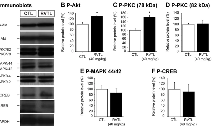
In order to get first insight about a possible signaling mechanism involved in the effects of RVTL on neuroplasticity, we looked at the relative levels of key signaling proteins known to be involved in regulating cell proliferation, survival and dendritic spine formation in the total pro-tein extracts of hippocampus (Fig 7A). Mice treated with RVTL (40 mg/kg) showed a signifi-cant increase in the phosphorylated form of protein kinase B (also known Akt; ~28%) in comparison to control treated mice (control: 100 ± 7.9% versus RVTL 40 mg/kg: 128 ± 4.85%; t
(*p<0.001). Data are expressed as the mean±S.E.M. of percentage of control. Dunnet’s post hoc test was performed after one-way ANOVA. (C)Cell proliferation determined by BrdU incorporation showed a significant decrease with 10μM RVTL (*p<0.001). Data are expressed as the mean±S.E.M. of percentage of total cells. Tukey’s post hoc test was performed after one-way ANOVA. (D)Representative images ofβ III-tubulin-, GFAP- or MAP2-positive cells are shown. Scale bar = 100μm. Cellular phenotypes were determined in adherent hippocampal precursor cells that underwent to differentiation with either the vehicle (CTL) or RVTL (0.001, 0.01, 0.1, 1 or 10μM) for 5 days. (E)βIII-tubulin-positive cells were significantly decreased with 0.1, 1 and 10μM RVTL (*p<0.001), whereas (F) MAP2-positive cells were significantly increased with 0.001, 0.01 and 10μM RVTL (**p= 0.02,*p= 0.047). (G)GFAP-positive cells derived from precursor cells showed that 10μM RVTL significantly increased its proportion (*p= 0.014). Data are expressed as the mean±S.E.M. of percentage of total Dapi cells. Tukey’s post hoc test was performed after one-way ANOVA. Experiments were done twice by quadruplicate.
Fig 5. Resveratrol increases dendritic spines in granular cells of the dentate gyrus in female Balb/C mice at six months of age.(A)Micrograph depicts granular cells with primary and secondary dendrites in the dentate gyrus (DG). Dendrite projections reach the molecular layer and show some protuberances corresponding to dendritic spines. Scale bar = 30μm. Number of dendritic spines along 10μm in secondary dendrites was higher in mice treated with resveratrol (RVTL; 40 mg/kg) than control treated mice (*p= 0.001). Data are expressed as the mean±S.E.M. and were analyzed applying an un-paired Student’st-test. (B)
Representative micrographs of dendritic spines of granular cells derived from control (CTL) or RVTL-treated mice are shown. Scale bar = 5μm. Proportion of the different types of dendritic spines showed significant increases in stubby- and mushroom-shape dendritic spines in RVTL-treated mice (**p= 0.011,*p= 0.026, respectively), whereas the thin-shape dendritic spines were higher in CTL treated mice (***p<0.001). Data are expressed as the mean±S.E.M. of the proportion for each dendritic spine-shape. Data were analyzed with the unpaired Student’st-test.n= 4–5 mice per group.
Fig 6. Resveratrol improves performance of female Balb/C mice at six months of age in the SDPA.(A) Different cohorts of mice were used to test their improve performance in the step down passive avoidance (SDPA). One day after the end of the treatment the SDPA was performed and tested 1.5 (day 15) and 24 hours (day 16) after training. Control (CTL) or resveratrol (RVTL; 40 mg/kg) treated mice were tested in the step-down passive avoidance test. Resveratrol treated mice showed significant increase in the retention latency to move into the dark side of the chamber than the CTL group at 1.5 (**p<0.001) and 24 hours (*p= 0.011) after basal test.n= 8 per group Along the exposure sessions mice treated with RVTL (40 mg/kg) showed an increase in the retention latency in comparison to the basal test (++p<0.001). However, control mice just showed significant increase latency at 24 hours in comparison to the basal test (&&p<0.001). Data are expressed as the mean±S.E.M. of the retention latency parameter. Tukey
’s post hoc test was performed after repeated measures two-way ANOVA. (B) Eight-radial arm maze (8-RAM) was performed after 14 days of treatment with RVTL (40 mg/kg) and the training was followed for 15 days (day 15–29). Also mice treated with RVTL (40 mg/kg) showed a decrease percentage of reference memory errors along the training in the eight-radial arm maze (8-RAM). Significant differences were observed at the day 12 (**p= 0.005) and 15 (*p= 0.015) in comparison to the first session (day 1); whereas control treated mice did not show significant differences (p= 1). In addition, differences between RVTL and the
respective session of CTL group were analyzed applying unpaired Student’st-test reflecting that at day 12 exist a significant difference (&
p= 0.041) between groups. (E) Percentage of working memory errors in both groups was not changed (p= 1). (F) The total number of entries to the eight arms of the maze was not different along the sessions within RVTL (p1), however the control group showed differences respective to the first session (day6**p= 0.008; day10 *p= 0.015, days11,14***p0.001), but not between treatment per sessions (p= 0.216). Data are expressed as the mean±S.E.M.n= 12 mice per group.
= -3.96;p= 0.017;Fig 7B). A similar effect was seen in the levels of one of the two identified phosphorylated forms of protein kinase C (PKC 78 kDa; control: 100 ± 7.13% versus RVTL 40 mg/kg: 161 ± 9.62%; t = -4.87;p= 0.002;Fig 7C). However, we did not find changes in the lev-els of the active isoform of PKC at 82 kDa; (t = -0.11,p= 0.91;Fig 7D), in the levels of the phos-phorylated forms of the mitogen activated protein kinases 44/42 (MAPK 44/42; t = 0.61,
p= 0.56;Fig 7E) and of cAMP response element-binding protein (CREB; t = -0.24,p= 0.81; Fig 7F). These results might suggest the involvement of Akt and PKC (78 kDa) in the mecha-nism by which RVTL modulates brain structural changes such as hippocampal neurogenesis and dendritic spines.
Discussion
In the present study, we have explored the complex effects of RVTL on neuroplasticity includ-ing in it the generation of new neurons and the density and morphology of dendritic spines on hippocampal granule cells in female Balb/C mice at six months of age. In addition, we found that RVTL-treated mice showed improvement performance in behavioral tests related to mem-ory formation.
Basic and preclinical studies have already provided information concerning the broad effects of RVTL on the organisms related to metabolism and obesity, cardiovascular function, cancer and, most notably, life-span [39–41].
Studies in animal models of Alzheimer’s disease showed that RVTL prevented cognitive-learning impairment [42] and hippocampal neurodegeneration [43]. Common among these Fig 7. Resveratrol increases the levels of phosphorylated- Akt and—PKC proteins in the hippocampus of female Balb/C mice at six months of age. (A)Representative immunoblots of the phosphoproteins analyzed in the hippocampal protein extracts of mice treated with resveratrol (RVTL, 40 mg/Kg) are shown. (A-C)Resveratrol treated mice showed significant increase in the levels of phospho-Akt (*p= 0.017) and phospho-PKC (78 kDa) (*p= 0.002), whereas the levels of phospho-PKC (82 kDa,p= 0.91;D), phospho-MAPK 44/42 (p= 0.56;E) and phospho-CREB (p= 0.82;F) did not show significant changes. Data were analyzed with the unpaired Student’st-test.n= 4–5 mice per group.
conditions is that aging is a key risk factor for the development of neurodegeneration and that RVTL may act as an anti-aging molecule to counteract age-related risks (see, for example, dis-cussion in [44]).
In this context, it is somewhat surprising that in this context adult neurogenesis has not attracted more attention. In a murine model of chronic fatigue, the oral administration of RVTL (40 mg/kg) rescued the population of proliferative cells [23], whereas in prenatally stressed rats the administration of RVTL (10 mg/kg) favored the recovery of the DCX-popula-tion [22].
Treating calcitonin gene related peptide (CGRP) knockout mice with 20 mg/kg RVTL reportedly caused an increase in cell proliferation; however, this result may also reflect the short-term survival effects after 5 days of cumulative BrdU injections. Moreover, RVTL increases BrdU/Calbindin colabeled cells in CGRP knockout mice [24]. By and large these results agree with our findings here, even though our data concerning the effects of RVTL on proliferation and, at lower doses, on DCX-cells do not allow for a definitive conclusion. Never-theless, in conjunction with the DCX and caspase 3 (active) findings, our observations also sup-port the view that RVTL acts on precursor cell stages in the course of adult neurogenesis to affect multiple aspects of development, including the promotion of cell survival.
However, there are contradictory findings in the literature as well. In one study, mice that were treated with RVTL by intraperitoneal administration (1 or 10 mg/kg) showed a decrease in cell proliferation, survival, and the numbers of DCX-positive cells and new neurons. In the same study, but at the precursor cell level, micromolar concentrations of RVTL (20 to 50μΜ) decreased proliferation of embryonic cerebral cortical and C17.2 precursor cells [20]. More-over, hippocampus-derived precursor cells that were cultured with different concentrations of RVTL (2 to 4μΜ) showed a decrease in the number ofβIII-tubulin neurons that were derived from differentiated adult neurospheres [21]. Similarly, our study revealed that the in vitro addi-tion of 10μΜof RVTL affected cell proliferation.
These discrepancies regarding the effects of RVTL on adult hippocampal neurogenesis may be due to several factors, such as the strain of mice, the duration of treatment, the timing and mode of labeling of proliferative cells and the age of mice at the time of analysis. Thus, depend-ing upon the dose, the route of administration and the concentration of RVTL that reaches the brain, it is possible to observe a positive regulation of the neurogenic process. Here, the survival of newborn cells in female Balb/C mice at six months of age was of central importance, particu-larly because our general hypothesis is that adult hippocampal neurogenesis provides a“ neuro-genic reserve”that allows for the maintenance of cognitive flexibility in older age. We showed that the oral administration of RVTL (40 mg/kg) decreased apoptosis and increased cell sur-vival, the population of DCX-cells and, consequently, increased hippocampal neurogenesis in female Balb/C mice at six month of age without affecting cell proliferation after 14 days of treatment. Interestingly cellular proliferation was not affected at the precursor level in adherent cells or in neurospheres that were derived from the dentate gyrus that were cultured with nano-molar concentrations of RVTL. Unfortunately, we did not analyze the presence of resveratrol in the lysates of dissected hippocampus, however the pharmacokinetic studies have reported a very low quantity of RVTL in the brains of rodents after 14 days of oral administration [16]. Thus, it is possible that nanomolar concentrations of RVTL favor hippocampal neurogenesis in female Balb/C mice at six months of age, whereas micromolar concentrations impair the neurogenic process in the hippocampus.
improved performance of female Balb/C mice at six months of age in tests that were related to learning and memory such as the SDPA and the 8-RAM. Interestingly, RVTL-treated mice showed better performance in the SDPA than in the 8-RAM. The differences in these results could be related to the type of learning and memory assessed with these tests because the SDPA and the 8-RAM recruit different behavioral, neural, and cognitive demands (i.e., [46]). Thus, in accordance with our results RVTL improved fear- and spatial- memories, the former more significantly than the latter. Thus, RVTL produced stronger effects in acute responses (SDPA) than in long-lasting acquisition procedures (8-RAM). Nevertheless, our data did not discard any effect on learning- and memory-related paradigms that could be produced by lon-ger treatment with RVTL. Nevertheless, our data demonstrated that concomitantly with cell survival and hippocampal neurogenesis, RVTL modulates neuroplasticity at the age of 6 months, a time when physiological levels of adult neurogenesis have declined to very low base-line levels (i.e., [27,28,30]).
One main focus of research has been to observe the effects of RVTL on the so-called longev-ity gene Sirt1, which may imply the promotion of epigenetic effects by RVTL [47]. Numerous other direct or indirect mechanisms have been proposed at several levels, which may ultimately yield an epigenetic cause. Initially, RVTL was promoted as an antioxidant; however despite growing insight into its molecular links, such as to the sirtuin genes, its exact mechanisms of action remain elusive. In a recent review, Kulkarni and Cantó concluded that“The intrinsic anti-oxidant capacity of the resveratrol molecule and its ability to trigger the activation/repres-sion of a wide range of membrane receptors,kinases and other enzymes have turned the quest for a molecular mechanism of action into an epic task.”[48].
Accordingly, the mechanisms by which RVTL may promote neuroplasticity are unknown; however, recent studies have proposed that AMP-activated protein kinase and Sirt-1 [17,18] have beneficial participation on RVTL in neurodegenerative diseases models [16]. Addition-ally, it is known that the neuroprotective effects of RVTL are partially mediated by the activa-tion of theγisoform of protein kinase C, which exhibits a molecular weight of approximately 78 kDa [49]. Here, we found an increased level of phospho-PKC (78 kDa) in the hippocampal lysates of mice that were treated with RVTL (40 mg/kg). Members of the protein kinase C fam-ily have been involved in several biological events, such as cell proliferation and survival, as well as in the reorganization of the cytoskeleton [50–53]. Moreover, we found an increase in the active form of the Akt protein that is involved in both the dendrite maturation and the pro-motion of cell survival [54]. Our study suggests that RVTL may involve phospho-PKC and phospho-Akt increases to induce neuroplastic changes that consequently improve performance in tests that are related to the learning and memory processes in female Balb/C mice of six months of age. Nevertheless, our results cannot discard the influence of RVTL on the epige-netic modifications, which may be relevant for the positive effects that were observed on neuro-plasticity and that could be modulated through Sirt-1, as was reported in other models (for review see [9]). The possible mechanism by which RVTL modulates neuroplasticity deserves further investigation.
reason, these results are difficult to use in the interpretation of the effect of RVTL alone on neu-roplasticity and memory.
Due to the complex nature of the effects of RVTL, it is necessary to perform controlled clini-cal studies to decipher the effect of this flavonoid on neuroplasticity [55]. Thus, our present data support the idea that RVTL is a compound that has interesting implications for strategies that seek to improve brain plasticity in the context of aging or brain disease. Nevertheless, our data indicate that the role of RVTL in the context of adult neurogenesis and other types of neu-roplasticity within an improvement in learning and memory is worth pursuing.
Acknowledgments
We are grateful to Clara Amaya-Castellanos, Liliana Soto-Gutiérrez and Kristin Kretzschmar for helpful administrative procedures. We thank Dr. Marco A. Meraz-Ríos (Centro de Investi-gación y de Estudios Avanzados, CINVESTAV, México) and Dr. Andres Navarrete-Castro (Facultad de Química, Universidad Nacional Autónoma de México, UNAM, México) for their support to perform additional experiments.
Author Contributions
Conceived and designed the experiments: GBRR GK. Performed the experiments: MTP LOL MI RITB NMVR AGS GBRR. Analyzed the data: MTP LOL MI RITB NMVR AGS GBRR. Contributed reagents/materials/analysis tools: MCG. Wrote the paper: GBRR GK.
References
1. Dias GP, Cavegn N, Nix A, do Nascimento Bevilaqua MC, Stangl D, Zainuddin MS, et al. The role of dietary polyphenols on adult hippocampal neurogenesis: molecular mechanisms and behavioural effects on depression and anxiety. Oxid Med Cell Longev. 2012; 2012:541971. Epub 2012/07/26. doi: 10.1155/2012/541971PMID:22829957; PubMed Central PMCID: PMC3395274.
2. Zainuddin MS, Thuret S. Nutrition, adult hippocampal neurogenesis and mental health. Br Med Bull. 2012; 103(1):89–114. Epub 2012/07/27. lds021 [pii] doi:10.1093/bmb/lds021PMID:22833570.
3. Stangl D, Thuret S. Impact of diet on adult hippocampal neurogenesis. Genes Nutr. 2009; 4(4):271–82. Epub 2009/08/18. doi:10.1007/s12263-009-0134-5PMID:19685256; PubMed Central PMCID: PMC2775886.
4. Xu Y, Ku B, Cui L, Li X, Barish PA, Foster TC, et al. Curcumin reverses impaired hippocampal neuro-genesis and increases serotonin receptor 1A mRNA and brain-derived neurotrophic factor expression in chronically stressed rats. Brain Res. 2007; 1162:9–18. Epub 2007/07/10. S0006-8993(07)01259-0 [pii] doi:10.1016/j.brainres.2007.05.071PMID:17617388.
5. An L, Zhang YZ, Yu NJ, Liu XM, Zhao N, Yuan L, et al. The total flavonoids extracted from Xiaobuxin-Tang up-regulate the decreased hippocampal neurogenesis and neurotrophic molecules expression in chronically stressed rats. Prog Neuropsychopharmacol Biol Psychiatry. 2008; 32(6):1484–90. Epub 2008/06/13. S0278-5846(08)00140-1 [pii] doi:10.1016/j.pnpbp.2008.05.005PMID:18547700.
6. van Praag H, Lucero MJ, Yeo GW, Stecker K, Heivand N, Zhao C, et al. Plant-derived flavanol (-)epica-techin enhances angiogenesis and retention of spatial memory in mice. J Neurosci. 2007; 27
(22):5869–78. Epub 2007/06/01. 27/22/5869 [pii] doi:10.1523/JNEUROSCI.0914-07.2007PMID: 17537957.
7. Giacosa A, Barale R, Bavaresco L, Faliva MA, Gerbi V, La Vecchia C, et al. Mediterranean Way of Drinking and Longevity. Crit Rev Food Sci Nutr. 2014. Epub 2014/09/11. PMID:25207479.
8. Russo MA, Sansone L, Polletta L, Runci A, Rashid MM, De Santis E, et al. Sirtuins and resveratrol-derived compounds: a model for understanding the beneficial effects of the Mediterranean diet. Endocr Metab Immune Disord Drug Targets. 2014; 14(4):300–8. Epub 2014/07/11. EMIDDT-EPUB-61310 [pii]. PMID:25008762.
9. Baur JA, Sinclair DA. Therapeutic potential of resveratrol: the in vivo evidence. Nat Rev Drug Discov. 2006; 5(6):493–506. Epub 2006/05/30. nrd2060 [pii] doi:10.1038/nrd2060PMID:16732220.
(23):7862–70. Epub 2014/06/06. 34/23/7862 [pii] doi:10.1523/JNEUROSCI.0385-14.2014PMID: 24899709.
11. Huang HM, Liang YC, Cheng TH, Chen CH, Juan SH. Potential mechanism of blood vessel protection by resveratrol, a component of red wine. Ann N Y Acad Sci. 2005; 1042:349–56. Epub 2005/06/21. 1042/1/349 [pii] doi:10.1196/annals.1338.032PMID:15965080.
12. Kang OH, Jang HJ, Chae HS, Oh YC, Choi JG, Lee YS, et al. Anti-inflammatory mechanisms of resver-atrol in activated HMC-1 cells: pivotal roles of NF-kappaB and MAPK. Pharmacol Res. 2009; 59 (5):330–7. Epub 2009/05/07. S1043-6618(09)00026-7 [pii] doi:10.1016/j.phrs.2009.01.009PMID: 19416633.
13. Oh YC, Kang OH, Choi JG, Chae HS, Lee YS, Brice OO, et al. Anti-inflammatory effect of resveratrol by inhibition of IL-8 production in LPS-induced THP-1 cells. Am J Chin Med. 2009; 37(6):1203–14. Epub 2009/11/26. S0192415X09007600 [pii] doi:10.1142/S0192415X09007600PMID:19938227.
14. Tsai SK, Hung LM, Fu YT, Cheng H, Nien MW, Liu HY, et al. Resveratrol neuroprotective effects during focal cerebral ischemia injury via nitric oxide mechanism in rats. J Vasc Surg. 2007; 46(2):346–53. Epub 2007/06/30. S0741-5214(07)00728-8 [pii] doi:10.1016/j.jvs.2007.04.044PMID:17600658.
15. Sonmez U, Sonmez A, Erbil G, Tekmen I, Baykara B. Neuroprotective effects of resveratrol against traumatic brain injury in immature rats. Neurosci Lett. 2007; 420(2):133–7. Epub 2007/05/29. S0304-3940(07)00506-X [pii] doi:10.1016/j.neulet.2007.04.070PMID:17531385.
16. Vingtdeux V, Giliberto L, Zhao H, Chandakkar P, Wu Q, Simon JE, et al. AMP-activated protein kinase signaling activation by resveratrol modulates amyloid-beta peptide metabolism. J Biol Chem. 2010; 285 (12):9100–13. Epub 2010/01/19. M109.060061 [pii] doi:10.1074/jbc.M109.060061PMID:20080969; PubMed Central PMCID: PMC2838330.
17. Canto C, Auwerx J. PGC-1alpha, SIRT1 and AMPK, an energy sensing network that controls energy expenditure. Curr Opin Lipidol. 2009; 20(2):98–105. Epub 2009/03/12. 00041433-200904000-00004 [pii]. PMID:19276888; PubMed Central PMCID: PMC3627054.
18. Um JH, Park SJ, Kang H, Yang S, Foretz M, McBurney MW, et al. AMP-activated protein kinase-defi-cient mice are resistant to the metabolic effects of resveratrol. Diabetes. 2010; 59(3):554–63. Epub 2009/11/26. db09-0482 [pii] doi:10.2337/db09-0482PMID:19934007; PubMed Central PMCID: PMC2828647.
19. Kempermann G. The neurogenic reserve hypothesis: what is adult hippocampal neurogenesis good for? Trends Neurosci. 2008; 31(4):163–9. Epub 2008/03/11. S0166-2236(08)00064-7 [pii] doi:10. 1016/j.tins.2008.01.002PMID:18329110.
20. Park HR, Kong KH, Yu BP, Mattson MP, Lee J. Resveratrol inhibits the proliferation of neural progenitor cells and hippocampal neurogenesis. J Biol Chem. 2012; 287(51):42588–600. Epub 2012/10/30. M112.406413 [pii] doi:10.1074/jbc.M112.406413PMID:23105098; PubMed Central PMCID: PMC3522260.
21. Saharan S, Jhaveri DJ, Bartlett PF. SIRT1 regulates the neurogenic potential of neural precursors in the adult subventricular zone and hippocampus. J Neurosci Res. 2013; 91(5):642–59. Epub 2013/02/ 14. doi:10.1002/jnr.23199PMID:23404532.
22. Madhyastha S, Sekhar S, Rao G. Resveratrol improves postnatal hippocampal neurogenesis and brain derived neurotrophic factor in prenatally stressed rats. Int J Dev Neurosci. 2013; 31(7):580–5. Epub 2013/07/16. S0736-5748(13)00107-X [pii] doi:10.1016/j.ijdevneu.2013.06.010PMID:23850968.
23. Moriya J, Chen R, Yamakawa J, Sasaki K, Ishigaki Y, Takahashi T. Resveratrol improves hippocampal atrophy in chronic fatigue mice by enhancing neurogenesis and inhibiting apoptosis of granular cells. Biol Pharm Bull. 2011; 34(3):354–9. Epub 2011/03/05. JST.JSTAGE/bpb/34.354 [pii]. PMID: 21372384.
24. Harada N, Zhao J, Kurihara H, Nakagata N, Okajima K. Resveratrol improves cognitive function in mice by increasing production of insulin-like growth factor-I in the hippocampus. J Nutr Biochem. 2011; 22 (12):1150–9. Epub 2011/02/08. S0955-2863(10)00245-7 [pii] doi:10.1016/j.jnutbio.2010.09.016PMID: 21295960.
25. Kodali M, Parihar VK, Hattiangady B, Mishra V, Shuai B, Shetty AK. Resveratrol prevents age-related memory and mood dysfunction with increased hippocampal neurogenesis and microvasculature, and reduced glial activation. Sci Rep. 2015; 5:8075. Epub 2015/01/30. srep08075 [pii] doi:10.1038/ srep08075PMID:25627672.
26. Kempermann G, Kuhn HG, Gage FH. Genetic influence on neurogenesis in the dentate gyrus of adult mice. Proc Natl Acad Sci U S A. 1997; 94(19):10409–14. Epub 1997/09/18. PMID:9294224.
28. Ben Abdallah NM, Slomianka L, Vyssotski AL, Lipp HP. Early age-related changes in adult hippocam-pal neurogenesis in C57 mice. Neurobiol Aging. 2010; 31(1):151–61. Epub 2008/05/06. S0197-4580 (08)00095-X [pii] doi:10.1016/j.neurobiolaging.2008.03.002PMID:18455269.
29. Clark PJ, Kohman RA, Miller DS, Bhattacharya TK, Brzezinska WJ, Rhodes JS. Genetic influences on exercise-induced adult hippocampal neurogenesis across 12 divergent mouse strains. Genes Brain Behav. 2011; 10(3):345–53. Epub 2011/01/13. doi:10.1111/j.1601-183X.2010.00674.xPMID: 21223504.
30. Ramirez-Rodriguez G, Vega-Rivera NM, Benitez-King G, Castro-Garcia M, Ortiz-Lopez L. Melatonin supplementation delays the decline of adult hippocampal neurogenesis during normal aging of mice. Neurosci Lett. 2012; 530(1):53–8. Epub 2012/10/10. S0304-3940(12)01291-8 [pii] doi:10.1016/j. neulet.2012.09.045PMID:23043890.
31. Toth LA, Kregel K, Leon L, Musch TI. Environmental enrichment of laboratory rodents: the answer depends on the question. Comp Med. 2011; 61(4):314–21. Epub 2012/02/15. PMID:22330246.
32. Kempermann G, Gast D, Kronenberg G, Yamaguchi M, Gage FH. Early determination and long-term persistence of adult-generated new neurons in the hippocampus of mice. Development. 2003; 130 (2):391–9. Epub 2002/12/06. PMID:12466205.
33. Babu H, Cheung G, Kettenmann H, Palmer TD, Kempermann G. Enriched monolayer precursor cell cultures from micro-dissected adult mouse dentate gyrus yield functional granule cell-like neurons. PLoS One. 2007; 2(4):e388. Epub 2007/04/27. doi:10.1371/journal.pone.0000388PMID:17460755.
34. Babu H, Claasen JH, Kannan S, Runker AE, Palmer T, Kempermann G. A protocol for isolation and enriched monolayer cultivation of neural precursor cells from mouse dentate gyrus. Front Neurosci. 2011; 5:89. Epub 2011/08/04. doi:10.3389/fnins.2011.00089PMID:21811434.
35. Ramirez-Rodriguez G, Gomez-Sanchez A, Ortiz-Lopez L. Melatonin maintains calcium-binding calreti-nin-positive neurons in the dentate gyrus during aging of Balb/C mice. Exp Gerontol. 2014; 60:147–52. Epub 2014/12/03. S0531-5565(14)00293-9 [pii] doi:10.1016/j.exger.2014.10.014PMID:25446980.
36. Ramirez-Rodriguez G, Ocana-Fernandez MA, Vega-Rivera NM, Torres-Perez OM, Gomez-Sanchez A, Estrada-Camarena E, et al. Environmental enrichment induces neuroplastic changes in middle age female Balb/c mice and increases the hippocampal levels of BDNF, p-Akt and p-MAPK1/2. Neurosci-ence. 2014; 260:158–70. Epub 2013/12/24. S0306-4522(13)01042-7 [pii] doi:10.1016/j.neuroscience. 2013.12.026PMID:24361917.
37. Maurice T, Privat A. SA4503, a novel cognitive enhancer with sigma1 receptor agonist properties, facili-tates NMDA receptor-dependent learning in mice. Eur J Pharmacol. 1997; 328(1):9–18. Epub 1997/06/ 05. S0014-2999(97)83020-8 [pii]. PMID:9203561.
38. Cleveland DW, Fischer SG, Kirschner MW, Laemmli UK. Peptide mapping by limited proteolysis in sodium dodecyl sulfate and analysis by gel electrophoresis. J Biol Chem. 1977; 252(3):1102–6. Epub 1977/02/10. PMID:320200.
39. Singh CK, Ndiaye MA, Ahmad N. Resveratrol and cancer: Challenges for clinical translation. Biochim Biophys Acta. 2015; 1852(6):1178–85. Epub 2014/12/03. S0925-4439(14)00334-2 [pii] doi:10.1016/j. bbadis.2014.11.004PMID:25446990.
40. Bhullar KS, Hubbard BP. Lifespan and healthspan extension by resveratrol. Biochim Biophys Acta. 2015; 1852(6):1209–18. Epub 2015/02/03. S0925-4439(15)00021-6 [pii] doi:10.1016/j.bbadis.2015. 01.012PMID:25640851.
41. Zordoky BN, Robertson IM, Dyck JR. Preclinical and clinical evidence for the role of resveratrol in the treatment of cardiovascular diseases. Biochim Biophys Acta. 2015; 1852(6):1155–77. Epub 2014/12/ 03. S0925-4439(14)00322-6 [pii] doi:10.1016/j.bbadis.2014.10.016PMID:25451966.
42. Sharma M, Gupta YK. Chronic treatment with trans resveratrol prevents intracerebroventricular strepto-zotocin induced cognitive impairment and oxidative stress in rats. Life Sci. 2002; 71(21):2489–98. Epub 2002/09/25. S0024320502020830 [pii]. PMID:12270754.
43. Kim YA, Kim GY, Park KY, Choi YH. Resveratrol inhibits nitric oxide and prostaglandin E2 production by lipopolysaccharide-activated C6 microglia. J Med Food. 2007; 10(2):218–24. Epub 2007/07/27. doi: 10.1089/jmf.2006.143PMID:17651055.
44. Donmez G. The neurobiology of sirtuins and their role in neurodegeneration. Trends Pharmacol Sci. 2012; 33(9):494–501. Epub 2012/07/04. S0165-6147(12)00087-9 [pii] doi:10.1016/j.tips.2012.05.007 PMID:22749331.
46. Myhrer T. Neurotransmitter systems involved in learning and memory in the rat: a meta-analysis based on studies of four behavioral tasks. Brain Res Brain Res Rev. 2003; 41(2–3):268–87. Epub 2003/03/ 29. S0165017302002680 [pii]. PMID:12663083.
47. Hubbard BP, Gomes AP, Dai H, Li J, Case AW, Considine T, et al. Evidence for a common mechanism of SIRT1 regulation by allosteric activators. Science. 2013; 339(6124):1216–9. Epub 2013/03/09. 339/ 6124/1216 [pii] doi:10.1126/science.1231097PMID:23471411.
48. Kulkarni SS, Canto C. The molecular targets of resveratrol. Biochim Biophys Acta. 2015; 1852 (6):1114–23. Epub 2014/10/16. S0925-4439(14)00311-1 [pii] doi:10.1016/j.bbadis.2014.10.005PMID: 25315298.
49. Menard C, Bastianetto S, Quirion R. Neuroprotective effects of resveratrol and epigallocatechin gallate polyphenols are mediated by the activation of protein kinase C gamma. Front Cell Neurosci. 2013; 7:281. Epub 2014/01/15. doi:10.3389/fncel.2013.00281PMID:24421757; PubMed Central PMCID: PMC3872731.
50. Catley MC, Cambridge LM, Nasuhara Y, Ito K, Chivers JE, Beaton A, et al. Inhibitors of protein kinase C (PKC) prevent activated transcription: role of events downstream of NF-kappaB DNA binding. J Biol Chem. 2004; 279(18):18457–66. Epub 2004/02/21. doi:10.1074/jbc.M400765200[pii]. PMID: 14976190.
51. Granados-Soto V, Kalcheva I, Hua X, Newton A, Yaksh TL. Spinal PKC activity and expression: role in tolerance produced by continuous spinal morphine infusion. Pain. 2000; 85(3):395–404. Epub 2000/ 04/27. S030439599900281X [pii]. PMID:10781912.
52. Hoshi N, Langeberg LK, Gould CM, Newton AC, Scott JD. Interaction with AKAP79 modifies the cellular pharmacology of PKC. Mol Cell. 2010; 37(4):541–50. Epub 2010/03/02. S1097-2765(10)00043-2 [pii] doi:10.1016/j.molcel.2010.01.014PMID:20188672; PubMed Central PMCID: PMC3014287.
53. Perkins GA, Wang L, Huang LJ, Humphries K, Yao VJ, Martone M, et al. PKA, PKC, and AKAP locali-zation in and around the neuromuscular junction. BMC Neurosci. 2001; 2:17. Epub 2001/11/22. PMID: 11716788; PubMed Central PMCID: PMC59892.
54. Kim JY, Duan X, Liu CY, Jang MH, Guo JU, Pow-anpongkul N, et al. DISC1 regulates new neuron development in the adult brain via modulation of AKT-mTOR signaling through KIAA1212. Neuron. 2009; 63(6):761–73. Epub 2009/09/26. S0896-6273(09)00617-5 [pii] doi:10.1016/j.neuron.2009.08. 008PMID:19778506; PubMed Central PMCID: PMC3075620.