Consequences of Low Dose Ionizing
Radiation Exposure on the Hippocampal
Microenvironment
Munjal M. Acharya, Neal H. Patel, Brianna M. Craver, Katherine K. Tran, Erich Giedzinski, Bertrand P. Tseng, Vipan K. Parihar, Charles L. Limoli*
Department of Radiation Oncology, University of California Irvine, Irvine, CA, 92697–2695, United States of
America
Abstract
The response of the brain to irradiation is complex, involving a multitude of stress inducible pathways that regulate neurotransmission within a dynamic microenvironment. While signif-icant past work has detailed the consequences of CNS radiotherapy following relatively high doses (45 Gy), few studies have been conducted at much lower doses (2 Gy), where the response of the CNS (like many other tissues) may differ substantially from that expected from linear extrapolations of high dose data. Low dose exposure could elicit radio-adaptive modulation of critical CNS processes such as neurogenesis, that provide cellular input into hippocampal circuits known to impact learning and memory. Here we show that mice deficient for chemokine signaling through genetic disruption of the CCR2 receptor ex-hibit a neuroprotective phenotype. Compared to wild type (WT) animals, CCR2 deficiency spared reductions in hippocampal neural progenitor cell survival and stabilized neurogen-esis following exposure to low dose irradiation. While radiation-induced changes in micro-glia levels were not found in WT or CCR2 deficient animals, the number of Iba1+ cells did differ between each genotype at the higher dosing paradigms, suggesting that blockade of this signaling axis could moderate the neuroinflammatory response. Interestingly, changes in proinflammatory gene expression were limited in WT animals, while irradiation caused significant elevations in these markers that were attenuated significantly after radioadaptive dosing paradigms in CCR2 deficient mice. These data point to the importance of chemokine signaling under low dose paradigms, findings of potential significance to those exposed to ionizing radiation under a variety of occupational and/or medical scenarios.
Introduction
The central nervous system (CNS) is sensitive to a variety of insults that include trauma, ische-mia, depression, chemotherapy and ionizing radiation. Exposure to ionizing radiation can lead to a wide spectrum of radiolytic lesions throughout different cellular compartments that elicits
OPEN ACCESS
Citation:Acharya MM, Patel NH, Craver BM, Tran KK, Giedzinski E, Tseng BP, et al. (2015) Consequences of Low Dose Ionizing Radiation Exposure on the Hippocampal Microenvironment. PLoS ONE 10(6): e0128316. doi:10.1371/journal. pone.0128316
Academic Editor:Roberto Amendola, ENEA, ITALY
Received:January 22, 2015
Accepted:April 24, 2015
Published:June 4, 2015
Copyright:© 2015 Acharya et al. This is an open access article distributed under the terms of the Creative Commons Attribution License, which permits unrestricted use, distribution, and reproduction in any medium, provided the original author and source are credited.
Data Availability Statement:All relevant data are within the paper.
Funding:This work for supported by the Office of Science (BER) U.S. Department of Energy grant no. DE-FG02-09ER64798 (CLL).
learning and memory. Other mechanisms regulate the inhibition and/or recovery of neurogen-esis and include a variety of stress responsive of signaling mechanisms that impact the level of neuroinflammation [5,8,9].
Less is known however, regarding the response of the CNS to low dose ionizing radiation (LDIR,2 Gy), typically encountered from environmental or occupational exposures, or from various medical procedures. Lower dose exposures have also been found to significantly per-turb metabolism leading to elevated reactive oxygen (ROS) and nitrogen (RNS) species, that can trigger changes in the redox balance of the CNS microenvironment [2,10]. Many of these alterations may elicit opposing or compensatory effects in the CNS that exhibit trends opposite to that observed after higher dose exposures. Precisely how LDIR impacts the CNS to compro-mise or facilitate functionality was a focus of the current investigation.
Our past data has shown that low dose exposure (1 Gy) to heavy ions was associated with varying degrees of cognitive dysfunction, suggesting that certain radiation types pose potential risks to the long-term cognition [3]. At similar low dose ranges, protons were found to elicit similar behavioral decrements and a persistent reduction in dendritic complexity and spine density, linking certain behavioral deficits to structural alterations of neurons throughout the hippocampus [11,12]. While studies with charged particles have pointed to potential detri-mental effects in the CNS, few studies have addressed the importance of inflammatory signal-ing in the brain subjected to low and/or adaptive doses of photon radiation.
A wealth of data has substantiated the importance of neuroinflammation in a wide range of CNS pathologies [13]. Chemokine receptors and ligands are important mediators of acute and chronic neuroinflammation that regulate immune cell responses following CNS injury [14]. Microglia, the resident immune cell of the CNS, express chemokine receptors, and the chemo-kine (C-C motif) receptor 2 (CCR2) has been shown to play an important role in modulating inflammatory responses in the irradiated CNS [8,15–17]. CCR2 expression is also found on hippocampal NSCs, granule cell neurons in the DG, and pyramidal cell neurons in the CA1 [19,20]. Past work measuring a number of CNS endpoints have suggested that CCR2 knockout (CCR2-KO) mice were protected against radiation alone or in combination with traumatic brain injury [15]. Compared to wild type (WT) mice, CCR-deficient animals showed reduced neuroinflammation, increased neurogenesis, and improved hippocampal based learning and memory [8,15].
Materials and Methods
Animals, Radiation Exposure and BrdU treatment
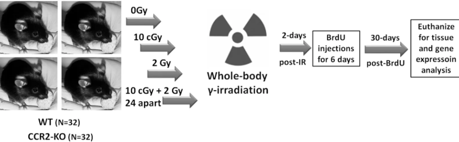
All animal procedures were carried out in compliance with the U.S. Department of Health and Human Services (DH&HS) Guide for the Care and Use of Laboratory Animals and this study was approved specifically by the University of California Institutional Animal Care and Use Committee (IACUC). Two months old, male transgenic CCR2-KO mice (stock 0004999) on a C57BL/6 background and WT C57BL/6 mice (stock 000664) were purchased from the Jackson Laboratories. Schematics of the research design are shown inFig 1. A total of 64 mice were used in this study. A group of 32 mice per genetic background (WT or CCR2-KO) were divided into 4 groups: 0 Gy, 10 cGy, 2 Gy and 10 cGy + 2Gy. For radioadaptive dosing, the initial prim-ing (10 cGy) and subsequent challenge (2 Gy) doses were separated by 24h. All irradiations were whole body and were conducted using137Cs irradiator (J. L. Shepherd Associates, Glen-dale, CA, USA) at a dose rate of 1.07 Gy/min. Mice (n = 4 at a time) were briefly restrained in a compartmentalized Plexiglas container with an aeration space. Sham animals (0 Gy dose) were treated identically without exposure. To assess the impact of LDIR on hippocampal neurogen-esis between transgenic CCR2-KO and WT mice, 5’-bromo-2’-deoxyuridine (BrdU; 50mg/kg i. p., Sigma-Aldrich, MO, USA) was injected for 6 consecutive days at 2 days post-irradiation. At 1-month post-irradiation, animals (n = 4 each group) were deeply anesthetized with isoflurane and euthanized via intracardiac perfusion using saline with heparin (10 U/ml, Sigma-Aldrich) followed by 4% paraformaldehyde (Acros Organics). Brains were cryoprotected in a serial gra-dient of buffered sucrose (10–30%, Sigma-Aldrich) and sectioned coronally (30μm thick) using
a cryostat.
Assessment of neurogenesis and microglia
To assess neurogenesis in the WT and CCR2-KO mice, free floating dual immunofluorescence staining procedure for BrdU and neuron-specific nuclear antigen (NeuN) was performed on representative sections from each experimental group as described previously [6,20]. This technique provides for a relative quantification of neurogenesis, since unbiased stereology was not performed in the present study. To determine the status of resident microglia following
Fig 1. Schematics of research design.WT and CCR2-KO mice were divided into 4 experimental groups (0, 10 cGy, 2Gy, 10 cGy + 2 Gy 24h apart). Mice were restrained briefly in plexiglass chambers and given whole body gamma radiation using137Cs irradiator at the dose rate of 1.07 Gy per minute. At 2 days
post-irradiation, animals received 6 consecutive BrdU injections (50mg/kg, i.p.) and were euthanized 30 days later for immunohistochemical and qPCR analyses.
Gene expression analysis for inflammatory markers
To assess the impact of LDIR on inflammatory signatures within WT and CCR2-KO mice, quantitative real time PCR (qPCR) was performed to measure the expression of inflammatory markers (Iba-1, CD68 and CD11c) in the neocortex and hippocampus from each group (n = 4 per group). These markers were selected based on past experience and on published reports an-alyzing the impact of ionizing radiation on inflammatory signaling [8,17]. Briefly, animals were deeply anesthetized with isoflurane and euthanized by decapitation. Brains were dissected using micro-scissors to separate hippocampal and cortical regions. Freshly dissected tissue samples were placed in RNAlater solution (Ambion, LifeTechnologies, CA, USA) and stored at -20°C. A maximum of 50mg of brain tissue was dissolved in RNA extraction buffer and Trizol (Ambion) for homogenization followed by treatment with BCP reagent (1-bromo-3-chloro-propane, Molecular Research Center, OH, USA) to separate the RNA layer from DNA and protein. The extract was centrifuged (12000 ×g, at 4°C for 15 minutes) and RNA was
precipi-tated using isopropanol, washed twice (75% ethanol) and dried (vacuum centrifugation). The RNA pellet was re-suspended in 20μL of nuclease free water (Ambion) prior to PCR analysis.
PCR was carried out on Applied Biosystems Step One RT-PCR machine with 48-well plate for-mat. Triplicates from each sample of mouse tissue were analyzed, normalized to control values for Iba-1, CD68, and CD11c expression.
Statistics
Statistical analyses were performed using one-way ANOVA to confirm overall significance (GraphPad Prism, v6.0, San Diego, CA). For comparisons between genotype (WT and
CCR2-KO) and control and irradiated groups, parametric two-tailed unpaired t-tests were per-formed with Welch’s correction. All data are shown as the mean ± SEM of 4 independent observations.
Results
Effects of LDIR on newly born cell survival in the hippocampal DG in WT
versus CCR2-KO animals
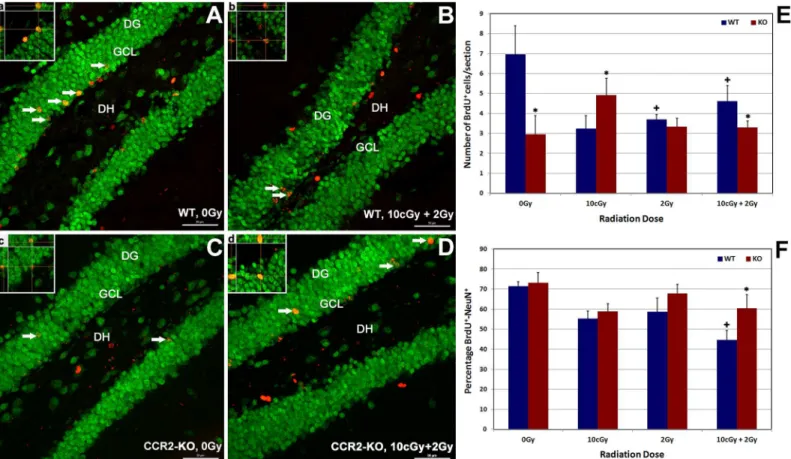
and CCR2-KO mice. Quantification of confocal micrographs indicated that compared to WT, CCR2 deficiency reduced significantly (48%, p = 0.05) the level of basal, newly born cell surviv-al (0 Gy group) in the hippocampus (Fig 2E). Exposure of WT animals led to significant reduc-tions (20–50%, p = 0.05) in the number of BrdU-positive cells 1-month post-irradiation, an effect that was relatively dose-independent under each of the dosing paradigms used (Fig 2E). In contrast, the numbers of BrdU-positive cells within the CCR2 deficient hippocampus were comparable to unirradiated controls (0 Gy) irrespective of the irradiation paradigm. Compared to 0 Gy controls, exposure of CCR2-KO animals to LDIR (10 cGy) led to a significant increase (60%, p = 0.05) in the number of surviving BrdU-positive cells (Fig 2E). Overall results showed that compared to WT animals, CCR2 deficiency protected animals against LDIR-induced re-ductions in cell survival, which may reflect changes in hippocampal proliferation.
Effects of LDIR on hippocampal neurogenesis in WT versus CCR2-KO
animals
To explore the effects of LDIR on neurogenesis within the WT and CCR2-KO brain, the per-centage of BrdU-NeuN dual-labeled cells was quantified in each group (Fig 2A–2D and 2F).
Fig 2. Effect of low dose ionizing radiation on hippocampal proliferation and neurogenesis within WT (A-B and a-b) and CCR2-KO (C-D and c-d) background.Dual immunofluorescence staining for BrdU positive cells (red) co-labeled with NeuN (green) was identified using laser scanning confocal microscopy (arrows). Exposure to low dose priming (10cGy) or challenge (2 Gy) irradiation lead to declined proliferation (BrdU positive cells) in the WT hippocampus significantly in comparison with CCR2-KO background (E) when analyzed 1 month post-irradiation. Formation and maturation of neurons (BrdU-NeuN) reduced significantly following 10 cGy + 2 Gy combination irradiation in the WT brain, whereas percentage of BrdU-NeuN was unchanged in the CCR2-KO hippocampus (F). Orthogonal reconstruction of 1 micro thick Z-stacks confirms presence of dual labeled cells (panela-d). DG, dentate gyrus; DH, dentate hilus and GCL, granule cell layer. Scales bars: 50μm (A-D) and, 10μm (a-d). Data are shown as mean±SEM of 4 independent observations.*, p = 0.05 indicates comparisons between WT and CCR2-KO genotypes. +, p = 0.05 indicates comparison with 0 Gy group.
treated under the same conditions (Fig 2F). These data provide further evidence that neuro-genesis following LDIR is relatively unaffected in CCR2 deficient animals.
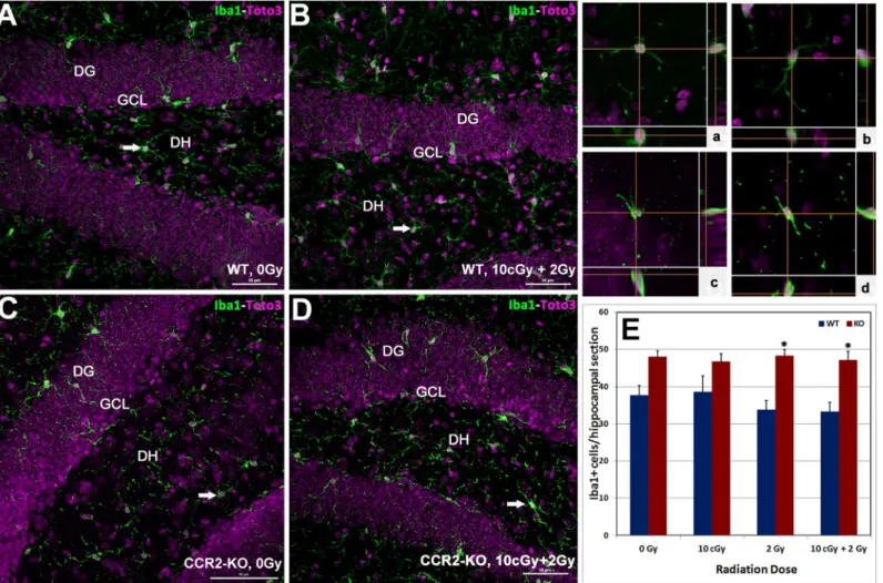
Quiescent microglia (Iba-1 positive) in WT and CCR2-KO animals
following LDIR
To further understand the impact of LDIR on the inflammatory status of the hippocampus in WT and CCR2-KO mice, the number of Iba-1 positive resting microglia were quantified. Immunoreactivity of Iba-1 (green) was evident within each hippocampal subfield (DG, CA1 and CA3) and quantified as the number of Iba-1 positive cells per section (Fig 3A–3Dand in-serts,a-d). Overall ANOVA for the number of Iba-1 positive resting microglia was significant (p = 0.001) between the WT and CCR2-KO mice. Compared to WT, trends toward higher numbers of Iba-1 positive microglia were observed in unirradiated CCR2 deficient animals as well as those exposed to 10 cGy (Fig 3E). Cohorts from either genotype exposed to irradiation did not exhibit significant changes in the number of Iba-1 positive microglia in the hippocam-pus (Fig 3E).
Gene expression status for pro-inflammatory markers following LDIR
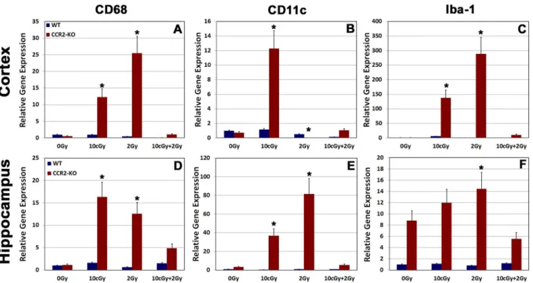
To further analyze the effects of LDIR on expression of pro-inflammatory markers within the WT and CCR2-KO brain, we carried out quantitative RT-PCR for Iba-1, CD68 and CD11c from freshly dissected cortical and hippocampal tissues (Fig 4). RT-PCR data revealed that ex-pression of Iba-1, CD68 and CD11c did not changed significantly in the WT brain following exposure to LDIR. In contrast, gene expression of all pro-inflammatory markers elevated sig-nificantly (p = 0.05) within the CCR2-KO brains when exposed to either single 10 cGy or 2 Gy radiation doses (Fig 4). The expression level of each pro-inflammatory marker was however, reduced significantly (p = 0.05) following radioadaptive dosing in CCR2-deficient animals (Fig 4).Discussion
Our results on neurogenesis and microglia (Figs2and3) show that CCR2 deficiency alters the cellular (NSC survival and/or proliferation, differentiation and Iba-1) and molecular (pro-inflammatory gene expression) response of the hippocampus following LDIR. While the num-ber of surviving (BrdU positive) cells in the CCR2-KO compared to the WT hippocampus were low, acute or radioadaptive irradiation paradigms did not cause further reductions in sur-vival in the CCR2 deficient background 1-month post-irradiation (Fig 2E). Moreover, the ca-pability of forming new neurons (BrdU-NeuN positive cells) remained intact within the CCR2-KO hippocampus following exposure to the radioadaptive dosing paradigm (Fig 2F), an effect not observed in WT mice when analyzed one month post-irradiation. While our neuro-genic measurements are limited in that they provided only relative rather than absolute quanti-fication of differentiated phenotypes, results did corroborate past data demonstrating a neuroprotective role of CCR2 blockade [8,15,17]. Constitutive expression of CCR2 receptors by hippocampal NSCs and mature neurons [18,19] makes them sensitive to radiation-induced changes in chemokine signaling. Thus, CCR2 deficiency may simply dampen the sensitivity of
Fig 3. Status of hippocampal microglia following low dose ionizing radiation exposure.Immunofluorescence staining for Iba-1 positive (green), resting microglia, with nuclear counter staining (Toto-2, pink) was identified using laser scanning confocal microscopy (A-D). Iba-1 positive cells were distributed throughout the hippocampus (DG, DH, CA3 and CA1 subfields) and orthogonal Z-stacks confirmed the microglial morphology (panela-d). In general, numbers of Iba-1 positive cells were higher in CCR2-KO hippocampus comparison with WT animals (E). Following exposure to 2 Gy alone or 10 cGy + 2 Gy combined irradiation, the number of Iba-1 positive cells elevated significantly in the CCR2-KO mice compared to WT when analyzed 1 month post-irradiation. DG, dentate gyrus; DH, dentate hilus and GCL, granule cell layer. Scales bars: 50μm (A-D) and, 10μm (a-d). Data are shown as mean±SEM of 4
independent observations.*, p = 0.05 indicates comparisons between WT and CCR2-KO genotypes.
the CCR2 signaling axis to LDIR that manifests as a neuroprotective phenotype over protracted post-irradiation intervals.
Microglial activation helps modulate the innate immune response in a number of neurode-generative conditions and during tissue remodeling following radiation injury [8,17,24–26]. In the CCR2 deficient background, data indicates that the number of resting microglia (Iba-1 positive) was relatively high in comparison to WT controls following each of the irradiation paradigms. Furthermore, the levels of resting microglia were not responsive to acute or radioa-daptive dosing, as the numbers of Iba-1 positive cells remained constant over the 1-month post-irradiation interval (Fig 3E). Analysis of mRNA (qPCR) within the CCR2-KO brain indi-cated a significant increase in the message levels of pro-inflammatory markers (CD68, CD11c and Iba-1) following 10 cGy and 2 Gy acute exposures in the hippocampus and neocortex (Fig 4). Interestingly, elevated proinflammatory markers after acute exposure were attenuated sig-nificantly in the brains of CCR2 deficient mice after radioadaptive dosing (Fig 4). Exposure to low priming doses of radiation (10 cGy) may reset the inflammatory tone within the
CCR2-KO hippocampal microenvironment, thereby preventing the upregulation of inflamma-tory markers following subsequent exposure to higher (2 Gy) challenge doses (Fig 4). In sum, our findings demonstrate that the low dose radiation response between WT and CCR2 defi-cient mice is different, possibly mediated by a CCR2 dependent signaling feedback.
Radiation exposure of the brain disrupts neurotransmission and elicits varying degrees of cognitive dysfunction. Multiple mechanisms are involved, including the inhibition of hippo-campal neurogenesis [6], the degradation of neuronal structure and the alteration of synaptic
Fig 4. Gene expression analysis for pro-inflammatory markers from freshly dissected cortex and hippocampus.Relative gene expression for pro-inflammatory markers (CD68, CD11c and Iba-1) was analyzed using quantitative real-time PCR (qPCR) in cortex (A-C) and hippocampus (D-F) within the WT and CCR2-KO background. The mRNAs for all pro-inflammatory markers elevated significantly following 10 cGy or 2 Gy irradiation within the CCR2-KO animals (A-F). In contrast, relative gene expression for these pro-inflammatory markers drops down following 10 cGy + 2 Gy irradiation paradigm (A-F). WT animals did not display alterations in gene expression profile following irradiation. Data are shown as mean±SEM of 4 independent observations.*, p = 0.05
in comparison with 0 Gy group.
integrity and neuronal plasticity [3,11,27]. Irradiation of the brain also triggers a complex temporal response of secondary reactive processes involving oxidative stress and inflammation [2,4–6,10]. Changes in redox metabolism promote early and recurrent inflammatory signa-tures that perturb the neurogenic microenvironment of the hippocampus. Pharmacologic modulation of inflammatory and oxidative signaling via peroxisome proliferating-activated re-ceptor (PPAR) agonists has been shown to be neuroprotective following whole-brain irradia-tion [28,29]. Past and current studies have now demonstrated that genetic disruption of chemokine signaling can alter the radioresponse of the CNS, mediated in part through changes in the levels of microglia [8,17]. Importantly, our quantitative measurements of neurogenesis and microglia corroborate past data collected at higher doses, and confirm that similar neuro-protective phenotypes transpire after LDIR. Our data supports the potential utility of attenuat-ing chemokine signalattenuat-ing to reduce CNS damage followattenuat-ing acute and/or protracted LDIR exposure encountered under a variety of occupational and medical scenarios.
Acknowledgments
This work for supported by the Office of Science (BER) U.S. Department of Energy grant no. DE-FG02-09ER64798 (CLL).
Author Contributions
Conceived and designed the experiments: CLL MMA VKP BPT. Performed the experiments: NHP BMC KKT EG VKP MMA. Analyzed the data: MMA VKP BPT CLL. Wrote the paper: MMA CLL.
References
1. Fike JR, Rola R, Limoli CL. Radiation response of neural precursor cells. Neurosurg Clin N Am. 2007; 18(1):115–27. Epub 2007/01/25. S1042-3680(06)00126-4. PMID:17244559.
2. Fike JR, Rosi S, Limoli CL. Neural precursor cells and central nervous system radiation sensitivity. Semin Radiat Oncol. 2009; 19(2):122–32. Epub 2009/03/03. S1053-4296(08)00085-4. PMID:
19249650; PMCID: PMC2683587. doi:10.1016/j.semradonc.2008.12.003
3. Tseng BP, Giedzinski E, Izadi A, Suarez T, Lan ML, Tran KK, et al. Functional consequences of radia-tion-induced oxidative stress in cultured neural stem cells and the brain exposed to charged particle ir-radiation. Antioxid Redox Signal. 2013; 20(9):1410–22. doi:10.1089/ars.2012.5134PMID:23802883 4. Chiang CS, Hong JH, Stalder A, Sun JR, Withers HR, McBride WH. Delayed molecular responses to
brain irradiation. Int J Radiat Biol. 1997; 72(1):45–53. PMID:9246193.
5. McBride WH, Pajonk F., Chiang C.S., Sun J.R. NF-kappa B, cytokines, proteasomes, and low-dose ra-diation exposure. Mil Med. 2002; 167(2):66–7. PMID:11873521
6. Parihar VK, Acharya MM, Roa DE, Bosch O, Christie LA, Limoli CL. Defining functional changes in the brain caused by targeted stereotaxic radiosurgery. Translational Cancer Res. 2014; 3(2):124–37. doi:
10.3978/j.issn.2218-676X.2013.06.02PMID:24904783.
7. Chen H, Yoshioka H, Kim GS, Jung JE, Okami N, Sakata H, et al. Oxidative stress in ischemic brain damage: mechanisms of cell death and potential molecular targets for neuroprotection. Antioxid Redox Signal. 2011; 14(8):1505–17. doi:10.1089/ars.2010.3576PMID:20812869.
8. Belarbi K, Jopson T, Arellano C, Fike JR, Rosi S. CCR2 deficiency prevents neuronal dysfunction and cognitive impairments induced by cranial irradiation. Cancer Res. 2013; 73(3):1201–10. Epub 2012/12/
18. doi:10.1158/0008-5472.CAN-12-2989PMID:23243025
9. Greene-Schloesser D, Moore E, Robbins ME. Molecular pathways: radiation-induced cognitive im-pairment. Clinical Cancer Res. 2013; 19(9):2294–300. doi:10.1158/1078-0432.CCR-11-2903PMID:
23388505.
10. Limoli CL, Giedzinski E, Rola R, Otsuka S, Palmer TD, Fike JR. Radiation response of neural precursor cells: Linking cellular sensitivity to cell cycle checkpoints, apoptosis and oxidative stress. Radiat Res. 2004; 161:17–27. PMID:14680400
18. Banisadr G, Queraud-Lesaux F, Boutterin MC, Pelaprat D, Zalc B, Rostene W, et al. Distribution, cellu-lar localization and functional role of CCR2 chemokine receptors in adult rat brain. J Neurochem. 2002; 81(2):257–69. PMID:12064472.
19. van der Meer P, Ulrich AM, Gonzalez-Scarano F, Lavi E. Immunohistochemical analysis of CCR2, CCR3, CCR5, and CXCR4 in the human brain: potential mechanisms for HIV dementia. Exp Mol Pathol. 2000; 69(3):192–201. doi:10.1006/exmp.2000.2336PMID:11115360.
20. Liao AC, Craver BM, Tseng BP, Tran KK, Parihar VK, Acharya MM, et al. Mitochondrial-targeted human catalase affords neuroprotection from proton irradiation. Radiat Res. 2013; 180(1):1–6. Epub
2013/05/16. doi:10.1667/RR3339.1PMID:23672429.
21. Elmore MR, Najafi AR, Koike MA, Dagher NN, Spangenberg EE, Rice RA, et al. Colony-stimulating fac-tor 1 recepfac-tor signaling is necessary for microglia viability, unmasking a microglia progenifac-tor cell in the adult brain. Neuron. 2014; 82(2):380–97. doi:10.1016/j.neuron.2014.02.040PMID:24742461. 22. Naert G, Rivest S. CC chemokine receptor 2 deficiency aggravates cognitive impairments and amyloid
pathology in a transgenic mouse model of Alzheimer's disease. J Neurosci. 2011; 31(16):6208–20. doi:
10.1523/JNEUROSCI.0299-11.2011PMID:21508244.
23. Semple BD, Kossmann T, Morganti-Kossmann MC. Role of chemokines in CNS health and pathology: a focus on the CCL2/CCR2 and CXCL8/CXCR2 networks. J Cereb Blood Flow Metab. 2010; 30 (3):459–73. doi:10.1038/jcbfm.2009.240PMID:19904283.
24. Linard C, Marquette C, Mathieu J, Pennequin A, Clarencon D, Mathe D. Acute induction of inflammato-ry cytokine expression after gamma-irradiation in the rat: effect of an NF-kappaB inhibitor. Int J Radiat Oncol Biol Phys. 2004; 58(2):427–34. PMID:14751512.
25. Ransohoff RM, Brown MA. Innate immunity in the central nervous system. J Clin Invest. 2012; 122 (4):1164–71. doi:10.1172/JCI58644PMID:22466658.
26. Ridet JL, Malhotra SK, Privat A, Gage FH. Reactive astrocytes: cellular and molecular cues to biologi-cal function. Trends Neurosci. 1997; 20(12):570–7. PMID:9416670.
27. Acharya MM, Christie LA, Lan ML, Giedzinski E, Fike JR, Rosi S, et al. Human neural stem cell trans-plantation ameliorates radiation-induced cognitive dysfunction. Cancer Res. 2011; 71(14):4834–45.
Epub 2011/07/16. doi:10.1158/0008-5472.CAN-11-0027PMID:21757460.
28. Schnegg CI, Greene-Schloesser D, Kooshki M, Payne VS, Hsu FC, Robbins ME. The PPARdelta ago-nist GW0742 inhibits neuroinflammation, but does not restore neurogenesis or prevent early delayed hippocampal-dependent cognitive impairment after whole-brain irradiation. Free Radic Biol Med. 2013; 61:1–9. doi:10.1016/j.freeradbiomed.2013.03.002PMID:23499837.