Jae Kyoung Kim, Trachette L. Jackson*
Department of Mathematics, University of Michigan, Ann Arbor, Michigan, United States of America
Abstract
The tumor suppressor p53 protein shows various dynamic responses depending on the types and extent of cellular stresses. In particular, in response to DNA damage induced byc-irradiation, cells generate a series of p53 pulses. Recent research has shown the importance of sustaining repeated p53 pulses for recovery from DNA damage. However, far too little attention has been paid to understanding how cells can sustain p53 pulses given the complexities of genetic heterogeneity and intrinsic noise. Here, we explore potential molecular mechanisms that enhance the sustainability of p53 pulses by developing a new mathematical model of the p53 regulatory system. This model can reproduce many experimental results that describe the dynamics of p53 pulses. By simulating the model both deterministically and stochastically, we found three potential mechanisms that improve the sustainability of p53 pulses: 1) the recently identified positive feedback loop between p53 and Roraallows cells to sustain p53 pulses with high amplitude over a wide range of conditions, 2) intrinsic noise can often prevent the dampening of p53 pulses even after mutations, and 3) coupling of p53 pulses in neighboring cells via cytochrome-c significantly reduces the chance of failure in sustaining p53 pulses in the presence of heterogeneity among cells. Finally, in light of these results, we propose testable experiments that can reveal important mechanisms underlying p53 dynamics.
Citation:Kim JK, Jackson TL (2013) Mechanisms That Enhance Sustainability of p53 Pulses. PLoS ONE 8(6): e65242. doi:10.1371/journal.pone.0065242
Editor:Toru Ouchi, University of Chicago, United States of America
ReceivedDecember 19, 2012;AcceptedApril 26, 2013;PublishedJune 3, 2013
Copyright:ß2013 Kim, Jackson. This is an open-access article distributed under the terms of the Creative Commons Attribution License, which permits unrestricted use, distribution, and reproduction in any medium, provided the original author and source are credited.
Funding:This work was supported by NSF DMS1026317 for computing resources, the Human Frontiers of Science Foundation (RPG 24/2012) and JSMF -220020079/SCS. The funders had no role in study design, data collection and analysis, decision to publish, or preparation of the manuscript.
Competing Interests:The authors have declared that no competing interests exist. * E-mail: tjacks@umich.edu
Introduction
p53 protein is one of the most important tumor suppressors that is mutated in more than half of all human cancers [1]. The p53 protein regulates key functions, such as DNA repair, cell cycle arrest and apoptosis, which prevent tumorigenesis in response to cellular stress (e.g. DNA damage and genomic instability) [2]. Recent studies have found that depending on the type of stresses, p53 protein exhibits different dynamical behaviors. While p53 protein levels are low in the absence of stress, transient DNA double-strand breaks (DSBs) that occur during a normal cell-cycle lead to spontaneous pulses of p53 [3]. A single pulse of p53 can also be triggered by UV irradiation [4]. The most dynamic behavior of p53 is induced by severe DNA damage. When severe DSBs are caused by c-irradiation or radiomimetic drugs, cells generate a series of p53 pulses [5,6,7,8]. Interestingly, these different dynamical behaviors appear to be highly correlated with appropriate responses of p53 to different types of stresses [3,4,9]. Thec-triggered p53 pulses have three notable features: 1) the pulse amplitudes are independent ofc-irradiation strength, 2) the pulses are sustained so long as DSBs persist (i.e. undamped pulses) and 3) while the period of the pulses is tightly regulated, the amplitude is highly variable. The molecular mechanisms under-lying these unique features of p53 pulses have been explored both experimentally and theoretically. Among many feedback loops regulating p53 [10], a negative feedback loop between p53 protein and E3 ubiquitin ligase Mdm2 is considered to be a core mechanism that generates p53 oscillations [11,12,13,14,15,16]. Recently, mathematical modeling and subsequent experiments have found an additional feedback loop between upstream kinase ATM and p53 through WIP1, which is also required to sustain
p53 pulses with amplitudes that are independent ofc-irradiation strength [5,17]. In addition to the molecular mechanisms underlying p53 pulses, the relationship between the dynamics of p53 and its output functions, such as DNA repair or cell cycle arrest have also been widely studied both theoretically and experimentally [3,9,18,19,20,21,22]. In light of the considerable theoretical and experimental focus on dynamics of p53 pulses and the role of these pulses, it is somewhat surprising that very little attention has been paid to how cells robustly sustain p53 pulses, even in the presence of perturbation (e.g. intrinsic noise or genetic perturbation) [14,22]. Given the importance of sustaining p53 pulses for cell fates in response to severe DNA damage [3,9], it is of considerable interest to understand how cells can sustain p53 pulses over a wide range of conditions.
robustly even in the presence of significant genetic perturbations [27,28]. Cellular coupling can also tightly regulate the periods and the amplitudes of c-AMP rhythms in Dictyostelium [29] and membrane potential spikes in neurons [30,31]. The coupling has been investigated as a mean to reduce the effects of noise on rhythms, but more recent studies have found that coupling and noise often synergistically enhance rhythms in calcium systems and circadian clocks [28,32].
The identification of mechanisms that enhance robustness of rhythms in various biological oscillatory systems has lead to the question of whether similar mechanisms that enhance robust c-triggered p53 pulses exist. Indeed, several positive feedback loops acting on p53 through PTEN, dapk1, c-Ha-Ras and DDR1 have been identified [33,34,35,36]. However, these feedback loops do not appear to be essential for sustaining p53 pulses [37]. A recent study identified a novel positive feedback loop between p53 and Rora, which may enhance the sustainability of p53 pulses [38]. In this positive feedback loop, p53 promotes gene expression of Rora in response to DNA damage, and increased RORa protein stabilizes p53. Together with additional feedback loops acting on p53, a recent study found radiation induced bystander effect (RIBE) [39], which may be a potential mechanism that couples p53 pulses of neighboring cells. RIBE refers to DNA damage of un-irradiatedcells induced by the molecular signals produced by their neighboring irradiated cells. Among various molecular signals involving RIBE, Cytochrome-c (cyt-c) can act as an excitatory signal for p53 pulses in neighboring cells [40]. More specifically, p53 activation induced by DNA damage stimulates the mitochon-drial release of cyt-c, which activates p53 in un-irradiated neighboring cells. However, whether cyt-c can couple p53 pulses inirradiatedcells has yet to be investigated.
Here, we develop a new mathematical model of p53 to explore molecular mechanisms that enhance sustainability of p53 pulses in the presence of DNA damage. Our new model is able to reproduce many key experimental observations that discern characteristics of p53 pulses. By simulating this model both deterministically and stochastically, we find that a positive feedback loop between p53 and Rora plays a pivotal role in sustaining p53 pulses over a wide range of conditions. Moreover, we find that noise can often prevent p53 pulses from dampening even after mutations of key molecular species that generate p53 pulses. Interestingly, we also found that p53 pulses in DNA-damaged cells have characteristics similar to Type II resonator neurons, which are prone to synchronize their spikes through excitatory couplings. Similar to Type II neurons, even a weak coupling via cyt-c can synchronize p53 pulses of cells and significantly increase the chance that p53 pulses will be sustained in response to DNA damage.
Results
Model Description
To explore mechanisms that enhance sustainability of p53 pulses, we have developed a new mathematical model that describes the dynamics of p53 pulses. Our model is based on the delay-differential equation (DDE) model of Batchelor et al. 2008, which made important contributions to understanding the mechanisms underlying p53 pulse generation [3,4,5,9]. In particular, their model found that DNA damage induced activation of ATM, an upstream signaling kinase for p53 phosphorylation, and that deactivation of ATM by p53 via WIP1 triggers a series of p53 pulses [5]. Batchelor’s model tracks the temporal changes of five molecular species: p53inactive,
p53active, Mdm2, Wip1 and ATM-P (Figure 1A). The interactions
among these five species can be described by three negative feedback loops:
1) ATM-P induced p53activepromotes production of Mdm2 and
Mdm2 induces ubiquitination of p53,
2) ATM-P induced p53active promotes production of Wip1
and Wip1 mediates dephosphorylation and inactivation of p53active, and
3) ATM-P induced p53activepromotes the production of Wip1
and Wip1 dephosphorylates and inactivates ATM-P [4,5].
In addition to these negative feedback loops, Mdm2 phosphor-ylation by ATM-P inactivates and destabilizes Mdm2.
We begin developing our new model by first converting the DDEs used in Batchelor’s model into ordinary differential equations (ODEs). Our rationale for this modification is that DDEs often generate rhythms in systems whose structures are not likely to produce rhythms naturally [25,41]. We, therefore, want to ensure that the p53 pulses in our model are not explicit delay-induced instabilities, which often occur in nonlinear feedback systems. Another reason for utilizing ODEs is that it is more difficult to perform stochastic simulations of DDEs than ODEs because methods to introduce stochasticity into the explicit time delays in DDEs have not been fully developed [42]. Two explicit time delays, 0.7 hours and 1.25 hours for p53active -dependent
production of Mdm2 and Wip1, respectfully were used in Batchelor’s model. We removed these explicit delays and introduced intermediate steps (mRNAs) required for the produc-tion of MDM2 protein and WIP1 protein (Figure 1B and Materials and Methods). To test whether the newly introduced intermediate steps without the explicit time delays are sufficient to generate sustained p53 pulses after 10 Gy irradiation, new parameters associated with the equations for the mRNA of Mdm2 and Wip1 were randomly searched until 500 parameters yielding oscillations were found (see Methods and Materials for details of parameter searching). Here, we defined sustained pulses as undamped oscillations (see Methods and Materials for details). However, the probability of rhythm occurrence was very low (,0.1%) (Figure 1D). Considering robustness as a design principle of biological systems whose essential functions are nearly independent of varying biochemical parameters [43,44,45], the low chance of rhythm occurrence implies that the current ODE model with intermediate steps for protein production might lack one or more essential components for generating rhythms.
One potential issue with the current ODE model is that it consists of only negative feedback loops. It is well known that an additional positive feedback loop often enhances the robustness of rhythms in other biological oscillatory systems [14,24,25]. Indeed, a recent study identified a positive feedback loop between Rora and p53, in which after c-irradiation, p53active induces the
p53 pulses: a large variation in amplitude, but little variation in periods [7]. Since recent studies have shown that the sustainability of p53 pulses is essential for the repair of DNA damage, our study further indicates that the positive feedback loop between p53 and Roracan play an important role in the appropriate response of p53 to DNA damage [9]. Indeed, Rora2/2cells failed to regulate apoptosis in response to DNA damage [38].
Finally, previous experimental studies found a nonlinear relationship between the amount of DNA damage and the activation rate of ATM [47,48]. We therefore changed the mechanisms for ATM activation induced by DSBs in Bachelor’s model, where ATM activation is linearly proportional to the extent of DNA damage [5]. In the new model, ATM activation becomes more sensitive to small amounts of DNA damage and becomes saturated for large amounts of DNA damage (Figure 1G and Materials and Methods). We found that this modification is critical to simulating the correct responses of p53 systems, such as period distributions in response to different strengths of c-irradiation (see below).
Model Validation
biochemical and genetic systems [11,50,51]. The lists of reactions together with the probability for their occurrence are described in Table S2. Hill equations and Michaelis-Menten equations of the deterministic model were also used in the stochastic simulation by assuming fast time scales for the elementary reactions underlying these equations [52,53]. This stochastic model also generated sustained p53 pulses after 5 Gy irradiation (Figure 2B).
First, with Gillespie’s algorithm, we simulated the period distributions of MDM2 oscillations in 500 cells after 0.3, 5, and 10 Gy irradiations. To consider the heterogeneity among cells [54], the original parameters of the model were randomly perturbed by multiplying eX (X , N(0, 0.2)). The simulations showed that the distributions of periods become narrower as c-irradiation strength increases, which is consistent with experimen-tal data (Figure 3A and 3B) [7]. With low strength c-irradiation (0.3 Gy), MDM2 rhythms showed a wide range of periods (e.g. 9 hr and 6 hr) (Figure 3C). Moreover, as the strength of c-irradiation becomes stronger, the fraction of cells oscillating with period 4–7 hours increases, matching experimental data (Figure 3D) [7]. Next, among oscillating cells, we measured the average amplitude, peak width of p53 and time delay between the peaks of p53 and Mdm2 for first five peaks of pulses (Figure 4A– C). All of these values were well matched with experimental data [7]. Moreover, the model simulated a significantly larger variation in the peak amplitudes than those in the peak width of p53 or delay between peak timing of p53 and Mdm2 (Figure 4D–F), as shown in previous experiments [7]. Taken together, the new model can successfully reproduce key features of p53 pulses induced byc-irradiation.
Noise can Enhance the Sustainability of p53 Pulses in Mutated Cells
To quantify the role of each molecular species in the model, we simulated various types of mutations. Given 10 Gy irradiation, the model with Wip1 knockout generates a single peak followed by a high steady state of p53 instead of sustained pulses of p53, matching a previous experimental study (Figure 5A) [5]. It is known that SNP309 (polymorphism of mdm2 promoter) increases
expression of Mdm2 mRNA about 10 times and induces the low stable state of p53 instead of oscillations afterc-irradiation [55,56]. We tested this in our model by increasing the transcription rate of Mdm2 by a factor of 10 and found that this causes the same behavior that was observed experimentally (Figure 5B). Finally, the model predicts that Rorais also essential to generate sustained pulses of p53 in the presence of 10 Gyc-irradiation (Figure 5C).
While deterministic simulations predict that sustained p53 pulses will not occur with these three types of mutations, stochastic simulations predict that the sustained pulses often occur even with these mutations (Figure 5D–F). In particular, the model showed clear oscillations of p53 in Rora2/2cells. Interestingly, previous studies also showed that noise could often induce rhythms in other biological oscillatory systems, such as the circadian clock, by moving the system away from its natural steady state [28]. To see how often noise can induce the sustained pulses with these mutations, we conducted a simulation of 200 cells with Gillespie algorithm. Here, we again assumed heterogeneity among cells as we did previously. Cells with any one of three mutations showed a lower chance of rhythm occurrence than WT cells (Figure 5G). In particular, Wip12/2cells had the lowest probability of sustaining pulses. Moreover, the amplitudes of pulses were also significantly reduced with these mutations (Figure 5H). Interestingly, a previous experimental study also showed that only small portion of cells can generate sustained p53 pulses with low amplitudes after treatment of Wip1 siRNA [5]. Our simulations indicate that while these mutated cells were unable to generate sustained pulses of p53 after c-irradiation at the cell population level (deterministic simulation), noise can often induce sustained rhythms at the single cell level (stochastic simulation).
Common Characteristics between the p53 Model and Type II Neurons
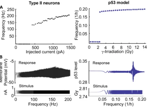
We noticed that the p53 regulatory system in cells behaves like an excitatory system similar to neurons. That is, while p53 levels remain at a steady state in the absence of DNA damage, transient DNA damage can induce spontaneous pulses of p53 [3]. Moreover, severe DNA damage induced by c-irradiation yields a series of p53 pulses with a small variation in period, but a large variation in amplitude (Figure 4D–E) [7]. Interestingly, these are also common features of type II neurons, which are also known as resonator neurons due to their preferred frequency of spiking [57]. When an external current greater than a certain threshold is applied, type II neurons begin generating spikes with a narrow range of frequencies, regardless of the strength of the external stimuli, while other types of neurons (e.g. type I neurons) yield spikes with a wide range of frequencies depending on the strength of the external currents [57,58,59].
We further explored whether pulses of p53 in cells and spikes of type II neurons have more common features in response to external stimuli. Given external constant currents with different strengths, type II neurons have a non-zero lower bound for frequency and a narrow range of frequency variation (Figure 6A) [57,59]. Similarly, p53 pulses also have a non-zero lower bound for their frequency and a narrow range of frequency variation for different strengths ofc-irradiation in the model (Figure 6A). When an oscillating current, as opposed to a constant current, with varying frequency and low amplitude is injected, type II neurons also show an interesting response. They respond sensitively to the injected current with a specific frequency [57,59] (Figure 6B). We tested whether p53 pulses would respond in a similar way to low amplitude c-irradiation with varying frequency,
2:78z0:02Sin(0:1pt1:15) Gy. Surprisingly, p53 levels showed a sensitive response to irradiation with a specific frequency (,0.16/
hr) similar to type II neurons (Figure 6B). Mathematically type II neurons are characterized by a Hopf bifurcation when the level of external current is varied. Indeed, our p53 model also shows a supercritical Hopf bifurcation when the strength ofc-irradiation is changed.
A Potential Coupling Mechanism of p53 Pulses among Neighboring Cells
We have shown that both p53 pulses in DNA-damaged cells and spikes of type II neurons have the characteristics of resonators. This raises the question of why DNA-damaged cells act as resonators or, in other words, what is the benefit of behaving as a resonator when p53 pulses are generated. One of the distinct characteristics of resonator neurons is that they can synchronize rhythms in purely excitatory networks, while integrator neurons cannot synchronize their rhythms [60]. This feature of the resonator neuron leads to the hypothesis that like resonator neurons, DNA-damaged cells are designed to synchronize p53 pulses.
Before exploring this hypothesis, we first examined whether there exists a coupling signal among cells similar to excitatory neurotransmitters among neurons. Interestingly, recent studies have found that neighboring cells communicate with each other afterc-irradiation through the ‘radiation induced bystander effect (RIBE)’ [39]. RIBE is characterized by DNA damage in un-irradiated cells that is induced by molecular signals produced by
their neighboring irradiated cells. While many molecular signals involving RIBE have been proposed, a recent study identified Cytochrome-c (cyt-c) as one of main signals inducing RIBE. Specifically, c-irradiation induces the p53 dependent release of mitochondrial cyt-c, which enters un-irradiated neighboring cells through gap junctions [61] and diffusion [40] (Figure 7A). The released cytochrome-c then causes DSBs or DNA damage that activates p53 in theun-irradiatedneighboring cells (Figure 7A) [62]. In this way, cyt-c can act as an excitatory neurotransmitter and provide a potential mechanism that couples p53 pulses of neighboring cells (Figure 7B). Similar couplings via diffusion of molecular signals have been identified in other biological oscillators, including the coupling of circadian rhythms of clock gene expressions via diffusion of VIP signal [63,64] and coupling ofDictosteliumcAMP oscillations via the diffusion of cAMP signal [29].
Given that previous studies of RIBE have explored only the effect onun-irradiatedcells neighboringirradiatedcells, the question remains whether the RIBE can activate p53 even inirradiatedcells. Regarding this question, a recent study has shown the promising result that DSBs induced by RIBE, persist for a longer period than those induced by direct irradiation [65]. This suggests that RIBE has a distinct pathway for the activation of p53 different from that of directc-irradiation. Thus, DSBs induced by cyt-c could activate p53 even in irradiated neighboring cells and provide a potential Figure 3. Period distributions of cells in response to different strengths ofc-irradiation. A. Simulated histograms of the characteristic
period of Mdm2 in 100 cells over the first 72 h after irradiation in response to different strengths of irradiation (see Materials and Methods for the details of period detection method).B. Experimentally measured histograms of the characteristic period of Mdm2-YFP signals in MCF-7 cells exposed to different strengths ofc-irradiation (Fig. 3 in [7]). Reprinted by permission from Macmillan Publishers Ltd: Molecular Systems Biology 2006 (doi: 10.1038/msb4100068).C.Examples of simulated Mdm2 oscillations with short (,6 hr) and long (,9 hr) periods after 0.3 Gy irradiation.D. Fractions of cells (out of the total number of cells) with a characteristic period of 4–7 h in response to different strengths of irradiation match experimental data [7].
coupling mechanism, although the strength of coupling may be weak (Figure 7B).
Coupling can Synchronize p53 Pulses and Enhance the Sustainability of p53 Pulses
To determine whether coupling via cyt-c can induce synchro-nization of p53 pulses among irradiated cells, we extended the current model to include cyt-c (See Table S3 and Methods and Materials for details). In the model, cyt-c is produced in proportion to activated p53 in each cell afterc-irradiation. Total cyt-c from all neighboring cells, representing the exogenous concentration of cyt-c, then activates ATM in all neighboring cells. Here, we assumed that cyt-c, released from each cell by 3 Gy irradiation, induces DSBs of neighboring cells similar to those induced by ,0.5 Gy irradiation, matching experimental data [40]. Although DSBs induced by cyt-c were significantly less than those induced by 3 Gy c-irradiation, the coupling via cyt-c can synchronize p53 pulses of four neighboring cells, all of which initially had different phases (Figure 8A). Here, we considered four cells since coupling through diffusion or gap junctions would work only locally.
This result leads to the question of what might be the potential benefit of synchronization through the coupling. Because previous studies have found that synchronization through a coupling can enhance rhythm occurrence in circadian clocks and Dictostelium cAMP oscillators [27,28,29,66], we tested whether the coupling can help to sustain p53 pulses. The sustainability of p53 pulses for 3 days in 4000 heterogeneous cells was simulated in response to different strengths ofc-irradiation. Here, parameters of every cell were perturbed by multiplying random numberseX(X,N(0, 0.2))
as we did previously. To test the role of coupling, we randomly coupled every four cells in the 4000 cells. Simulations showed a clear role of the coupling in sustaining p53 pulses after c-irradiation. That is, as the strength of coupling becomes stronger, more cells can maintain their p53 pulses (Figure 8B). For instance, while two cells are unable to generate sustained p53 pulses, coupling can restore their pulses (Figure 8D). This is also seen when only one of two cells oscillates (Figure 8E). Moreover, when both cells can sustain p53 pulses with a similar period, coupling increases the amplitude of rhythms (Figure 8F). In this way, coupling enhances the occurrence of p53 rhythms, but the effect of coupling is reduced as the strength of c-irradiation increases (Figure 8B). This is because ATM activation induced by coupling is too small to compete with activation induced by strong c-irradiation (Figure 8C). Moreover, when both cells sustain p53 pulses with significantly different periods, coupling cannot synchronize p53 pulses and increase the amplitude, but instead coupling reduces the period difference (Figure 8G), indicating that the coupling could also play a role in tight regulation of the periods of p53 pulses. Taken together, the coupling of cells via cyt-c can synchronize p53 pulses and enhance the sustainability of the pulses. The effect of coupling becomes even more remarkable when the strength of c-irradiation becomes weaker and the heterogeneity of periods is smaller.
Discussion
model by modifying and extending Batchelor’s model [5]. While most mathematical models of p53 pulses are based on a single
negative feedback loop between p53 and Mdm2
[11,12,13,14,15,16], recent experimental studies have found that additional negative feedback loops are required to generate p53 pulses. While Batchelor’s model includes these newly identified negative feedback loops, it also includes explicit time delays (,1 hr), which might be biologically unrealistic and often induce oscillation in systems whose structures are not likely to produce rhythms naturally [25,41]. Thus, rather than including the explicit time delays, we included the intermediate steps associated with Wip1 and Mdm2 production (Figure 1A). During this process, we found that a network structure consisting of three interlocked negative feedback loops, could not sustain p53 pulses over a wide range of conditions (Figure 1B and 1D). Interestingly, when we added a recently identified positive feedback loop between p53 and Rora, both sustainability and amplitude of the p53 pulses were significantly improved, which is consistent with previous studies showing the potential role of additional positive feedback loops [14,22,25] (Figure 1D and 1E). Furthermore, our new model with the additional positive feedback loop was able to reproduce many key features of p53 pulses (Figure 3–5). Our model proposes a new network structure, which can generate more robust p53 pulses: three interlocked negative feedback loops with an
additional positive feedback loop. Moreover, when we included noise through stochastic simulations, we found that noise can often prevent p53 pulses from dampening even after mutations of essential species of p53 oscillatory systems, such as Wip12/2, SNP309, and Rora2/2 (Figure 5D–F), although the overall chances of sustained p53 pulse occurrence are significantly reduced (Figure 5G). Finally, we found that when p53 pulses in neighboring cells are coupled via cyt-c signals (Figure 7), p53 pulses are synchronized and their sustainability is enhanced unless the difference of periods were significant (Figure 8). The coupling effect becomes more significant as the strength of c-irradiation becomes weaker (Figure 8B). Interestingly, a similar influence of coupling has been found in other biological oscillatory systems, such as circadian clocks and cAMP oscillators [27,28,29]. In summary, we found three potential mechanisms that enhance the sustainability of p53 pulses: 1) an additional positive feedback loop between p53 and Rora, 2) intrinsic noise, and 3) the intercellular coupling through cyt-c.
Biological oscillatory systems can be categorized according to the driving sources of their rhythms. One category consists of endogenous oscillators (e.g. circadian clocks and sino-atrial node), which can generate rhythms without external stimuli [67,68]. The p53 regulatory system belongs to the other category, exogenous oscillators, which require external stimuli to sustain oscillations. One of the most widely studied exogenous oscillators in cell biology is the neuron. Depending on their responses to external stimuli, most neurons can succinctly be classified as type I or type II neurons [57,58,59]. Type I neurons behave like an integrator, which accumulates various external current inputs that generate rhythms. Type II neurons behave like a resonator, which generate rhythms when an external current with a specific frequency is applied. We found that DNA-damaged cells behave like type II neurons (Figure 6). The fact that type II neurons easily synchronize their spikes when they are coupled through excitatory signals [60] led to the question of whether neighboring cells also have a coupling mechanism that synchronizes p53 pulses. Indeed, a recent experimental study found a potential coupling signal (cyt-c) for p53 pulses among neighboring cells (Figure 7) [40]. When we included this intercellular coupling in the model, we found that coupling through cyt-c can synchronize p53 pulses unless the difference in the periods of coupled cells is significant (Figure 8). Moreover, the coupling significantly enhances the sustainability of p53 pulses (Figure 8B).
Regarding the synchronization of p53 pulses, a high correlation between the distance among cells and their phase relationship would be an indicator of the presence of local coupling that synchronizes p53 pulses of neighboring cells. Unfortunately, most previous studies measured only the time courses of p53 pulses in individual cells without keeping track of spatial information [4,5,7]. We did, however, find one set of experimental data that recorded the time courses of p53 pulses among five neighboring cells [7]. Interestingly, when we analyzed this data, we found that the five cells could be categorized into two groups, each with the same peak timing of p53 pulses. This indicates that closer cells may have more similar phases or that they may synchronize p53 pulses. However, to derive a significant conclusion about the relationship between p53 phases and distance among cells, a data set much larger than five cells is required. Another interesting experiment would be to test whether cyt-c can act as a coupling mechanism of p53 pulses. For this, we first need to study whether cyt-c increases DNA damage even in irradiated neighboring cells since cyt-c induced DNA damage has been studied in only un-irradiated neighboring cells (Figure 7). If cyt-c could induce DNA damage even inirradiatedneighboring cells, the next step would be to test Figure 5. Simulations of knockout or overexpression
muta-tions. A–C. Deterministic simulations ofWip12/2,Mdm2
overexpres-sion (10 fold), andRora2/2showed dampened p53 pulses after 10 Gy
irradiation.D–F. Stochasticity or intrinsic noise was often able to induce sustained p53 pulses even with these mutations after 10 Gy irradiation.
G. Probability of sustained p53 pulse occurrence with noise for 500 cells in response to 10 Gy irradiation. See Materials and Methods for details regarding the method used to determine whether simulated time-courses are oscillating. H. Mean relative amplitude of p53 pulses of oscillating cells among 500 cells (normalized to average relative amplitudes in WT cells).
whether cyt-c can synchronize p53 pulses and enhance their sustainability. The direct test for this would involve studying the effect of the inhibition of cyt-c via cyclosporine A on the p53 pulses. More specifically, future research could test whether the inhibition of cyt-c yields a wider distribution of phases and periods of p53 pulses and lower chance of sustained p53 pulses occurrence. Moreover, additional future modeling could consider a spatio-temporal modeling approach to study the role of cyt-c in depth [69].
We also found that Rora may be an essential component in sustaining c-triggered p53 pulses. That is, sustainability and amplitudes of p53 pulses are significantly reduced in Rora2/2 cells (Figure 1D–E and 5G–H), although noise can often induce sustained p53 pulses even with Rora2/2(Figure 5F). It would be a
worthwhile future experiment to test the role of Rora on c-triggered p53 pulses by knocking out Rora. Interestingly, Rorais one of core circadian (,24 hr) clock genes, whose gene expression shows 24 hr periodic rhythms [70]. Since p53 also exhibits circadian rhythms at both the mRNA and protein level, various candidate pathways underlying p53 circadian rhythms have been proposed, such as c-myc [71,72]. Our study proposed another potential mechanism that generates the circadian rhythms of p53 protein: positive feedback between p53 and Rora, which could be an important target for chronotherapy. This hypothesis can be tested by considering the effect of Rora2/2on circadian rhythms of p53. A previous study suggested that noise in protein production rate with a slow correlation time (10,20 h) could be a reason for the variability observed inc-triggered p53 pulses [7]. Circadian Figure 6. p53 pulses in the model and spikes of type II neurons show similar responses to the external stimuli with different strengths (A) and with varying frequencies (B). A. Both oscillating spikes in type II neurons and sustained p53 pulses of model have a non-zero lower bound for frequencies and a narrow range of frequencies in response to different strengths of constant external stimuli.B. Both membrane potential in type II neurons and p53 in the model respond sensitively to oscillating stimuli with a specific frequency. The figures of type II neurons are reproduced with Figures 7.3 and 7.17 in [73] by permission from The MIT Press.
doi:10.1371/journal.pone.0065242.g006
Figure 7. Radiation-induced bystander effects (RIBE) can be a potential mechanism that couples p53 pulses.c-irradiation stimulates the p53-dependent release of cytochrome-c. The released cytochrome-c can stimulate the upper signal of p53 pathway of neighboring cells. This can be a potential excitatory coupling mechanism of p53 pulses among neighboring cells in response toc-irradiation.
rhythms of Roragene expression or p53 gene expression could be the source of the slowly varying noise in the protein production rates, causing a large variability in the amplitude of p53 pulses.
Materials and Methods
Modifications and Extensions of Model
Here, we describe new aspects of our mathematical model. The complete system of ODEs is provided in the Text S1.
(1) Intermediate steps instead of explicit time delays. We include intermediate steps (mRNA) for Wip1 and Mdm2 instead of explicit time delays that have been previously used in the production rates of WIP1 protein and MDM2 protein (Figure 1A and 1B). The two new ODEs that describe dynamics of Mdm2 and Wip1 mRNA are given below.
d½Mdm2
dt ~bmm½p53activezbmi{amm½Mdm2 d½Wip1
dt ~bim½p53active{aim½Wip1
(2) Saturating production rate for active ATM in response to c-irradiation. To match experimental data [47,48], we modified production rate of active ATM so that it is more sensitive to a weak IR and saturates for strong IR (Figure 1G).
IR?bs IRNg TgzIRNg
(3) Additional positive feedback between p53 and Rora. A recently found positive feedback between p53 and Rora is included in our model (Figure 1C). This adds two new ODEs to describe dynamics of RoramRNA and Roraprotein.
d½Rora
dt ~brm½p53activezbrmi{arm½Rora d½RORa
dt ~br½Rora{ar½RORa
Moreover, since the MDM2-dependent degradation rate for p53 is inhibited by Rora, the MDM2-dependent degradation rate of p53 is modeled as follows:
ampi½Mdm2½p53inactive?ampi½Mdm2½p53inactive Trr Trrz½Rora
ampa½Mdm2½p53active?ampa½Mdm2½p53active Trr Trrz½Rora
(4) Coupling via cytochrome-c. To include cyt-c, one ODE is added per cell (Figure 7). That is, in each cell, active p53 Figure 8. Coupling enhances the sustainability of p53 pulses. A. After 3 Gy irradiation, coupling through cyt-c synchronizes p53 pulses with different initial phases of four cells.B. As the strength of coupling becomes stronger, the fraction of cells that can sustain p53 pulses increases. Here, a heterogeneous mixture of 4000 cells is considered, with coupling of four randomly selected cells. p53 pulses were simulated for 72 hours after c-irradiation with different strengths (1, 3, 5 and 7 Gy). For the different strength of coupling, 6, 3, 1.5 and 0 were used asbcf,a coupling strength
parameter (Table S3).C. The effect of the cyt-c signal on ATM activation becomes weaker as the strength ofc-irradiation becomes stronger. Here, bcf= 6 (blue), 3 (red), 1.5 (green) and 0 (purple).D–G.Comparison of p53 pulses in two neighboring cells without coupling and with coupling in the
promotes production of cyt-c and cyt-c is degraded at rate proportional to its own concentration.
d½Cyt{c{i
dt ~bc
½p53active{inc
½p53active{inczTcnc
{ac½Cyt{c{i
Here, sub-index i represents the ith cell. To describe, cyt-c
induced ATM activation, the production rate of active ATM is given by:
bs IR
Ng
TgzIRNg?bs
(IRzbcf n
Pn i~1
Cyt{c{i)Ng Tgz(IRzbcf
n Pn i~1
Cyt{c{i)Ng
Here, the average of cyt-c produced in each cell represent exogenous concentration of cyt-c.
Oscillation and Period Detection
To determine whether stochastically simulated time courses of p53 or Mdm2 are oscillating (Figure 3 and 5), we used autocorrelation method, which is used to detect pitch in speech. This method is also used in the analysis of the experimental data, which we used to compare with our simulations (Figure 3B) [7]. The stochastically simulated time courses are sampled every 0.05 hr and then sampled signals are smoothed through Gaussian smoothing. The autocorrelation of signals are then calculated and if the autocorrelation value is higher than 0.2, we concluded that the time courses are oscillating. Moreover, the detected pitch period is used as the period of time courses. To determine whether pulses of p53 are sustained in deterministic simulations (Figure 1 and 8), we simulated 500 hrs (,100 cycles) in the presence of DNA damage and tested for amplitude damping.
Parameter Search
In Figure 1D and E, parameters are randomly drawn from the uniform distributions of parameters similar to a previous study [24]. The ranges or supports of uniform distributions are described in Table S4.
Model Simulation
All the deterministic and stochastic simulations were done with 15068 Ghz CPU using MATHEMATICA 8.0 (Wolfram Re-search).
Supporting Information
Figure S1 Parameters with relative sensitivity higher than 0.1. If there exists a dominant feedback loop in the biological oscillatory system, most parameters with high sensitiv-ities are related to the core feedback loop [1,2]. However, in our p53 model, parameters with sensitivity higher than 0.1 are associated with all species, indicating that all species and feedback loops in the model play important roles in regulating p53 pulses. Furthermore, the period of p53 pulses shows the most sensitive response to the perturbations of degradation rates of Wip1 mRNA and protein, which propose interesting future experiments. (TIF)
Table S1 Deterministic model parameters. Cs = simu-lated concentration units. Newly added parameters are highlighted in bold. The name of parameters follows the original model [3,4]. Sensitivity was calculated by dlog(period)=dlog(parameter) in response to 5 Gy irradiation. Minimum and maximum factor of parameters between 0 and 10 that can generate sustained p53 pulses were also calculated.
(DOCX)
Table S2 Reaction steps and probabilities of reactions in stochastic simulations. The parameter V represents the number of molecules in the system. Here, we assumed that V~100as did in previous studies [5,6].
(PDF)
Table S3 Parameters that describe coupling through Cytochrome-C.
(DOCX)
Table S4 Ranges of the random parameter sets. The random parameters were drawn from the uniform distributions in Figure 1D–E. The wider supports or ranges of uniform distributions were used for production rates than degradation rates.
(DOCX)
Text S1 Supplementary information. (DOCX)
Acknowledgments
We thank Daniel Forger, Victoria Booth, and Gait Lahav for valuable discussions.
Author Contributions
Analyzed the data: JKK TLJ. Contributed reagents/materials/analysis tools: JKK. Wrote the paper: JKK TLJ. Designed the research: JKK TLJ. Performed numerical simulations: JKK.
References
1. Jin S, Levine AJ (2001) The p53 functional circuit. Journal of cell science 114: 4139–4140.
2. Meek DW (2009) Tumour suppression by p53: a role for the DNA damage response? Nature reviews Cancer 9: 714–723.
3. Loewer A, Batchelor E, Gaglia G, Lahav G (2010) Basal dynamics of p53 reveal transcriptionally attenuated pulses in cycling cells. Cell 142: 89–100. 4. Batchelor E, Loewer A, Mock C, Lahav G (2011) Stimulus-dependent dynamics
of p53 in single cells. Molecular systems biology 7: 488.
5. Batchelor E, Mock CS, Bhan I, Loewer A, Lahav G (2008) Recurrent initiation: a mechanism for triggering p53 pulses in response to DNA damage. Molecular cell 30: 277–289.
6. Hamstra DA, Bhojani MS, Griffin LB, Laxman B, Ross BD, et al. (2006) Real-time evaluation of p53 oscillatory behavior in vivo using bioluminescent imaging. Cancer research 66: 7482–7489.
7. Geva-Zatorsky N, Rosenfeld N, Itzkovitz S, Milo R, Sigal A, et al. (2006) Oscillations and variability in the p53 system. Molecular systems biology 2: 2006 0033.
8. Lahav G, Rosenfeld N, Sigal A, Geva-Zatorsky N, Levine AJ, et al. (2004) Dynamics of the p53-Mdm2 feedback loop in individual cells. Nature genetics 36: 147–150.
9. Purvis JE, Karhohs KW, Mock C, Batchelor E, Loewer A, et al. (2012) p53 dynamics control cell fate. Science 336: 1440–1444.
10. Harris SL, Levine AJ (2005) The p53 pathway: positive and negative feedback loops. Oncogene 24: 2899–2908.
11. Ouattara DA, Abou-Jaoude W, Kaufman M (2010) From structure to dynamics: frequency tuning in the p53-Mdm2 network. II Differential and stochastic approaches. Journal of theoretical biology 264: 1177–1189.
12. Hunziker A, Jensen MH, Krishna S (2010) Stress-specific response of the p53-Mdm2 feedback loop. BMC systems biology 4: 94.
13. Ma L, Wagner J, Rice JJ, Hu W, Levine AJ, et al. (2005) A plausible model for the digital response of p53 to DNA damage. Proceedings of the National Academy of Sciences of the United States of America 102: 14266–14271. 14. Ciliberto A, Novak B, Tyson JJ (2005) Steady states and oscillations in the p53/
15. Vogelstein B, Lane D, Levine AJ (2000) Surfing the p53 network. Nature 408: 307–310.
16. Lev Bar-Or R, Maya R, Segel LA, Alon U, Levine AJ, et al. (2000) Generation of oscillations by the p53-Mdm2 feedback loop: a theoretical and experimental study. Proceedings of the National Academy of Sciences of the United States of America 97: 11250–11255.
17. Toettcher JE, Mock C, Batchelor E, Loewer A, Lahav G (2010) A synthetic-natural hybrid oscillator in human cells. Proceedings of the National Academy of Sciences of the United States of America 107: 17047–17052.
18. Sun T, Yang W, Liu J, Shen P (2011) Modeling the basal dynamics of p53 system. PloS one 6: e27882.
19. Iwamoto K, Hamada H, Eguchi Y, Okamoto M (2011) Mathematical modeling of cell cycle regulation in response to DNA damage: exploring mechanisms of cell-fate determination. Bio Systems 103: 384–391.
20. Li Z, Ni M, Li J, Zhang Y, Ouyang Q, et al. (2010) Decision making of the p53 network: Death by integration. Journal of theoretical biology.
21. Sun T, Chen C, Wu Y, Zhang S, Cui J, et al. (2009) Modeling the role of p53 pulses in DNA damage- induced cell death decision. BMC bioinformatics 10: 190.
22. Zhang T, Brazhnik P, Tyson JJ (2007) Exploring mechanisms of the DNA-damage response: p53 pulses and their possible relevance to apoptosis. Cell cycle 6: 85–94.
23. Kim JK, Forger DB (2012) A mechanism for robust circadian timekeeping via stoichiometric balance. Molecular systems biology 8: 630.
24. Tsai TY, Choi YS, Ma W, Pomerening JR, Tang C, et al. (2008) Robust, tunable biological oscillations from interlinked positive and negative feedback loops. Science 321: 126–129.
25. Novak B, Tyson JJ (2008) Design principles of biochemical oscillators. Nature reviews Molecular cell biology 9: 981–991.
26. Pomerening JR, Kim SY, Ferrell JE, Jr. (2005) Systems-level dissection of the cell-cycle oscillator: bypassing positive feedback produces damped oscillations. Cell 122: 565–578.
27. Liu AC, Welsh DK, Ko CH, Tran HG, Zhang EE, et al. (2007) Intercellular coupling confers robustness against mutations in the SCN circadian clock network. Cell 129: 605–616.
28. Ko CH, Yamada YR, Welsh DK, Buhr ED, Liu AC, et al. (2010) Emergence of noise-induced oscillations in the central circadian pacemaker. PLoS biology 8: e1000513.
29. Kim J, Heslop-Harrison P, Postlethwaite I, Bates DG (2007) Stochastic noise and synchronisation during dictyostelium aggregation make cAMP oscillations robust. PLoS computational biology 3: e218.
30. Moortgat KT, Bullock TH, Sejnowski TJ (2000) Gap junction effects on precision and frequency of a model pacemaker network. Journal of neurophysiology 83: 984–997.
31. Herzog ED, Aton SJ, Numano R, Sakaki Y, Tei H (2004) Temporal precision in the mammalian circadian system: a reliable clock from less reliable neurons. Journal of biological rhythms 19: 35–46.
32. Li Q, Wang Y (2007) Coupling and internal noise sustain synchronized oscillation in calcium system. Biophysical chemistry 129: 23–28.
33. Mayo LD, Dixon JE, Durden DL, Tonks NK, Donner DB (2002) PTEN protects p53 from Mdm2 and sensitizes cancer cells to chemotherapy. The Journal of biological chemistry 277: 5484–5489.
34. Martoriati A, Doumont G, Alcalay M, Bellefroid E, Pelicci PG, et al. (2005) dapk1, encoding an activator of a p19ARF-p53-mediated apoptotic checkpoint, is a transcription target of p53. Oncogene 24: 1461–1466.
35. Deguin-Chambon V, Vacher M, Jullien M, May E, Bourdon JC (2000) Direct transactivation of c-Ha-Ras gene by p53: evidence for its involvement in p53 transactivation activity and p53-mediated apoptosis. Oncogene 19: 5831–5841. 36. Ongusaha PP, Kim JI, Fang L, Wong TW, Yancopoulos GD, et al. (2003) p53 induction and activation of DDR1 kinase counteract p53-mediated apoptosis and influence p53 regulation through a positive feedback loop. The EMBO journal 22: 1289–1301.
37. Lahav G (2008) Oscillations by the p53-Mdm2 feedback loop. Advances in experimental medicine and biology 641: 28–38.
38. Kim H, Lee JM, Lee G, Bhin J, Oh SK, et al. (2011) DNA damage-induced RORalpha is crucial for p53 stabilization and increased apoptosis. Molecular cell 44: 797–810.
39. Little JB (2006) Cellular radiation effects and the bystander response. Mutation research 597: 113–118.
40. He M, Zhao M, Shen B, Prise KM, Shao C (2011) Radiation-induced intercellular signaling mediated by cytochrome-c via a p53-dependent pathway in hepatoma cells. Oncogene 30: 1947–1955.
41. Mackey MC, Glass L (1977) Oscillation and chaos in physiological control systems. Science 197: 287–289.
42. Josic K, Lopez JM, Ott W, Shiau L, Bennett MR (2011) Stochastic delay accelerates signaling in gene networks. PLoS computational biology 7: e1002264.
43. Alon U, Surette MG, Barkai N, Leibler S (1999) Robustness in bacterial chemotaxis. Nature 397: 168–171.
44. Barkai N, Leibler S (1997) Robustness in simple biochemical networks. Nature 387: 913–917.
45. Savageau MA (1989) Are There Rules Governing Patterns of Gene-Regulation. Theoretical Biology: 42–66.
46. Wang Y, Solt LA, Kojetin DJ, Burris TP (2012) Regulation of p53 stability and apoptosis by a ROR agonist. PloS one 7: e34921.
47. Bakkenist CJ, Kastan MB (2004) Initiating cellular stress responses. Cell 118: 9– 17.
48. Huang LC, Clarkin KC, Wahl GM (1996) Sensitivity and selectivity of the DNA damage sensor responsible for activating p53-dependent G1 arrest. Proceedings of the National Academy of Sciences of the United States of America 93: 4827– 4832.
49. Gillespie DT (1977) Exact Stochastic Simulation of Coupled Chemical-Reactions. Journal of Physical Chemistry 81: 2340–2361.
50. Gonze D, Halloy J, Goldbeter A (2002) Deterministic versus stochastic models for circadian rhythms. Journal of Biological Physics 28: 637–653.
51. Forger DB, Peskin CS (2005) Stochastic simulation of the mammalian circadian clock. Proceedings of the National Academy of Sciences of the United States of America 102: 321–324.
52. Rao CV, Arkin AP (2003) Stochastic chemical kinetics and the quasi-steady-state assumption: Application to the Gillespie algorithm. Journal of Chemical Physics 118: 4999–5010.
53. Barik D, Paul MR, Baumann WT, Cao Y, Tyson JJ (2008) Stochastic simulation of enzyme-catalyzed reactions with disparate timescales. Biophysical journal 95: 3563–3574.
54. Loewer A, Lahav G (2011) We are all individuals: causes and consequences of non-genetic heterogeneity in mammalian cells. Current opinion in genetics & development 21: 753–758.
55. Hu W, Feng Z, Ma L, Wagner J, Rice JJ, et al. (2007) A single nucleotide polymorphism in the MDM2 gene disrupts the oscillation of p53 and MDM2 levels in cells. Cancer research 67: 2757–2765.
56. Bond GL, Hu W, Bond EE, Robins H, Lutzker SG, et al. (2004) A single nucleotide polymorphism in the MDM2 promoter attenuates the p53 tumor suppressor pathway and accelerates tumor formation in humans. Cell 119: 591– 602.
57. Izhikevich EM (2002) Resonance and selective communication via bursts in neurons having subthreshold oscillations. Bio Systems 67: 95–102.
58. Hodgkin AL (1948) The local electric changes associated with repetitive action in a non-medullated axon. The Journal of physiology 107: 165–181.
59. Izhikevich EM, Desai NS, Walcott EC, Hoppensteadt FC (2003) Bursts as a unit of neural information: selective communication via resonance. Trends in neurosciences 26: 161–167.
60. Hansel D, Mato G, Meunier C (1995) Synchrony in excitatory neural networks. Neural computation 7: 307–337.
61. Peixoto PM, Ryu SY, Pruzansky DP, Kuriakose M, Gilmore A, et al. (2009) Mitochondrial apoptosis is amplified through gap junctions. Biochemical and biophysical research communications 390: 38–43.
62. Banaz-Yasar F, Lennartz K, Winterhager E, Gellhaus A (2008) Radiation-induced bystander effects in malignant trophoblast cells are independent from gap junctional communication. Journal of cellular biochemistry 103: 149–161. 63. Brown TM, Colwell CS, Waschek JA, Piggins HD (2007) Disrupted neuronal
activity rhythms in the suprachiasmatic nuclei of vasoactive intestinal polypeptide-deficient mice. Journal of neurophysiology 97: 2553–2558. 64. Aton SJ, Colwell CS, Harmar AJ, Waschek J, Herzog ED (2005) Vasoactive
intestinal polypeptide mediates circadian rhythmicity and synchrony in mammalian clock neurons. Nature Neuroscience 8: 476–483.
65. Ojima M, Furutani A, Ban N, Kai M (2011) Persistence of DNA double-strand breaks in normal human cells induced by radiation-induced bystander effect. Radiation research 175: 90–96.
66. Kim JR, Shin D, Jung SH, Heslop-Harrison P, Cho KH (2010) A design principle underlying the synchronization of oscillations in cellular systems. Journal of cell science 123: 537–543.
67. Ko CH, Takahashi JS (2006) Molecular components of the mammalian circadian clock. Human Molecular Genetics 15: R271–R277.
68. Rosenblum M, Pikovsky A (2003) Synchronization: from pendulum clocks to chaotic lasers and chemical oscillators. Contemporary Physics 44: 401–416. 69. Sturrock M, Terry AJ, Xirodimas DP, Thompson AM, Chaplain MAJ (2011)
Spatio-temporal modelling of the Hes1 and p53-Mdm2 intracellular signalling pathways. Journal of Theoretical Biology 273: 15–31.
70. Sato TK, Panda S, Miraglia LJ, Reyes TM, Rudic RD, et al. (2004) A functional genomics strategy reveals rora as a component of the mammalian circadian clock. Neuron 43: 527–537.
71. Antoch MP, Kondratov RV (2010) Circadian Proteins and Genotoxic Stress Response. Circulation Research 106: 68–78.
72. Filipski E, Innominato PF, Wu MW, Li XM, Iacobelli S, et al. (2005) Effects of light and food schedules on liver and tumor molecular clocks in mice. Journal of the National Cancer Institute 97: 507–517.