• Nenhum resultado encontrado

Braz. oral res. vol.26 número3

N/A
N/A
Protected

Academic year: 2018

Share "Braz. oral res. vol.26 número3"

Copied!
7
0
0

Texto

(1)

Priya Kumar(a) Shubhada Kane(b) Govind Pomaji Rathod(c)

(a) Department of Oral and Maxillofacial Pathology, Maulana Azad Institute of Dental Sciences, New Delhi - 110002, Delhi, India.

(b) Department of Pathology, Tata Memorial Hospital, Lower Parel, Mumbai, India.

(c) Department of Oral and Maxillofacial Pathology, Rama Dental College, Kanpur, India.

Corresponding author: Priya Kumar

E-mail: drpri_kumar@yahoo.com

Received for publication on Oct 07, 2011 Accepted for publication on Mar 09, 2012

Coexpression of p53 and Ki 67 and lack of

c-erbB2 expression in oral leukoplakias in

India

Abstract: Oral cancer is commonly preceded by premalignant lesions and con-ditions. The clinician’s ability to identify lesions at an increased risk of cancer development is critical for its control. The purpose of this study was to com-pare the expression of tumor suppressor gene p53, proliferation marker Ki-67, and oncogene c-erbB2 and to evaluate the relevance of their co-expression in the diagnosis of, and prognosis for, oral leukoplakia. In the present study, the expression of biomarkers was studied immunohistochemically in 55 cases of leukoplakia (26 without dysplasia, 29 with dysplasia) and 10 cases of normal epithelia. The Labeling Indices (LI) of p53 and Ki-67 were found to increase

signiicantly with an increase in the grade of dysplasia. A signiicant correla -tion was also found between the LI of p53 and that of Ki-67. It was also ob-served that c-erbB2 expression was only cytoplasmic, indicating incomplete receptor degradation. Hence, it can be concluded from the present study that the increased expression of p53 and Ki-67 with an increase in the grade of

dysplasia suggests that their co-expression may be used for the identiication

of high-risk lesions. Also, c-erbB2 has no pathogenetic role in early carcino-genesis in the studied population, although incomplete receptor degradation, as evidenced by cytoplasmic staining, may indicate an early change.

Descriptors: Leukoplakia; Immunohistochemistry; Mouth Neoplasms; Oncogenes; Tumor Suppressor Protein p53.

Introduction

Oral cancer is most commonly associated with clinically deinable prema -lignant lesions and conditions. In India, the most prevalent are leukoplakia and

oral submucous ibrosis.1 The presence of epithelial dysplasia is generally

ac-cepted as one of the most important predictors of malignant development in premalignant lesions. However, the diagnosis of epithelial dysplasia is based on a static “snapshot”. A better understanding of the fundamental molecular biology of the process of cancer development in the oral cavity through stages,

conventionally deined as epithelial dysplasia / carcinoma in situ, may be the only way to improve our possibilities for predicting malignant development from precursor lesions.2

Activation of oncogenes, inactivation of tumor suppressor genes, and in-creased cellular proliferation are some of the key events in the multistep pro-cess of carcinogenesis. Located on chromosome 17p13.1 in the human genome, and encoding a 53-kDa nuclear phosphoprotein, the p53 gene has been aptly termed the “Guardian of the Genome”.3 It is referred to as a tumor

suppres-Declaration of Interests: The authors certify that they have no commercial or associative

interest that represents a conlict of interest in

(2)

sor gene because its normal function is to suppress the development of tumors by detecting genetic mistakes in G1 cells resulting in arrested cell growth (cell cycle arrest) or destruction (programmed cell death) of those cells. Alterations in the p53 tumor suppressor gene are among the most common genetic aberrations in the spectrum of oral cancers.4

Ki-67 is a nuclear non-histone protein expressed maximally in cells in the G2 and M phases of the cell cycle, but is absent in resting cells. Hence, Ki-67 can be employed to measure the growth fraction of normal tissues, as well as premalignant and malignant lesions.5

The c-erbB2 proto-oncogene is a member of the Erb B family of transmembrane tyrosine kinase receptors. It can be activated by hetero-oligomerization with oth-er memboth-ers of the Erb family. This oncogene has been

suggested to be signiicant with regard to inducing and

maintaining oncogenesis in several human

malignan-cies, and ampliication of the c-erbB2 oncogene cor -relates with the immunohistochemical detection of the oncoprotein (p185) in primary carcinomas of the breast, ovary, stomach, pancreas, urinary bladder, kidney, oral

cavity, and lungs, often with a signiicant association between c-erbB2 ampliication and poor prognosis.6

This study was designed for the simultaneous eval-uation and comparison of the expression of the tumor suppressor gene, p53, proliferation marker Ki-67, and oncogene c-erbB2, in oral leukoplakias and their

com-bined eficacy as predictors of high-risk lesions during

screening procedures.

Methodology

The present study was done by a single investigator at the Government Dental College and Hospital, Mum-bai (Protocol #GDCHM 1-2006). The specimens were removed surgically from 55 patients (51 men and four women; ages ranging from 21 to 76 years, with a me-dian of 47 years) having oral leukoplakia. Ten samples of normal oral epithelia were also evaluated as controls. Informed consent was obtained from all patients. The samples were subjected to routine histological process-ing followed by H & E stainprocess-ing. Based on the WHO cri-teria for histologic typing of cancer and precancer of the oral mucosa, they were then divided into lesions which exhibited keratosis or hyperplasia without dysplasia and dysplastic lesions with mild, moderate, and severe

dys-plasia. Lesions having uniform color and surface char-acteristics were labeled “homogeneous”, and those

hav-ing mixed (red and white) and/or irregular surfaces were

labeled as “non-homogeneous”. Immunohistochemistry

Sections of 5-µm thickness were prepared for each specimen, and immunohistochemical (IHC) analy-sis was carried out for each of the 3 primary

antibod-ies. The sections were de-parafinized and incubated in

0.6% methanol H2O2 for 15-20 min to block endogenous peroxidase activity. Subsequently, slides were washed in 0.1 M citric acid solution and irradiated in a microwave oven for 25 min, allowed to cool for 20 min, washed with Tris buffer, and prepared for immunohistochemi-cal staining. Sections were covered with optimally di-luted primary antibody anti-p53 (DO 7, Dako, Glostrup, Denmark), anti-Ki-67 (MiB-1, Dako), anti-c-erbB2 (CB 11, Novocastra, New Castle, UK), and negative reagent and incubated overnight at 4°C, washed gently in buffer, and then incubated with biotinylated rabbit anti-mouse IgG (1:400 dilution) for 30 min. Enough drops of strep-tavidin peroxidase (1:800 dilution) were applied to cover the specimen, which was then incubated for 45 min, fol-lowed by the application of freshly prepared chromogen solution substrate and incubation at room temperature for 5–6 min. Sections were then counterstained with haematoxylin, dehydrated, and mounted.

Quantitative analysis of IHC staining

The expression of p53, Ki-67, and c-erbB2 was eval-uated as follows:

• (for p53 and Ki-67) Brown nuclear staining was considered positive regardless of staining intensity. In the positive specimens, the Labeling Indices (LI) were calculated by manual counting of 500 cells in

5 different ields and the number of positive cells in each ield. The percentage of positive cells was then calculated. The counting was done at 20× magniica -tion.

(3)

The following grading criteria were used: • (1) None - No staining

• (2) 1+ - < 10% cells stained • (3) 2+ - 10-50% cells stained • (4) 3+ - > 50% cells stained

Statistical analysis

All data were analyzed with SPSS (Statistical Pack-age for the Social Sciences) software, version 16.0 (IBM, Chicago, USA). We applied the Kruskal-Wallis

and Mann Whitney tests to assess the signiicance of

differences within each group. We used Spearman’s

correlation coeficient to assess the correlation between

the LI of p53 and that of Ki-67. The p-values of less than

0.05 were considered signiicant.

Results

Of the 55 cases examined, dysplasia was found in 29 (52.7%). Mild dysplasia was observed in 19 (34.54%), moderate in 7 (12.7%), and severe in 3 (5.4%) cases. The clinical types and sites of lesions did not correlate with the degree of dysplasia. The expression of both p53 and Ki-67 was observed as brown granular nuclear staining. The distribution of both biomarkers was limited to bas-al and parabasbas-al layers in cases of leukoplakia without

dysplasia, but extended to the suprabasal layers in cases with dysplasia. Distinct membranous c-erbB2 immu-nostaining was not seen in any of the cases. Although cytoplasmic staining was observed in many cases, it was considered negative. Positive controls were stained appropriately (Figures 1 through 4).

A statistically signiicant increase was observed in

p53 LI (p = 0.003) and Ki-67 LI (p = 0.002) with an in-crease in the grade of dysplasia. Also, based on

Spear-man’s correlation coeficient, the mean p53 LI correlated signiicantly with the mean Ki-67 LI (p = 0.011) (Figure

5). However, when the LI were correlated with clinical

types and sites, no statistically signiicant correlation

could be found (Table 1).

Discussion

The clinician’s ability to identify oral leukoplakia pa-tients at increased risk of cancer development is critical for improving control of oral cancer. Although dysplas-tic changes are often found adjacent to foci of cancerous transformation, and long-term studies have shown that epithelial dysplasia almost invariably antedates the ap-pearance of cancer, the term “dysplasia” without

quali-ication does not indicate cancerous transformation, and

dysplasias do not necessarily progress to cancer.6 Also,

diagnosis of epithelial dysplasia is subjective, and thus

Number p53 Signiicance Ki 67 Signiicance

Dysplasia* Normal epithelium 10 1.88 ± 1.69

P < 0.0001

6.38 ± 2.32

P < 0.0001

No dysplasia 26 4.47 ± 5.00 9.802 ± 6.3691

Mild dysplasia 19 8.75 ± 6.40 13.559 ± 6.3171

Moderate dysplasia 7 9.88 ± 7.38 17.836 ± 1.9871

Severe dysplasia 3 24.54 ± 16.60 21.100 ± 7.0612

Site*

Buccal mucosa 45 7.004 ± 8.1387

0.093

11.5 ± 6.5879

0.086

Palate 2 9.415 ± 8.9025 18.71 ± 4.3982

Labial mucosa 4 14.775 ± 5.4920 18.17 ± 7.1969

Tongue 3 10.763 ± 2.8789 18.597 ± 3.0554

Alveolar mucosa 1 0 17.230 ± 0

Clinical Type#

Homogeneous 37 26.43

0.293 27.09 0.548

Non-homogeneous 18 31.22 29.86

*Kruskal-Wallis test; #Mann-Whitney U-test; p < 0.05.

(4)

Figure 1 - Leukoplakia showing moderate epithelial dysplasia. Original magniication 200×.

Figure 2 - Strong p53 positivity with distribution of positive cells from basal layer to supericial epithelial layers in a case with moderate epithelial dysplasia. Original magniication 200×.

Figure 4 - Negative expression of c-erbB2 in the same case. Diffuse cytoplasmic staining is observed. Original magniica -tion 200×.

Figure 3 - Strong Ki67 positivity seen in the basal layer with few positive cells in the supericial layers. Original magniica -tion 200×.

(5)

the inclusion of molecular biology techniques in the de-tection of premalignant lesions may ostensibly improve the detection of alterations that are invisible to the mi-croscope.7 Out of the vast array of molecular markers

available, markers for tumor growth and those for tu-mor suppression have been found to have the most prog-nostic relevance.8 We studied the expression of c-erbB2

and Ki-67 (markers for tumor growth), the expression of p53 (a marker of tumor suppression under normal con-ditions), and the relevance of their co-expression in the diagnosis of, and prognosis for, oral leukoplakia.

Under normal circumstances, p53 protein prevents the propagation of genetically damaged cells by assist-ing in DNA repair, and alterations in this gene have been found to be the most common event in most human malignancies, including oral cancer.9 In this study, p53

was found to be positive in 40 of the 55 (72.7%) cases of leukoplakia. Similar observations have been made in many other studies.10-12 The inding of p53 mutations as

-sociated with an invasive phenotype in oral squamous cell carcinoma suggests the possibility that this change can be used for the molecular screening of oral prema-lignant lesions. This approach may provide an opportu-nity for the better monitoring of patients in whom the development of oral cancer is unpredictable and the ma-jority of whom, despite similar histology, will not devel-op progressive disease.13 The immunohistochemical

ex-pression of p53 in smokeless tobacco users, for example, has been found to be discordant in various studies.

Since the present study involved subjects indulg-ing in various forms of tobacco abuse—i.e., smokeless, smoking, and a combination of both forms—the study of the mutational status of p53 may be of more prognostic

signiicance. This especially holds true for p53 immu

-nostaining, wherein it is dificult to determine whether

positive staining is a consequence of p53 mutation or an elevated “wild” p53 expression, where p53 protein is bound to another protein and retained in the tissue but in an inactive state.14 At the same time, it is important to

emphasize that immunohistochemical identiication of

p53 almost certainly implies a protein with a defective function because of either mutation or inactivation.15

Along with the activity of tumor suppressor genes, tumor differentiation and proliferative activity are also important predictors of biologic behavior, and cell pro-liferation markers may be helpful in assessing the types

and degrees of oral premalignant and malignant lesions. The “S-phase fraction” correlates with the growth frac-tion and is indicative of the proliferative capacity of the cell, which can be measured by immunohistochemical staining with Ki-67.16 In our study, we observed Ki-67

positivity in 49 of the 55 cases.

Ampliication of the c-erbB2 gene has been seen

in many human malignancies, but the relevance of its expression in head and neck squamous cell carcinoma is controversial.17,18 While c-erbB2 membrane

expres-sion has been correlated with gene ampliication, the signiicance of cytoplasmic c-erbB2 expression, which

was largely observed in our cases, is not exactly known but has been postulated to represent incomplete recep-tor degradation.19 In the literature, the results of c-erbB2

immunostaining have been reported to be extremely variable, since both cytoplasmic staining and membra-nous staining are considered positive.18

In this study, distinct c-erbB2 membrane staining was not observed in any case. Although cytoplasmic staining was noted in many cases, it was not considered positive, since the manufacturer’s instructions for inter-pretation recommended that cases with only cytoplas-mic staining should be considered negative.16 Our ind

-ings are similar to those of Liu et al.20

However, Hou21et al. and Wilkman et al.22 reported

a progressive increase in c-erbB2 expression with an increase in the grade of epithelial dysplasia. This posi-tive staining reaction could be due to differences in their criteria, i.e., their inclusion of cytoplasmic staining as a positive reaction.

The staining patterns of p53 and Ki67 were found

to be similar. A statistically signiicant increase in the

LI of p53 and Ki67 was noted with increasing grades of dysplasia in oral leukoplakias. Also, the staining was restricted to the basal cell layer in non-dysplastic leuko-plakias, with an increase in suprabasal positivity as the grade of dysplasia increased. Such a pattern of staining was also observed by Cruz et al.,23,24 Kerdpon et al.,11

Vered et al.15 and Nasser et al.25 These investigators also

found that the p53 expression pattern was signiicantly

related to the development of carcinoma. Many previous studies have reported similar observations.26-28 Nasser et

al.25 found p53 and Ki-67 to have low positive predictive

(6)

non-dysplastic leukoplakias. Gouvea et al.29 found p53

and Ki-67 expression in only a few cases of mild and moderately dysplastic proliferative verrucous leukopla-kias (PVL), and their immunoexpression did not corre-late with progressive epithelial alterations. This discrep-ancy may be explained by the fact that PVL is a distinct lesion predominating in females and having an etiology other than the use of tobacco.

Under normal circumstances, p53 regulates the cell cycle by inducing G1 arrest or apoptosis in cells that are genetically damaged by ultraviolet light or chemical carcinogens.30 Mutations of p53 lead to uncontrolled cell

growth. Thus, the signiicant correlation between p53

and Ki-67 in our study indicated that alterations in p53 may lead to increased cellular proliferation in premalig-nant lesions. Also, cytoplasmic and granular membra-nous expression of c-erbB2 may be an early indicator of oncogene activation in premalignancy.

Conclusion

The expression of p53 and Ki-67 increased with increasing grades of dysplasia, indicat-ing that they may be used as predictive markers in oral cancer development. Also, the expression of

p53 correlated signiicantly with Ki-67, suggest -ing that altered p53 gene product promoted cellular proliferation. Lack of c-erbB2 expression in oral leu-koplakia suggested that it does not play a pathogenetic

role in the early stages of carcinogenesis in this Indian population. However, incomplete receptor degradation, as evidenced by cytoplasmic staining, may indicate an early change in oral cancer development.

References

1. Mehta FS, Gupta PC, Daftary DK, Pindborg JJ, Choksi SK. An epidemiologic study of oral cancer and precancerous conditions among 101,761 villagers in Maharashtra, India. Int J Cancer. 1972 Jul;10(1):134-41.

2. Reibel J. Prognosis of oral premalignant lesions: Significance of clinical, histopathological, and molecular biological characteris-tics. Crit Rev Oral Biol Med. 2003 Jan;14(1):47-62.

3. Lane DP. p53, guardian of the genome. Nature. 1992 Jul;358(6381):15-6.

4. Sarnath D, Tandle AT, Teni TR, Dedhia PM, Borges AM, Parikh D, et al. p53 inactivation in chewing tobacco-induced oral cancers and leukoplakias from India. Oral Oncol. 1999 May;35(3):242-50. 5. Iamaroon A, Khemaleelakul U, Pongsiriwet S, Pintong J. Co-expression of p53 and Ki 67 and lack of EBV Co-expression in oral squamous cell carcinoma. J Oral Pathol Med. 2004 Jan;33(1):30-6. 6. Hynes NE. Amplification and overexpression of the erbB-2 gene in

human tumors: its involvement in tumor development, significance as a prognostic factor, and potential as a target for cancer therapy. Semin Cancer Biol. 1993 Feb;4(1):19-26.

7. Chimenos-Kustner E, Font-Costa I, Lopez-Lopez J. Oral cancer risk and molecular markers. Med Oral Patol Oral Cir Bucal. 2004 Nov-Dec;9(5):377-84.

8. Schliephake H. Prognostic relevance of molecular markers of oral cancer - a review. Int J Oral Maxillofac Surg. 2003 Jun;32(3):233-45.

9. Levine AJ. p53, the cellular gate keeper for growth and division. Cell. 1997 Feb;88(3): 323-31.

10. Agarwal S, Mathur M, Srivastava A, Ranju R. MDM2/p53

co-expression in oral premalignant and malignant lesions: potential prognostic implications. Oral Oncol. 1999 Mar;35(2):209-16. 11. Kerdpon D, Rich AM, Reade PC. Expression of p53 in oral mucosal

hyperplasia, dysplasia and squamous cell carcinoma. Oral Dis. 1997 Mar;3(2):86-92.

12. Murti PR, Warnakulasuriya KAAS, Johnson NW, Bhonsle RB, Gupta PC, Daftary DK, et al. p53 expression in oral precan-cer: a marker for malignant potential. J Oral Pathol Med. 1998 May;27(5):191-6.

13. van Oijen MGCT, van de Craats JG, Slootweg PJ. p53 overex-pression in oral mucosa in relation to smoking. J Pathol. 1999 Mar;187(4):469-74.

14. Warnakulasuriya KAAS, Ralhan R. Clinical, pathological and molecular lesions caused by oral smokeless tobacco – a review. J Oral Pathol Med. 2007 Feb;36(2):63-77.

15. Vered M, Allon I, Dayan D. Maspin, p53, and Ki- 67 in epithelial lesions of the tongue: from hyperplasia to dysplasia to carcinoma. J Oral Pathol Med. 2009 Mar;38(3):314-20.

16. Girod SC, Pfeiffer P, Ries J, Pape HD. Proliferative activity and loss of function of tumour suppressor genes as biomarkers in di-agnosis and prognosis of benign and preneoplastic oral lesions and oral squamous cell carcinoma. Br J Oral Maxillofac Surg. 1998 Aug;36(4):252-60.

17. Yamamoto T, Ikawa S, Akiyama T, Semba K, Nomura N, Mi-yajima N, et al. Similarity of protein encoded by the human c-erb-B-2 gene to epidermal growth factor receptor. Nature. 1986 Jan;319(6050):230-4.

(7)

19. Ibrahim SO, Lillehaug JR, Johannessen AC, Liavaag PG, Nilsen R, Vasstrand EN, et al. Expression of biomarkers (p53, transforming

growth factor alpha, epidermal growth factor receptor, c-erbB-2/

neu and the proliferative cell nuclear antigen) in oropharyngeal squamous cell carcinoma. Oral Oncol. 1999 May;35(3):302-13. 20. Liu X, Han Z, Wang Y. Xing jiao yuan he c-erbb-2 zai kou qiang

nian zhu jun bai ban zhong de biao da gao yan. Zhonghua Kou Qiang Yi Xue Za Zhi. 1999 Nov.;34(6):325-7.

21. Hou L, Shi D, Tu SM, Zhang HZ, Hung MC, Ling D. Oral cancer

progression and c-erb-B2/neu proto-oncogene expression. Cancer

Lett. 1992 Aug;65(3):215-20. Complement.

22. Wilkman TSE, Hietanen JPH, Malmstrom MJ, Konttinen YT. Immunohistochemical analysis of the oncoprotein c-erb-B2 expres-sion in oral benign and malignant leexpres-sions. Int J Oral Maxillofac Surg. 1998 Jun;27(3):209-12.

23. Cruz I, Napier SS, van der Waal I, Snijders PJF, Walboomers JMM, Lamey PJ, et al. Suprabasal p53 immunoexpression is strongly associated with high grade dysplasia and risk for malignant trans-formation in potentially malignant oral lesions from Northern Ireland. J Clin Pathol. 2002 Feb;55(2):98-104.

24. Cruz IB, Snijders PF, Meijer CJ, Braakhuis BJ, Snow BG, Wal-bloomers JM, et al. p53 expression above the basal cell layer in oral mucosa is an early event of malignant transformation and has predictive value for developing oral squamous cell carcinoma. J Pathol. 1998 Apr;184(4):360-8.

25. Nasser W, Flechtenmacher C, Holzinger D, Hofele C, Bosch FX. Aberrant expression of p53, p16INK4a and Ki-67 as basic biomarker for malignant progression of oral leukoplakias. J Oral Pathol Med. 2011 Sep;40(8):629-35.

26. Kurokawa H, Matsumoto S, Murata T, Yamashita Y, Tomoyose T, Zhang M et al. Immunohistochemical study of syndecan-1 down-regulation and expression of p53 protein or Ki-67 antigen in oral leukoplakia with or without epithelial dysplasia. J Oral Pathol Med. 2003 Oct;32(9):513-21.

27. Girod SC, Krueger G, Pape HD. p53 and Ki-67 expression in pre-neoplastic and pre-neoplastic lesions of the oral mucosa. Int J Oral Maxillofac Surg. 1993 Oct;22(5):285-8.

28. Takeda T, Sugihara K, Hirayama Y, Hirano M, Tanuma JT. Immu-nohistological evaluation of Ki67, p63, CK 19 and p53 expression in oral epithelial dysplasias. J Oral Pathol Med. 2006 Jul;35(6):369-75. 29. Gouvea AF, Vargas PA, Coletta RD, Jorge J, Lopes MA. Clinico-pathological features and immunohistochemical expression of p53, Ki-67, Mcm-2 and Mcm-5 in proliferative verrucous leukoplakia. J Oral Pathol Med. 2010 Jul;39(6):447-52.

Imagem

Table 1 - Relationship between  clinicopathologic features and  labeling indices of p53 protein  and Ki-67 antigen.
Figure 1 - Leukoplakia showing moderate epithelial dysplasia.

Referências

Documentos relacionados

Considerando que aspectos morais são atribuições racistas ao outro “diferente”, e que são estes aspectos que congregam a possibilidade de discriminações de toda ordem, a

The probability of attending school four our group of interest in this region increased by 6.5 percentage points after the expansion of the Bolsa Família program in 2007 and

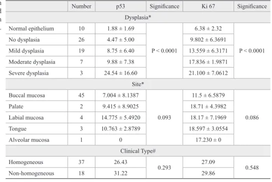
The aim of this study was to analyze the expression of H3K9ac and H4K12ac in oral leukoplakia and its association with cell proliferation marker Ki-67 and

[...] o Estado que emergiu no pós-30, durante a crise que afetou os setores fundamentais da economia brasileira, ligados à agricultura de exportação, foi criando

From the 12 studies included (13 entries) in this meta-analysis, they all started from the same research assumption, in which the elderly with classifications according to the

This log must identify the roles of any sub-investigator and the person(s) who will be delegated other study- related tasks; such as CRF/EDC entry. Any changes to

Além disso, o Facebook também disponibiliza várias ferramentas exclusivas como a criação de eventos, de publici- dade, fornece aos seus utilizadores milhares de jogos que podem

A partir dos conceitos de documentos eletrˆonicos, seguranc¸a, das lingua- gens em uso e das necessidades apresentadas para garantir a validade jur´ıdica e confi- dencialidade de