Highly and Broad-Spectrum
In Vitro
Antitumor Active
cis
-Dichloridoplatinum(II)
Complexes with 7-Azaindoles
PavelŠtarha, Zdeněk Dvořák, Zdeněk Trávníček*
Regional Centre of Advanced Technologies, Division of Biologically Active Complexes and Molecular Magnets, Faculty of Science, Palacký University, Olomouc, Czech Republic
Abstract
Thecis-[PtCl2(naza)2] complexes (1–3) containing monosubstituted 7-azaindole
halogeno-derivatives (naza), showed significantly higher activity thancisplatintowards ovarian carci-noma A2780, itscisplatin-resistant variant A2780R, osteosarcoma HOS, breast carcinoma MCF7 and cervix carcinoma HeLa cell lines, with the IC50values of 3.8, 3.5, 4.5, 2.7, and
9.2μM, respectively, obtained for the most active complex3. As for4and5having disubsti-tuted 7-azaindoles in their molecule, the significant cytotoxicity was detected only for4 against A2780 (IC50= 4.8μM), A2780R (IC50= 3.8μM) and HOS (IC50= 4.3μM), while5
was evaluated as having only moderate antiproliferative effect against the mentioned can-cer cell lines with IC50= 33.4, 24.7 and 46.7μM, respectively. All the studied complexes1–
5effectively avoided the acquired resistance of ovarian carcinoma cell line. On the other hand, the complexes did not reveal any inhibition activity on the purified 20S proteasome from the A2780 cells. The representative complexes3and5showed low ability to be hydro-lysed, but their stability was markedly lowered in the presence of physiological sulphur-con-taining biomolecule glutathione (GSH), as proved by the1H NMR spectroscopy and mass spectrometry studies. A rate of interaction of the studied complexes with GSH was affected by an addition of another mechanistically relevant biomolecule guanosine monophosphate. The differences in interactions of3and5with GSH correlate well with their different cytotox-icity profiles.
Introduction
A clinically successful metal-based anticancer drugcisplatin,cis-[Pt(NH3)2Cl2] [1,2], was
fol-lowed by a lot of platinum complexes, which entered or even completed the clinical trials with the aim to become an improvedcisplatinanalogue in the treatment of cancer diseases [3,4]. Interestingly, the general structural characterization of the mononuclear platinum(II) analogues ofcisplatinis quite strict and can be generalized as follows: 1/ the leaving groups are either two chloride anions (e.g.NSC 170898[5] orpicoplatin[6]) or one bidentate (e.g. world-wide clinically
OPEN ACCESS
Citation:Štarha P, Dvořák Z, Trávníček Z (2015) Highly and Broad-SpectrumIn VitroAntitumor Active
cis-Dichloridoplatinum(II) Complexes with 7-Azaindoles. PLoS ONE 10(8): e0136338. doi:10.1371/journal.pone.0136338
Editor:Swati Palit Deb, Virginia Commonwealth University, UNITED STATES
Received:June 3, 2015
Accepted:July 31, 2015
Published:August 26, 2015
Copyright:© 2015Štarha et al. This is an open access article distributed under the terms of the Creative Commons Attribution License, which permits unrestricted use, distribution, and reproduction in any medium, provided the original author and source are credited.
Data Availability Statement:All relevant data are within the paper and its Supporting Information files.
Funding:The authors gratefully thank the National Program of Sustainability I (LO1305) of the Ministry of Education, Youth and Sports of the Czech Republic for financial support (www.msmt.cz).
usedcarboplatin[7] oroxaliplatin[8]) or two monodentate (aroplatin[9]) carboxylates, with an exception ofspiroplatin[10] andProLindac[11]; 2/ the carrier ligands are either two ammines (e.g.carboplatin) or one bidentate (e.g.oxaliplatin) or two monodentate (e.g.NSC 170898) amines;NSC 170898=cis-dichlorido-bis(cyclopentylamine)platinum(II),picoplatin= cis-ammine-dichlorido-(2-methylpyridine)platinum(II),carboplatin = diammine-(cyclobutane-1,1-dicarboxylato)platinum(II),oxaliplatin= 1R,2R-cyclohexanediamineoxalatoplatinum(II), aroplatin= 1R,2R-diaminocyclohexane-bis(neodecanaoto)platinum(II),spiroplatin= cyclohex-ane-1,1-dimethylamine-sulfatoplatinum(II),ProLindac= a polymeric prodrug of 1R,2R-cyclo-hexanediamineplatinum(II) bound to hydroxypropylmethacrylamide. As for the carrier ligands, there can be also found the exceptions, namely the mentionedpicoplatinormiboplatin (R-2-aminomethylpyrrolidine-(cyclobutane-1,1-dicarboxylato)platinum(II)), which involve the heterocyclicN-donor carrier ligands [12]. The mentionedpicoplatininvolving, in comparison withcisplatin, one bulky 2-methylpyridineN-donor ligand instead of one ammine, showed very promising results on a wide spectrum of tumour types including those resistant tocisplatinand evenoxaliplatin[13,14]. Introducing of the mentioned 2-methylpyridine into the structure of picoplatinresults in slower hydrolysis rate and different pKavalues as compared withcisplatin
[15], and causes the steric hindrance of the Pt(II) atom which consequently hinders approach of nucleophiles (e.g. glutathione) to the Pt(II) atom.Picoplatinfailed in the Phase II clinical trials against both small and non-small cell lung cancer, but it is currently investigated against colorec-tal and prostate cancers.
The pharmacological perspective of the platinum(II) complexes involvingN-donor hetero-cyclic ligands can be also highlighted by a very recently published monofunctional platinum (II) complexphenanthriplatin(cis-diammine-chlorido-phenanthridineplatinum(II) nitrate) [16,17]. This compound, which monofunctionally interacts with DNA and more efficiently binds nucleobases than sulphur-containing biomolecules (50
-deoxyguanosine monophosphate andN-acetyl methionine were used as the model systems), showed extraordinary anticancer effectivity and no correlation with any other platinum-based anticancer drug at NCI-60 DTP Human Tumor Cell Line Screen, which favoursphenanthriplatinfor the future clinical testing.
In our laboratory, we deal with platinum(II) complexes involving variousN-donor hetero-cyclic ligands, such asN6-benzyladenine or 7-azaindole derivatives (e.g. [18,19]), for more than ten years. In the case of the latter ones, we recently reported a series of cis-dichloridoplati-num(II) complexes involving 3-chloro-7-azaindole (3Claza), 3-iodo-7-azaindole (3Iaza) and 5-bromo-7-azaindole (5Braza) together with their significantin vitrocytotoxicity against a panel of human cancer cell lines, mechanistic studies and promisingin vivoanticancer activity on the mouse model of L1210 lymphocytic leukaemia [19–21]. Some details regarding the
mentioned biological aspects will be discussed below within the present paper.
Herein we report a new series of thecis-dichloridoplatinum(II) complexes containing halo-geno-derivatives of 7-azaindole different from the above mentioned ones (namely 4-chloro-7-azaindole (4Claza), 3-bromo-4-chloro-7-azaindole (3Braza), 4-bromo-4-chloro-7-azaindole (4Braza), 3-iodo-5-bromo-7-azaindole (3I5Braza) and 3-chloro-3-iodo-5-bromo-7-azaindole (3Cl5Braza);Fig 1). The complexes were prepared and studied with an aim to clarify an effect of the type (chloro, bromo and iodo), position (3, 4 and 5) and rate (mono- and disubstituted derivatives) of the 7-azaindole ring derivatization by the mentioned halogens to the resultingin vitrocytotoxicity. Additionally to our recently reported studies of the analogical complexes with differently substituted 7-azaindole moiety [19–21], we 1) studied more deeply the interaction with the
bio-molecules relevant from the mechanistic point of view (reduced glutathione (GSH) and guano-sine 5'-monophosphate disodium salt hydrate (GMP)) on the selected representative
the inhibition activity of the described complexes on the purified 20S proteasome extracted from the A2780 human ovarian carcinoma cell line.
Materials and Methods
Chemicals
The chemicals (K2[PtCl4], 4-chloro-7-azaindole (4Claza), 3-bromo-7-azaindole (3Braza),
4-bromo-7-azaindole (4Braza), 3-iodo-5-bromo-7-azaindole (3I5Braza), 3-chloro-5-bromo-7-azaindole (3Cl5Braza),cisplatin, reduced glutathione (GSH), guanosine 5'-monophosphate disodium salt hydrate (GMP)) and solvents (ethanol,N,N`-dimethylformamide, acetone, methanol, diethyl ether) were purchased from Sigma-Aldrich (Prague, Czech Republic) and Acros Organics (Pardubice, Czech Republic).
Synthesis
Complexes were prepared as recently described for their analogues containing differently substituted 7-azaindole derivatives [19]. Briefly, the water solution of 0.5 mmol of K2[PtCl4]
was mixed together with the ethanolic solution of 1.0 mmol of the appropriate 7-azaindole derivative (4Claza for1,3Braza for2,4Braza for3,3I5Braza for4and3Cl5Braza for5) (Fig 1). The products, which formed during 24 h of stirring at 50°C, were filtered off, washed (2 × 5 mL of distilled water, 2 × 5 mL of ethanol and 2 × 5 mL of diethyl ether), dried under vacuum and stored without any further purification.
cis-[PtCl2(4Claza)2] (1):Anal. Calcd. for C14H10N4Cl4Pt: C, 29.44; H, 1.76; N, 9.81%; found:
C, 29.39; H, 1.79; N, 9.80%.1H NMR (DMF-d7, ppm):δ13.52 (br, N1H, 1H), 8.88 (d, 6.2,
C6H, 1H), 7.94 (t, 3.2, C2H, 1H), 7.32 (d, 5.9, C5H, 1H), 6.67 (m, C3H, 1H).13C NMR (DMF-d7, ppm):δ147.8 (C7a), 145.8 (C6), 137.7 (C4), 128.9 (C2), 122.1 (C3a), 117.3 (C5), 100.2
(C3).15N NMR (DMF-d7, ppm):δ145.8 (N1), 168.0 (N7).195Pt NMR (DMF-d7, ppm): δ−2113.2. ESI+ MS (methanol,m/z): 609.0 (calc. 608.9; 30%; {[PtCl2(4Claza)2]+K}+), 593.1
(calc. 592.9; 10%; {[PtCl2(4Claza)2]+Na}+), 153.1 (calc. 153.0; 5%; {4Claza+H}+). ESI–MS
(methanol,m/z): 568.8 (calc. 568.9; 15%; {[PtCl2(4Claza)2]–H}–, 417.1 (calc. 416.9; 100%;
{[PtCl2(4Claza)]–H}–), 151.2 (calc. 151.0; 10%; {4Claza–H}–).
cis-[PtCl2(3Braza)2] (2):Anal. Calcd. for C14H10N4Br2Cl2Pt: C, 25.48; H, 1.53; N, 8.49%;
found: C, 25.28; H, 1.53; N, 8.63%.1H NMR (DMF-d7, ppm):δ13.54 (br, N1H, 1H), 9.02
(d, 5.8, C6H, 1H), 8.10 (d, 2.3, C2H, 1H), 7.96 (d, 8.0, C4H, 1H), 7.30 (m, C5H, 1H).13C NMR
Fig 1. Structural formula of the studied complexescis-[PtCl2(naza)2] (1–5) and 7-azaindole derivatives
used for their preparation.The general structural formulas of used 4-chloro-7-azaindole (4Claza; complex
1), 3-bromo-7-azaindole (3Braza;2), 4-bromo-7-azaindole (4Braza;3), 3-iodo-5-bromo-7-azaindole (3I5Braza;4) and 3-chloro-5-bromo-7-azaindole (3Cl5Braza;5) are given together with their atom numbering scheme.
(DMF-d7, ppm):δ146.7 (C6), 146.5 (C7a), 129.9 (C2), 127.6 (C4), 122.4 (C3a), 117.8 (C5), 89.0
(C3).15N NMR (DMF-d7, ppm):δ143.9 (N1), 172.8 (N7).195Pt NMR (DMF-d7, ppm):δ−2126.2. ESI+ MS (methanol,m/z): 698.8 (calc. 698.8; 100%; {[PtCl2(3Braza)2]+K}+), 625.0 (calc. 624.9;
20%; [PtCl(3Braza)2]+), 197.1 (calc. 197.0; 5%; {3Braza+H}+). ESI–MS (methanol,m/z): 658.9
(calc. 658.8; 100%; {[PtCl2(3Braza)2]–H}–), 461.1 (calc. 460.9; 50%; {[PtCl2(3Braza)]–H–}), 195.1
(calc. 195.0; 10%; {3Braza–H}–).
cis-[PtCl2(4Braza)2] (3):Anal. Calcd. for C14H10N4Br2Cl2Pt: C, 25.48; H, 1.53; N, 8.49%;
found: C, 25.20; H, 1.59; N, 8.01%.1H NMR (DMF-d7, ppm):δ13.53 (s, N1H, 1H), 8.79 (d,
6.5, C6H, 1H), 7.96 (t, 3.3, C2H, 1H), 7.46 (d, 6.3, C5H, 1H), 6.60 (m, C3H, 1H).13C NMR (DMF-d7, ppm):δ146.9 (C7a), 145.5 (C6), 128.9 (C2), 127.2 (C3a), 124.5 (C4), 120.4 (C5),
101.8 (C3).15N NMR (DMF-d7, ppm):δ145.8 (N1), 168.4 (N7).195Pt NMR (DMF-d7, ppm): δ−2116.2. ESI+ MS (methanol,m/z): 698.7 (calc. 698.8; 10%; {[PtCl2(4Braza)2]+K}+), 682.8
(calc. 682.8; 20%; {[PtCl2(4Braza)2]+Na}+), 197.0 (calc. 197.0; 5%; {4Braza+H}+). ESI–MS
(methanol,m/z): 658.7 (calc. 658.8; 35%; {[PtCl2(4Braza)2]–H}–), 460.9 (calc. 460.9; 50%;
{[PtCl2(3Braza)]–H}–).
cis-[PtCl2(3I5Braza)2] (4):Anal. Calcd. for C14H8N4Br2Cl2I2Pt: C, 18.44; H, 0.88; N, 6.14%;
found: C, 18.29; H, 1.01; N, 6.17%.1H NMR (DMF-d7, ppm):δ13.88 (br, N1H, 1H), 9.34 (s,
C6H, 1H), 8.17 (d, 2.5, C4H, 1H), 7.99 (s, C2H, 1H).13C NMR (DMF-d7, ppm):δ146.7 (C7a),
146.7 (C6), 134.4 (C4), 133.7 (C2), 126.9 (C3a), 111.1 (C5), 55.1 (C3).15N NMR (DMF-d7, ppm): δ150.1 (N1), 177.4 (N7).195Pt NMR (DMF-d7, ppm):δ−2117.7. ESI+ MS (methanol,m/z): 950.5 (calc. 950.6; 30%; {[PtCl2(3I5Braza)2]+K}+), 323.1 (calc. 322.9; 35%; {3I5Braza+H}+). ESI–
MS (methanol,m/z): 910.5 (calc. 910.6; 60%; {[PtCl2(3I5Braza)2]–H}–), 587.0 (calc. 586.8; 100%;
{[PtCl2(3I5Braza)]–H}–), 321.1 (calc. 320.9; 10%; {3I5Braza–H}–).
cis-[PtCl2(3Cl5Braza)2] (5):Anal. Calcd. for C14H8N4Br2Cl4Pt: C, 23.07; H, 1.11; N, 7.69%;
found: C, 23.12; H, 1.15; N, 7.83%.1H NMR (DMF-d7, ppm):δ13.37 (s, N1H, 1H), 8.91 (d, 1.9,
C6H, 1H), 8.45 (d, 1.9, C4H, 1H), 8.15 (d, 2.8, C2H, 1H).13C NMR (DMF-d7, ppm):δ149.5
(C6), 144.7 (C7a), 131.4 (C4), 127.2 (C2), 121.8 (C3a), 110.3 (C5), 103.7 (C3).15N NMR (DMF-d7, ppm):δ142.4 (N1), 165.9 (N7).195Pt NMR (DMF-d7, ppm):δ−2183.0. ESI+ MS (methanol,m/z): 231.1 (calc. 230.9; 70%; {3Cl5Braza+H}+). ESI–MS (methanol,m/z): 496.7
(calc. 496.8; 40%; {[PtCl2(3Cl5Braza)]–H}–).
Physical Measurements
Elemental analysis (C, H, N) was performed on a Flash 2000 CHNS Elemental Analyzer (Thermo Scientific). Electrospray ionization (ESI) mass spectra (methanol solutions) were obtained by an LCQ Fleet ion trap spectrometer (Thermo Scientific; QualBrowser software, version 2.0.7) in both positive (ESI+) and negative (ESI–) ionization modes.1H,13C and195Pt
NMR spectra and1H–1H gs-COSY,1H–13C gs-HMQC,1H–13C gs-HMBC and1H–15N
gs-HMBC two dimensional correlation experiments (gs = gradient selected, COSY = correlation spectroscopy, HMQC = heteronuclear multiple quantum coherence, HMBC = heteronuclear multiple bond coherence) of the DMF-d7solutions were measured at 300 K on a Varian 400
device at 400.00 MHz (1H), 100.58 MHz (13C), 86.00 MHz (195Pt) and 40.53 MHz (15N).1H and13C NMR spectra were calibrated against the residual DMF-d61H NMR (8.03, 2.92 and
2.75 ppm) and13C NMR (163.15, 34.89 and 29.76 ppm) signals.195Pt spectra were adjusted against K2[PtCl6] in D2O found at 0 ppm.1H–15N gs-HMBC experiments were obtained at
Nicolet) using the ATR technique. Raman spectra (150 and 3750 cm–1; except for3and5,
which burnt under laser beam) were recorded by an NXR FT-Raman Module (Thermo Nicolet).
Studies of stability and interaction with biomolecules. The representative complexes3
and5were dissolved in the DMF-d7/D2O mixture (1:1v/v) and their1H NMR spectra were
recorded on the fresh solution (0 h) and after 24 h and 48 h of standing at laboratory tempera-ture. The same1H NMR experiments, but with an addition of two molar equivalents of GSH (the experiments and spectra labeled as3+GSH and5+GSH), GMP (3+GMP and5+GMP) or GSH/GMP mixture (3+GSH/GMP and5+GSH/GMP) were carried out as well.
Complex3(10μM final concentration) or the mixture of3(10μM final concentration) with GSH (6μM final concentration [22]), both dissolved in the methanol/water mixture (1:1 v/v) were analysed by flow-injection analysis ESI-MS in both the positive and negative ioniza-tion mode immediately after the preparaioniza-tion (fresh soluioniza-tion) and after 2 and 24 h of standing at laboratory temperature. The mobile phase (90% methanol and 10% of 10 mM ammonium acetate) was pumped (0.2 mL/min) by the quaternary pump of Dionex Ultimate 3000 HPLC System. The samples were injected directly into the mobile phase flow (HPLC autosampler).
Biological Studies
Cell culture andin vitrocytotoxicity testing. In vitrocytotoxic effect of1–5andcisplatin
was assessed by an MTT assay [MTT = 3-(4,5-dimethylthiazol-2-yl)-2,5-diphenyl-tetrazolium bromide] against A2780 ovarian carcinoma, A2780Rcisplatin-resistant ovarian carcinoma, HOS osteosarcoma, G361 malignant melanoma, MCF7 breast carcinoma, A549 lung carci-noma, HeLa cervix epithelia carcinoma and LNCaP prostate carcinoma human cancer cell lines, which were purchased from European Collection of Cell Cultures (ECACC). The cell lines were cultured according to the ECACC instructions and they were maintained at 37°C and 5% CO2in a humidified incubator. The cells were treated with the solutions of1–5and
cis-platinfor 24 h, using multi-well culture plates of 96 wells. In parallel, the cells were treated with vehicle (DMF; 0.1%, v/v) and Triton X-100 (1%, v/v) to assess the minimal (100% viability) and maximal (0% viability) cell damage, respectively. The MTT assay was measured spectro-photometrically at 540 nm (TECAN, Schoeller Instruments LLC).
Analogical testing was carried out for3towards A2780 cancer cells with addition of L-buthionine sulfoximine (L-BSO). L-BSO was independently added to each well to give the 5.0μM final concentration known to be non-toxic and optimal for the experiments focusing on the modulation of anticancer activity. The experiments with L-BSO were performed with two negative controls (DMF and 5.0μM L-BSO) with no statistically different results obtained between both the controls.
The data from the cancer cells were acquired from three independent experiments (con-ducted in triplicate) using cells from three consecutive passages. The resulting IC50values (μM)
were calculated from viability curves and the results are presented as arithmetic mean±SD. The significance of the differences between the obtained results (p<0.05 considered to be
signifi-cant) was assessed by the ANOVA analysis (QC Expert 3.2, Statistical software, TriloByte Ltd.).
Inhibition of proteasome activity in purified 20S proteasome. A2780 cancer cell line was cultured in the complete RPMI 1640 medium (Sigma-Aldrich), supplemented with 10% heat-inactivated fetal-bovine serum (Sigma-Aldrich), 5 mL penicillin/streptomycin solution (PAA-cell culture company) and 2 mM L-glutamine (Sigma-Aldrich) at 37°C in a 5% CO2
buffer (100 mM Tris, 5 mM MgCl2, 5 mM ATP, 0.5 mM DTT, 20% glycerol (pH 7.8)). The
sol-uble protein fraction was isolated by centrifugation at 20,000 g for 30 min at 4°C. The pellet was discarded, and the supernatant fraction was centrifuged again at 100,000 g for 6 h at 4°C. The supernatant fraction was discarded, and the pellet was washed carefully in fresh buffer (without glycerol), which was also discarded. The washed pellets were re-suspended in 1 mL of buffer (per pellet) and stored at -80°C in 0.1 mL aliquots. The chymotrypsin-like activity of purified 20S proteasome was determined as follows: 5μg of purified proteasome was pre-incu-bated with or without compounds for 20 min in 90μL of assay buffer [30 mm Tris-HCl (pH 8.0)] at 37°C, followed by the incubation with 20μm of fluorogenic peptide substrate (Suc-LLVY-AMC) (Sigma-Aldrich), at 37°C for 1 h. After the incubation, the fluorescence of the hydrolysed (AMC) groups in reaction mixtures was measured at 380/460 nm (TECAN, Infinite M200PRO).
Results
Chemistry
Thecis-[PtCl2(naza)2] complexes containing4Claza (1),3Braza (2),4Braza (3),3I5Braza (4),
3Cl5Braza (5) (Fig 1) were prepared by a simple synthetic strategy recently reported for the analogues involving different 7-azaindoles (3Claza (I),3Iaza (II) and5Braza (III); [19]). K2[PtCl4] was used as the starting platinum(II) compound directly reacting with a
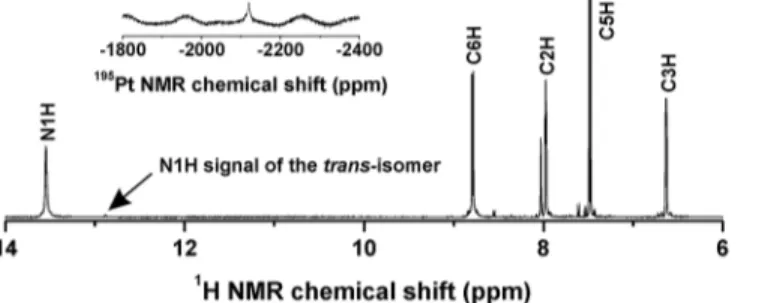
stoichiomet-ric amount of the appropriate 7-azaindole derivative (naza). The products were collected inca. 80% yields and their chemical purity was determined by means of the results of elemental anal-ysis and multinuclear NMR spectroscopy (see sectionSynthesis). The isomeric purity of the complexes1–5, as determined from the ratio of N1H signals of both thecis-[PtCl2(naza)2] and
trans-[PtCl2(naza)2] isomers observed in the1H NMR spectra, was found to be>95% (Fig 2).
The ESI+ mass spectrometry did not detect the molecular peaks of the studied complexes, but their adducts with the sodium or potassium ions were found according to their mass and isotopic distribution. The {naza+H}+fragments were observed in the spectra recorded in the positive ionization mode as well. The ESI–mass spectra contain the molecular peaks
corre-sponding to the {[PtCl2(naza)2]–H}−species (except for5) (S1 Fig). A release of one
7-azain-dole-based ligand from the structure of the studied complexes led to the detection of the {[PtCl2(naza)]–H}−and {naza–H}−fragments within ESI–mass spectra of the studied
complexes.
All the1H,13C and15N signals of free 7-azaindole derivatives were found in appropriate spectra of the studied complexes. The anticipated N7-coordination mode of the 7-azaindole derivatives was unambiguously proved by the15N coordination shifts (Δδ, ppm; calculated as
Δδ=δcomplex−δligand), whose values equal 2.5–3.6 ppm for N1 and−114.6–(–101.0) ppm for
Fig 2. The1H NMR and195Pt NMR (inset) spectra of 3 in DMF-d7.The spectra show the signals of the
corresponding atoms and point to chemical and isomeric purity of3.
N7 (S1 Table). The atoms adjacent to the N7 coordination site shifted by 2.1–4.9 ppm
down-field (for C6 in the13C NMR spectrum), 0.5–2.0 ppm upfield (for C7a in the13C NMR
spec-trum) and 0.52–0.99 ppm downfield (C6–H in the1H NMR spectrum) (S1 Table). The195Pt
chemical shift values ranged from−2126 to−2113 ppm for1–4, while195Pt NMR chemical
shift for5was detected at−2183 ppm.
Biological Activities Testing
In vitrocytotoxicity. The prepared complexes1–5andcisplatin(for comparative
pur-poses) were tested by the commonly used MTT assay (e.g. [25]) for theirin vitroantitumor activity against eight human cancer cell lines. The results are summarized inTable 1.
The IC50values of1–3(involving monosubstituted 7-azaindole derivatives) against A2780,
A2780R, HOS, G361, MCF7 and LNCaP were found to be lower than 7.5μM (Table 1,Fig 3). In vitrocytotoxicity of4and5with disubstituted 7-azaindole derivatives is mutually different.
4is, similarly to1–3, highly effective on the A2780, A2780R, HOS and G361 cancer cells
(IC50= 3.0–4.8μM), while5is significantly (p<0.05),ca. 1 order of magnitude less effective
Table 1. The results ofin vitrocytotoxicity of 1–5 andcisplatin(CDDP) against eight human cancer cell lines.Cells were treated with tested
com-pounds for 24 h, measurements were performed in triplicate, and cytotoxicity experiment was repeated in three different cell passages. Data are expressed as IC50±SD (μM).
1 2 3 4 5 CDDP
A2780 3.8±0.2* 4.1±0.4* 3.8±0.5* 4.8±1.0* 33.4±3.3 21.8±3.9
A2780R 3.6±0.7* 3.6±0.7* 3.5±1.1* 3.8±1.0* 24.7±1.0 32.0±9.6
HOS 7.5±2.4* 5.3±2.1* 4.5±2.7* 4.3±0.5* 46.7±4.0 25.4±8.5
G361 3.2±0.5 2.9±0.4 2.7±0.4 3.0±0.5 >50.0a 5.8±2.4
MCF7 3.5±1.0* 5.3±0.8* 2.7±1.2* >10.0a >50.0a 18.1±5.1
A549 11.6±4.2 19.0±5.6 11.1±0.3 >10.0a >50.0a >50.0a
HeLa 11.9±1.2* 17.1±0.8* 9.2±2.0* >10.0a >50.0a 39.9±4.6
LNCaP 3.9±0.1 4.9±0.1 4.0±0.6 >10.0a >50.0a 3.8±1.5
RFb 0.95 0.88 0.92 0.79 0.74 1.47
asterisks (*), significantly different values (p<0.05) between1–5andcisplatin a) IC
50were not reached up to the given concentration b) RF = resistance factor calculated as IC
50(A2780R)/IC50(A2780)
doi:10.1371/journal.pone.0136338.t001
Fig 3.In vitrocytotoxicity of 1–5.Graphically depicted comparison ofin vitrocytotoxicity of the studied
complexes against ovarian carcinoma (A2780),cisplatin-resistant ovarian carcinoma (A2780R), osteosarcoma (HOS), breast carcinoma (MCF7) and cervix carcinoma (HeLa), where the significant differences (p<0.05; assigned with the asterisks) between the obtained IC50values (μM) of1–5were observed.
against A2780, A2780R and HOS (IC50>24.7μM) as compared with1–4(Fig 3). The
resis-tance factors (RF), calculated as a IC50(A2780R)/IC50(A2780) ratio, equalled 0.74–0.95 for1–5
and 1.47 forcisplatin(Table 1). Further it has been found for the representative complex3that itsin vitrocytotoxicity against A2780 increased from IC50= 3.8±0.5μM (without L-BSO) to
IC50= 2.6±0.1μM with an addition of 5.0μM L-buthionine sulfoximine (L-BSO), a
well-known inhibitor ofγ-glutamylcysteine synthetase.
Studies of stability and interactions with GSH and GMP. The representative complexes
3and5(dissolved in the DMF-d7/D2O mixture, 1:1v/v) were studied by1H NMR at different
time points (0, 24 and 48 h) for its stability in the mentioned water-containing solvent. Except the signals (e.g. N1–H and C6–H) of3detected at 13.34 and 8.80 ppm (andtrans-3impurity at
12.91 and 8.58 ppm), new signals rose in time at 12.70 and 8.45 ppm in connection with a for-mation of new species within the studied water-containing solution of3(Fig 4). The {[Pt(H2O)
(4Braza)2Cl]}+species, whose intensity increased in time, was detected by ESI-MS experiments
performed on3dissolved in the methanol/H2O mixture, 1:1v/v(S2 Fig). In the case of5, any
new N1–H peak was not detected in the1H NMR spectra even after 48 h, however, its ability to
hydrolyse (generally said, very low as in the case of3) can be anticipated based on the new C6–
H signal, whose intensity increased in time (S3 Fig).
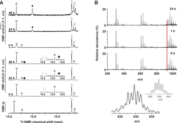
Analogical experiments (1H NMR, ESI-MS) were carried out for3and5with an addition of GSH.1H NMR spectrum of3contained a new set of signals of the4Braza ligand, represented Fig 4. Time-dependent studies of stability of 3 (1H NMR) and its interaction with GSH (1H NMR and ESI-MS).(A)1H NMR spectra ofcis
-[PtCl2(4Braza)2] (3) dissolved in DMF-d7or DMF-d7/H2O mixture (1:1,v/v) without or with glutathione (GSH) measured at different time points (0, 24 and
48 h).= N1–H,cis-[PtCl2(4Braza)2];*= N1–H,trans-[PtCl2(4Braza)2];●= N1–H, hydrolysis product,cis-[Pt(4Braza)2(H2O)Cl]+;♦= N1–H, GSH adduct of 3; ^ = C6–H,cis-[PtCl2(4Braza)2]; # = C6–H,trans-[PtCl2(4Braza)2];^= N–H of glycine and cysteine of GSH. (B) ESI–mass spectra (200–1050m/zrange) of
the mixture of3with GSH (dissolved in the methanol/H2O, 1:1,v/v) as detected at different time points (0, 1 and 24 h). The region of the {[Pt(4Braza)2Cl
(GSH)]–2H}−species is highlighted by red colour. The detail of the experimental peak of {[Pt(4Braza)
2Cl(GSH)]–2H}–, as observed after 24 h, is given below
(bottom left, given in black) together with the simulated isotope distribution (bottom right, given in grey).
by the N1–H signal at 12.03 ppm (Fig 3A). The ratio of the integral intensities of the N1–H
sig-nals of the starting complex and its analogue with GSH was approximately 1:1 after 48 h (note: an opacity observed in the solution after standing in the laboratory conditions belong to the trans-isomer of the starting complex, because its signals were not detected after centrifugation in the1H NMR spectra after 24 and 48 h). A peak, whose mass and isotopic distribution corre-sponded to {[Pt(4Braza)2Cl(GSH)]+} was detected by ESI-MS in the mixture of3and GSH (Fig
4). Complex5interacted with GSH as well, butca. 1:1 integral intensity ratio of the N1–H
sig-nals of5(12.65 ppm) and its adduct with GSH (11.88 ppm) was reached after 24 h. After 48 h, the5+GSH spectrum contained only the signals of the adduct of5with GSH (Fig 5). The1H NMR experiments were also performed on the mixtures of the studied complexes (3or5) with GMP. We did not observe any new4Braza peaks in the spectra of3mixed with GMP even after 48 h (N1–H signal of3was not detected;S4 Fig). On the other hand, the spectrum of5
contained several new peaks at 8.89, 8.23 and 7.74 ppm after 24 and 48 h of standing at labora-tory temperature, belonging to C6–H, C4–H, and C2–H of the adduct of5with GMP,
respec-tively (again, N1–H signal of5or its adduct with GMP was not detected;S5 Fig).
Opposing results obtained from the interaction experiments led us to investigation of behaviour of the studied complexes (3or5) mixed with both the used biomolecules,i.e. GSH and GMP. In the case of3+GSH/GMP, a ratio of the N1–H signals of3and its adduct with
GSH wasca. 20:1 after 48 h (S4 Fig). Any new peaks, as compared with3+GSH and3+GMP experiments, were not detected in the3+GSH/GMP spectra. The results found for5showed Fig 5. Time-dependent1H NMR studies of 5 and its interaction with GSH.1H NMR spectra ofcis
-[PtCl2(3Cl5Braza)2] (5) dissolved in the DMF-d7/H2O mixture (1:1,v/v) without or with glutathione (GSH)
measured at different time points (0, 24 and 48 h).= N1–H,5;●= N1–H, GSH adduct of5;□= C6–H,5;■=
C6–H, GSH adduct of5;Δ= C4–H,5;▲= C4–H, GSH adduct of5;^= C2–H,5;♦= C2–H, GSH adduct of5; # = N–H of glycine and cysteine of GSH.
that almost all the starting complex interacted with GSH after 48 h with the ratio of the N1–H
signals of5and its GSH adduct being about 1:10 (S5 Fig). However, no signals of5and GMP adduct, as observed by the experiment without GSH addition (i.e.5+GMP, as discussed above), were detected in the spectra recorded on5+GSH/GMP.
Proteasome inhibition activity. The ability of1–4to inhibit 20S proteasome activity,
which was assayed in purified proteasome obtained from A2780 cancer cell line, was studied as well. The results are summarized inS2 Tableand depicted inFig 6. Concentrations of the tested compounds were applied in decimal scale, up to 20μM, due to the limitations in solubility. None of the compounds tested did significantly inhibit a catalytic activity of 20S proteasome, in the whole concentration range.
Discussion
The studiedcis-[PtCl2(naza)2] complexes (1–5;Fig 1) were prepared in good yields and high
chemical and isomeric purity (as proved by1H NMR studies;Fig 2) by one-step reactions directly from K2[PtCl4], as recently reported for the analogues with different 7-azaindoles
[19,26]. The composition of1–5was proved by elemental analysis, ESI-MS spectrometry (S1 Fig) and multinuclear NMR spectroscopy. Since this work lacks results of a single crystal X-ray analysis (crystals suitable for X-ray analysis were not obtained yet), NMR spectroscopy was crucial technique for this work in terms of the structure description of the studied complexes
1–5. Previously, anN7-coordination mode of the 7-azaindole (aza) and its
halogeno-deriva-tives (e.g.3Claza) was proved by a single crystal X-ray analysis forcis-[PtCl2(aza)2] [26] and
cis-[PtCl2(3Claza)2] [19]. Moreover, these complexes showed considerable difference between
the15N NMR coordination shifts (Δδ) of both the nitrogen atoms involved within the 7-azain-dole moiety, in particular 2.3 ppm (N1) vs.−102.8 ppm (N7) forcis-[PtCl2(aza)2] [26], and
2.7 ppm (N1) vs.−101.5 ppm (N7) forcis-[PtCl2(3Claza)2] [19]. In the case of herein reported
complexes1–5, the same conclusion regarding a coordination mode of the used 7-azaindoles
can be done based on the obtained15N NMR results (Experimental section andS1 Table). The changes in the electron density distribution, caused by the coordination of the 7-azaindole derivatives to the central Pt(II) atom through the N7 atom, led besides highΔδs of the N7 atoms also to the typical changes in the1H and13C chemical shift values, especially those ones adjacent to the N7 coordination site (S1 Table). The195Pt NMR chemical shifts of1–4are in
good agreement with recently reportedcis-dichloridoplatinum(II) complexes involving Fig 6. The results (CT-like activity (%±SD) of the studies of the ability of the complexes 1–4 to inhibit
20S proteasome activity assayed in purified proteasome obtained from A2780 cancer cell line
7-azaindole (–2120 ppm) [26] or its differently substituted halogeno-derivatives (from−2112 to−2126 ppm) [19]. Interestingly,195Pt NMR chemical shifts differ between1–4and5byca.
60 ppm, which has to be highlighted here with respect to the described differences in thein vitrocytotoxicity results obtained for1–4in comparison to5.
Regardingin vitrocytotoxicity of the studied complexes1–5, it has to be emphasized, that 1–3(involving monosubstituted 7-azaindole derivatives) and4and5(involving disubstituted
7-azaindole derivatives) differ in the degree of substitution (Fig 1). In the case of1–3, their
cytotoxic effect can be generalized as follows: these complexes showed acutein vitro cytotoxic-ity against A2780, A2780R, HOS, G361, MCF7 and LNCaP (Table 1). A comparison of the results obtained for1–3withcisplatinshowed that all these substances are significantly more
effective (p<0.05) against A2780, A2780R, HOS, MCF7 and HeLa cancer cells than the
men-tioned clinically used platinum-based anticancer drug (Table 1). A quite different situation has to be discussed for the complexes4and5with disubstituted 7-azaindole derivatives, whosein vitrocytotoxicity is mutually different. Complex4is, similarly to1–3, highly effective against
A2780, A2780R, HOS and G361, and in the case of A2780, A2780R and HOS cells even signifi-cantly more active as compared withcisplatin(p<0.05;Table 1). A comparison of IC50values
of highly effective1–4emphasized that these substances did not differ in theirin vitro
cytotox-icity against A2780, A2780R, HOS, G361, A549 and LNCaP. In the case of MCF7 and HeLa cells, some differences were found. Particularly, IC50of3is significantly lower as compared
with2against breast cancer cells MCF7, and IC50values of complexes1and3were found to be
significantly lower than that of2against HeLa cell line (Fig 3).
Complex4showed lower solubility (to the 10.0μM concentration only), and thus bioavail-ability, in the medium used (0.1% DMF), as compared with the other studied complexes1–3
and5. Contrary to4, complex5was well-soluble (up to the upper tested concentration,i.e. 50.0μM) but its IC50values were significantly higher (p<0.05) against A2780, A2780R and
HOS as compared with1–4. Moreover,5was ineffective up to the upper tested concentration
(IC50>50.0μM) against G361, MCF7, A549, HeLa and LNCaP (Table 1). In other words, an
implementation of the second substituent to the 7-azaindole moiety negatively affected either solubility (in the case of4) or cytotoxicity (in the case of5).
The well-establishedin vitromodel of acquiredcisplatin-resistance usescisplatin-sensitive (A2780) andcisplatin-resistant (A2780R) ovarian carcinoma human cancer cell lines, numeri-cally expressed as resistance factor (RF). The calculated RFs of1–5were considerably lower in
comparison withcisplatin(Table 1). In other words, all the complexes1–5effectively overcome
acquiredcisplatin-resistance on the human ovarian carcinoma model.
Recently we reported thein vitroantitumor activity of three similarcis-dichloridoplatinum (II) complexes with halogeno-derivatives of 7-azaindole, namely3Claza (I),3Iaza (II) and 5Braza (III), against eight cancer cell lines used also in this work [19,21]. Comparison of their in vitrocytotoxic activity of all eightcis-dichloridoplatinum(II) complexes with halogeno-derivatives of 7-azaindole (1–5andI–III) showed previously reportedIIIas the highest active
one with the IC50values beingca. 6.7-, 12.9-, 13.7-, 5.7-, 9.8-, 5.3- and 2.5-times lower than
those ofcisplatinagainst A2780, A2780R, HOS, G361, MCF7, A549 and LNCaP, respectively. In the case of this work, the highest active complex3exhibited IC50values about 5.7-, 9.1-,
5.6-, 2.2- and 6.7-times lower as compared withcisplatinagainst A2780, A2780R, HOS, G361 and MCF7, respectively. HeLa was the only cell line, wherein vitrocytotoxicity ofIII(ca. 2.3-fold ofcisplatin) was exceeded by its analogues, namely1(ca. 3.4-fold ofcisplatin) and3
(ca. 2.3-fold ofcisplatin). It should be mentioned here as well thatIIIshowed the best and com-parable withcisplatin in vivoresults on mice from the tested complexesI–III[21]. Taken
together all eight complexes with halogeno-derivatives of 7-azaindole (1–5andI–III), it can be
of view, this position is among the studied 3, 4 and 5 substituted derivatives the closest one to the coordination site of 7-azaindole ring (N7 atom;Fig 1). This phenomenon, that the distance of the substituent from the coordination site of the carrier ligand affects the biological proper-ties of the complex involving such ligand, is known forpicoplatinand its less active isomer involving 3-methylpyridine [15]. Other conclusions, which could be made based on the obtained results, is that: 1/ cytotoxicity is independent from the type and position of the stituent in the case of 3- and 4-substituted 7-azaindole derivatives, and 2/ the degree of the sub-stitution could be more decisive than its type and position.
Interactions of the cytotoxic platinum(II) complexes, such as1–5or the clinically used
cis-platin, with sulphur-containing biomolecule GSH is known to be one of the crucial mechanistic steps of their action [3,27]. It is connected with intracellular transport of the platinum(II) spe-cies to the target DNA molecule and, more importantly, with their inactivation and eflux from the cancer cells, known as one of the mechanisms of the cancer cell resistance. Because it has been found that the studied complexes, represented by3and5, interact with GSH, it was of the great interest to evaluate, whether thein vitrocytotoxicity of the studied complexes can be modulated by reduction of the intracellular GSH level by co-application of3(selected as a rep-resentative complex) with an inhibitor ofγ-glutamylcysteine synthetase L-BSO. L-BSO has a direct effect on the synthesis of GSH and its application reduces the cellular level of GSH. The reduction of the GSH level should lead to higher cytotoxicity of such compounds, which are effectively inactivated by GSH [28] or whose mechanism of action is connected with the redox processes [29]. It has been found for3that itsin vitrocytotoxicity against A2780 increased about 1.5-times. This observation is very important for further studies of herein reported com-plexes (e.g.in vivostudies of their anticancer activity), because it indicated that co-administra-tion of these complexes with L-BSO can improve their biological effect against the cancer cells, as previously proved for platinum-based chemotherapeuticscisplatinandoxaliplatin[30,31].
The time-dependent1H NMR and ESI-MS studies of stability of the representative com-plexes3and5dissolved in the water-containing solvent (DMF-d7/D2O mixture, 1:1v/v)
indi-cated very low ability of3and5to hydrolyse. Regarding highlyin vitrocytotoxic complex3, new set of signals was detected in the1H NMR spectra recorded 24 and 48 h after the prepara-tion of the soluprepara-tion, as compared to the fresh soluprepara-tion (Fig 4). Since the chemical shifts of the new signals do not correspond to those ofcis-[PtCl2(4Braza)2],trans-[PtCl2(4Braza)2] or the
free4Braza molecule, it can be concluded, that they belong to the new platinum-containing species, most probably to thecis-[Pt(4Braza)2(H2O)Cl]+orcis-[Pt(H2O)2(4Braza)2]2+or
prod-ucts of their protolytic reactions. To get evidence regarding the composition of new species, we performed the ESI-MS experiments on3dissolved in the methanol/H2O mixture, 1:1v/v. We
detected a peak of {[Pt(H2O)(4Braza)2Cl]}+with an intensity increasing in time (S2 Fig). As for
5, whosein vitrocytotoxicity was considerably lower as compared with3, its very low ability to hydrolyse was proved by analogical1H NMR studies (S3 Fig).
The results of1H NMR clearly indicated that3gradually interacted with GSH withca. 1:1 ratio of the integral intensities of the N1–H signals after 48 h observed for the starting complex 3and its adduct with GSH, whose composition, as suggested by ESI-MS experiments, corre-sponds to {[Pt(4Braza)2Cl(GSH)]+} (Fig 4). Interestingly,5interacted with GSH markedly
faster than3, which could be suggested as one of the reasons connected with significantly lower antiproliferative effect of5as compared with1–4(represented by3within the stability and
interaction studies) (Fig 5), because it is well-known fact that platinum complexes interact with GSH and other sulphur-containing biomolecules, which is connected with their inactivation [3,6,32].
3towards GMP was detected by1H NMR (S4 Fig), while moderately active5interacted with the mentioned biomolecule, as proved by formation of new peaks of3Cl5Braza detected in the spectra (S5 Fig). This finding is very interesting from the mechanistic point of view, because it is well-known that interaction of thein vitrocytotoxic platinum(II) complexes, and especially cisplatinanalogues such as herein reportedcis-dichloridoplatinum(II) complexes1–5, with
nucleobases is crucial from the mechanistic point of view, because activity of such substances is based on their covalent interaction with nuclear DNA. Moreover, an ability of similar com-plexes with differently substituted 7-azaindoles to platinate nuclear DNA was clearly proved in one of our previous works [20].
With respect to the observations that5interacts with GMP, interacts faster than3with GSH and has lowerin vitrocytotoxicity than3, it was of great interest to investigate behaviour of3and5in the presence of the GSH/GMP mixture. These experiments could show which bio-molecule binds preferentially to the studied complexes and whether one biobio-molecule can replace another one within the inner coordination sphere on the pharmacologically relevant timescale (48 h). It was found that the ratio of the N1–H signals of3and its adduct with GSH
differs markedly after 48 h with (approximately 20:1) or without (1:1) addition of GMP, which means that the presence of GMP affects either the process of interaction of3with GSH or the stability of the GSH adduct with3(S4 Fig). Further, any new peaks, as compared with3+GSH and3+GMP experiments, were not detected in the3+GSH/GMP spectra. In other words an interaction of the complex3with GMP was not observed even by this1H NMR experiments. In the case of5, an interaction of this complex with GMP (as observed by5+GMP experi-ments) was suppressed by an addition of GSH, because only the peaks of the adduct of5and GSH were detected in the5+GSH/GMP spectra after 48 h (S5 Fig).
Taken together, the results of interaction experiments could prove that 1) affinities of GSH and GMP to5are higher as compared with3; 2) rate of interaction with GSH (or stability of the GSH adducts) is reduced by an addition of GMP in the case of3, which is not the case of5; 3) an interaction of5and GMP is suppressed in the presence of GSH.
The results of the mechanistic studies performed on the analogues (I–III) of the herein
studied complexes1–5were discussed in our previous papers [19,20]. Therein reported
com-plexes showed thecisplatin-like mechanism of action with some differences fromcisplatin, such as higher cell-uptake and DNA platination. In an effort to bring in new mechanistic knowledge about thecis-dichloridoplatinum(II) complexes with 7-azaindole derivatives, we studied the ability of1–4to inhibit 20S proteasome activity assayed in purified proteasome
from A2780, as described in the Experimental section (S2 Table,Fig 6). However, any inhibi-tion of 20S proteasome was not observed for the studied complexes1–4.
To conclude this work,cis-dichloridoplatinum(II) complexes involving 7-azaindole deriva-tives were studied for theirin vitrocytotoxicity against a panel of eight human cancer cell lines. Complexes1–3with monosubstitutedN-donor ligands showed significantly (p<0.05) higher
in vitrocytotoxic effect against A2780, A2780R, HOS, MCF7 and HeLa as compared with cis-platin, with the IC50values of1–3towards the mentioned cell lines ranging from 2.7 to
17.1μM. An insertion of the second halogeno-substituent on the 7-azaindole moiety led to either lower solubility (4) or lower biological effect (5) of such complexes. The obtained results also indicate the ability of1–5to overcome intrinsiccisplatin-resistance on the ovarian
derivatives were recently reported, we did not perform them on the herein reported complexes. However, in an effort to bring in new mechanistic finding regarding the cis-dichloridoplati-num(II) complexes with 7-azaindoles, we performed a study of their ability to inhibit the activ-ity on the 20S proteasome from the A2780 cells, because the proteasome represents a highly promising target for anticancer drugs [33], but we did not observed any inhibition effect. This work, together with our previous papers dealing withcis-dichloridoplatinum(II) complexes containing 7-azaindoles, indicated that the complexes with 5-substituted derivatives of 7-azain-dole exceedin vitrocytotoxicity of the complexes with the derivatives substituted at the posi-tions 3 or 4. The discussed observaposi-tions are quite challenging for the future work on the platinum complexes involving 7-azaindole derivatives with different 5-substituted derivatives and newly with the preparation and biological evaluation of hopefully even more pharmacolog-ically perspective 6-substituted derivatives of 7-azaindole with the mentioned heterocyclic compound substituted next to its coordination site. Regarding the future biological experi-ments, the selected representatives of the dichloridoplatinum(II) complexes containing halo-geno-derivatives of 7-azaindole, including those reported in this work, should be studied for their potency in the National Cancer Institute (NCI) NCI-60 cell-line screen, known to predict the mechanisms of action of the studied compounds. Moreover, the obtained NCI-60 results should also mechanistically distinguish the studied compounds from the clinically used ones, such ascisplatinoroxaliplatin, which is one of the crucial requirements for the followingin vivoexperiments, and preclinical and clinical studies.
Supporting Information
S1 Fig. ESI–mass spectrum of 3.The spectrum (200–1050m/zrange) is given with the
assigned (
) peek of the {[PtCl2(3Braza)2]–H}−species, whose experimental and simulated
iso-topic distribution is inserted. (TIF)
S2 Fig. ESI+ mass spectra of 3 at different time points.ESI+ mass spectra (400–800m/z
range) of the solution of3in methanol/H2O mixture (1:1,v/v) at different time points (0, 2 and
12 h) showing the peaks of the {[Pt(4Braza)2(H2O)Cl]}+species (●), overlapped peaks of the
{[Pt(4Braza)2(CH3OH)Cl]}+and {[PtCl2(4Braza)2]+H}+species (), and overlapped peaks of
the {[PtIIICl2(4Braza)2(H2O)]}+and {[PtCl2(4Braza)2]+Na}+species (^). The simulated mass
spectrum with the above-mentioned species given in the ratio 4: 3: 1: 10: 8 is also depicted for comparative purposes (top). The experimental and simulated (red tringles) isotopic distribu-tion of the {[Pt(4Braza)2(H2O)Cl]}+species is inserted on the right side.
(TIF)
S3 Fig. Time-dependent1H NMR spectra of 5.1H NMR spectrum of5dissolved in the DMF-d7/H2O mixture (1:1,v/v) (up) and part of the time-dependent1H NMR spectra of5dissolved
in the DMF-d7/H2O mixture (1:1,v/v) measured at different time points (0, 24 and 48 h)
(down). (TIF)
S4 Fig.1H NMR studies on the interaction of 3 with GSH and GMP.1H NMR spectra of3
(bottom spectrum) dissolved in the DMF-d7/H2O mixture (1:1,v/v) with guanosine
5'-mono-phosphate disodium salt hydrate (GMP) or the GMP mixture with glutathione (GSH) mea-sured at different time points (0, 24 and 48 h).= N1–H,3;●= N1–H, GSH adduct of3;□= C6–H,3;■= C6–H, GSH adduct of3;Δ= C2–H,3;▲= C2–H, GSH adduct of3;^= C5–H,3;
♦= C5–H, GSH adduct of3; # = N–H of glycine and cysteine of GSH;
= C8–H of GMP.
S5 Fig.1H NMR studies on the interaction of 5 with GSH and GMP.1H NMR spectra of5
(bottom spectrum) dissolved in the DMF-d7/H2O mixture (1:1,v/v) with guanosine
5'-mono-phosphate disodium salt hydrate (GMP) or the GMP mixture with glutathione (GSH) mea-sured at different time points (0, 24 and 48 h).= N1
–H,5;●= N1–H, GSH adduct of5;□=
C6–H,5;■= C6–H, GSH adduct of5;*= C6–H, GMP adduct of5;Δ= C4–H,5;▲= C4–H,
GSH adduct of5; × = C4–H, GMP adduct of5;^= C2–H,5;♦= C2–H, GSH adduct of5;^=
C2–H, GMP adduct of5; # = N–H of glycine and cysteine of GSH;
= C8–H of GMP.
(TIF)
S1 Table. The1H,13C and15N NMR coordination shifts (Δδ=δcomplex−δligand; ppm) of 1–5.
(PDF)
S2 Table. The results of the studies of the ability of the tested compounds to inhibit 20S proteasome activity assayed in purified proteasome obtained from A2780 cancer cell line.
(PDF)
S1 Text. The results of FTIR and Raman spectroscopy.
(PDF)
Acknowledgments
The authors gratefully thank the National Program of Sustainability I (LO1305) of the Ministry of Education, Youth and Sports of the Czech Republic for financial support. The authors also thank Ms. Kateřina Kubešová and Mr. Jindřich Sedláček for performing the biological experi-ments, Dr. Igor Popa and Dr. Radka Křikavová for performing the NMR, IR and Raman exper-iments, Dr. Ján Vančo and Dr. Bohuslav Drahošfor carrying out of the ESI-MS experiments, Dr. Tomáš Šilha for CHN analysis measurements and Mr. Alexandr Popa for his help with the synthesis.
Author Contributions
Conceived and designed the experiments: PS ZD ZT. Performed the experiments: PS ZD ZT. Analyzed the data: PS ZD ZT. Contributed reagents/materials/analysis tools: PS ZD ZT. Wrote the paper: PS ZD ZT.
References
1. Rosenberg B, VanCamp L, Krigas T. Inhibition of cell division in Escherichia coli by electrolysis prod-ucts from a platinum electrode. Nature. 1965; 205: 698–699. PMID:14287410
2. Rosenberg B, VanCamp L, Trosko JE, Mansour VH. Platinum compounds: a new class of potent antitu-mour agents. Nature. 1969; 222: 385–386. PMID:5782119
3. Kelland L. The resurgence of platinum-based cancer chemotherapy. Nature Rev Cancer. 2007; 7: 573–584.
4. Wheate NJ, Walker S, Craig GE, Oun R. The status of platinum anticancer drugs in the clinic and in clin-ical trials. Dalton Trans. 2010; 39: 8113–8127. doi:10.1039/c0dt00292ePMID:20593091
5. Akamatsu K, Endo K, Matsumoto T, Kamisango K, Morikawa K, Koizumi M, et al. Potent antitumour activity of (-)-(R)-2-aminomethylpyrrolidine (1,1-cyclobutanedicarboxylato)platinum(II) monohydrate (DWA2114R) against advanced L1210 leukaemia in mice. Br J Cancer. 1992; 66: 827–832. PMID: 1419625
6. Holford J, Sharp SY, Murrer BA, Abrams M, Kelland LR. In vitro circumvention of cisplatin resistance by the novel sterically hindered platinum complex AMD473. Br J Cancer. 1998; 77: 366–373. PMID: 9472630
8. Kidani Y, Inagaki K, Iigo M, Hoshi A, Kuretani K. Antitumor activity of 1,2-diaminocyclohexaneplatinum complexes against Sarcoma-180 ascites form. J Med Chem. 1978; 21: 1315–1318. PMID:722741
9. Perez-Soler R, Yang LY, Drewinko B, Lauterzstain J, Khokhar AR. Increased cytotoxicity and reversal of resistance tocis-diamminedichloro-platinum(II) with entrapment ofcis-bis-neodecanoato-trans-R,R -diaminocyclohexaneplatinum(II) in multilamellar lipid vesicles. Cancer Res. 1988; 48: 4509–4512. PMID:3396003
10. de Jong WH, Steerenberg PA, Vos JG, Butten EJ, Verbeek F, Kruizinga W, et al. Antitumor activity, induction of cross-resistance, and nephrotoxicity of a new platinum analogue,cis -1,1-diaminomethylcy-clohexaneplatinum(II) sulfate, and ofcis-diamminedichloroplatinum(II) in an immunocytoma model in the LOU/M rat. Cancer Res. 1983; 43: 4927–4934. PMID:6683993
11. Rice JR, Gerberich JL, Nowotnik D, Howell SB. Preclinical efficacy and pharmacokinetics of AP5346, a novel diaminocyclohexane-platinum tumor-targeting drug delivery system. Clin Cancer Res. 2006; 12: 2248–2254. PMID:16609041
12. Kobayashi H, Takemura Y, Miyachi H, Ogawa T. Antitumor activities of new platinum compounds, DWA2114R, NK121 and 254-S, against human leukemia cells sensitive or resistant to cisplatin. Invest New Drugs. 1991; 9: 313–319. PMID:1804804
13. Holford J, Raynaud F, Murrer BA, Grimaldi K, Hartley JA, Abrams M, et al. Chemical, biochemical and pharmacological activity of the novel sterically hindered platinum co-ordination complex, cis-[amminedi-chloro(2-methylpyridine)] platinum(II) (AMD473). Anticancer Drug Des. 1998; 13: 1–18. PMID: 9474239
14. Sharp SY, O’Neill CF, Rogers PM, Boxall FE, Kelland LR. Retention of activity by the new generation platinum agent AMD0473 in four human tumour cell lines possessing acquired resistance to oxaliplatin. Eur J Cancer. 2002; 38: 2309–2315. PMID:12441268
15. Chen Y, Guo Z, Parsons S, Sadler PJ. Stereospecific and kinetic control over the hydrolysis of a steri-cally hindered platinum picoline anticancer complex. Chem Eur J. 1998; 4: 672–676.
16. Park GY, Wilson JJ, Song Y, Lippard SJ. Phenanthriplatin, a monofunctional DNA-binding platinum anticancer drug candidate with unusual potency and cellular activity profile. Proc Natl Acad Sci USA. 2012; 109: 11987–11992. doi:10.1073/pnas.1207670109PMID:22773807
17. Johnstone TC, Park GY, Lippard SJ. Understanding and improving platinum anticancer drugs– phenan-thriplatin. Anticancer Res. 2014; 34: 471–476. PMID:24403503
18. Trávníček Z,Štarha P, Popa I, Vrzal R, Dvořák Z. Roscovitine-based CDK inhibitors acting as N-donor
ligands in the platinum(II) oxalato complexes: Preparation, characterization and in vitro cytotoxicity. Eur J Med Chem. 2010; 45: 4609–4614. doi:10.1016/j.ejmech.2010.07.025PMID:20692737
19. Štarha P, Trávníček Z, Popa A, Popa I, Muchová T, Brabec V. How to modify 7-azaindole to form
cyto-toxic Pt(II) complexes: highly in vitro anticancer effective cisplatin derivatives involving halogeno-substi-tuted 7-azaindole. J Inorg Biochem. 2012; 115: 57–63. doi:10.1016/j.jinorgbio.2012.05.006PMID: 22922312
20. Muchová T, Prachařová J,Štarha P, Olivová R, Vrána O, Benešová B, et al. Insight into the toxic effects
of cis-dichloridoplatinum(II) complexes containing 7-azaindole halogeno derivatives in tumor cells. J Biol Inorg Chem. 2013; 18: 579–589. doi:10.1007/s00775-013-1003-7PMID:23674329
21. Štarha P, Hošek J, Vančo J, Dvořák Z, Suchý P, Popa I, et al. Pharmacological and molecular effects of platinum(II) complexes involving 7-azaindole derivatives. PLoS ONE. 2014; 9: e90341. doi:10.1371/ journal.pone.0090341PMID:24603594
22. Salemi G, Gueli MC, D’Amelio M, Saia V, Mangiapane P, Aridon P, et al. Blood levels of homocysteine, cysteine, glutathione, folic acid, and vitamin B12 in the acute phase of atherothrombotic stroke. Neurol Sci. 2009; 30: 361–364. doi:10.1007/s10072-009-0090-2PMID:19484186
23. Hoffman L, Pratt G, Rechsteiner M. Multiple forms of the 20 S multicatalytic and the 26 S ubiquitin/ATP-dependent proteases from rabbit reticulocyte lysate. J Biol Chem. 1992; 267: 22362–22368. PMID: 1331052
24. Hirano Y, Murata S, Tanaka K. Large‐and small‐scale purification of mammalian 26S proteasomes. Methods Enzymol. 2005; 399: 227–240. PMID:16338359
25. Pichler V, Heffeter P, Valiahdi SM, Kowol CR, Egger A, Berger W, et al. Unsymmetric mono- and dinuc-lear platinum(IV) complexes featuring an ethylene glycol moiety: Synthesis, characterization, and bio-logical activity. J Med Chem. 2012; 55: 11052–11061. doi:10.1021/jm301645gPMID:23194425
26. Štarha P, Marek J, Trávníček Z. Cisplatin and oxaliplatin derivatives involving 7-azaindole: Structural
characterisations. Polyhedron. 2012; 33: 404–409.
28. Marverti G, Cusumano M, Ligabue A, Di Pietro ML, Vainiglia PA, Ferrari A, et al. Studies on the anti-pro-liferative effects of novel DNA-intercalating bipyridyl–thiourea–Pt(II) complexes against cisplatin-sensi-tive and-resistant human ovarian cancer cells. J Inorg Biochem. 2008; 102: 699–712 PMID:18082268
29. Romero-Canelón I, Sadler PJ. Next-generation metal anticancer complexes: Multitargeting via redox modulation. Inorg Chem. 2013; 52: 12276−12291 doi:10.1021/ic400835nPMID:23879584
30. Lewandowicz GM, Britt P, Elgie AW, Williamson CJ, Coley HM, Hall AG, et al. Cellular glutathione con-tent, in vitro chemoresponse, and the effect of BSO modulation in samples derived from patients with advanced ovarian cancer. Gynecol Oncol. 2002; 85:298–304 PMID:11972391
31. Laurent A, Nicco C, Chereau C, Goulvestre C, Alexandre J, Alves A, et al. Controlling tumor growth by modulating endogenous production of reactive oxygen species. Cancer Res. 2005; 65: 948–956 PMID:15705895
32. Ohmichi M, Hayakawa J, Tasaka K, Kurachi H, Murata Y. Mechanisms of platinum drug resistance. Trends in Pharmacological Sciences. 2005; 26: 113–116 PMID:15749154
![Fig 1. Structural formula of the studied complexes cis-[PtCl 2 (naza) 2 ] (1–5) and 7-azaindole derivatives used for their preparation](https://thumb-eu.123doks.com/thumbv2/123dok_br/18295938.347132/3.918.300.733.787.967/structural-formula-studied-complexes-ptcl-azaindole-derivatives-preparation.webp)

![Fig 5. Time-dependent 1 H NMR studies of 5 and its interaction with GSH. 1 H NMR spectra of cis- cis-[PtCl 2 (3Cl5Braza) 2 ] (5) dissolved in the DMF-d 7 /H 2 O mixture (1:1, v/v) without or with glutathione (GSH) measured at different time points (0, 24 a](https://thumb-eu.123doks.com/thumbv2/123dok_br/18295938.347132/9.918.301.702.114.562/dependent-studies-interaction-spectra-dissolved-glutathione-measured-different.webp)

