E
.
adenophorum
Induces Cell Cycle and
Apoptosis of Renal Cells through
Mitochondrial Pathway and Caspase
Activation in Saanen Goat
Yajun He1☯, Weihong Chen1☯, Yanchun Hu1
*, Biao Luo1, Lei Wu1, Yan Qiao1, Quan Mo1, Ruiguang Xu1, Yancheng Zhou1, Zhihua Ren1, Zhicai Zuo1, Junliang Deng1,
Guangneng Peng1, Wei He2, Yahui Wei2*
1Key laboratory of Animal Disease and Human Health of Sichuan Province, College of Veterinary Medicine, Sichuan Agricultural University, Wenjiang, Sichuan Province, China,2Key Laboratory of Resource Biology and Biotechnology in Western China, School of Life Science, Northwest University, Xi’an, China
☯These authors contributed equally to this work.
*[email protected](YH);[email protected](YW)
Abstract
The cytotoxicity effects ofE.adenophorumon cell cycle and apoptosis of renal cells in Saa-nen goat was evaluated by TUNEL, DAPI, AO/EB staining, DNA fragmentation assay, Cas-pase activity, Western-blot, qRT-PCR and flow cytometry analysis. 16 saanen goats randomly divided into four groups were fed on 0%, 40%, 60% and 80%E.adenophorum
diets. The Results showed thatE.adenophoruminduced typical apoptotic features of renal cells.E.adenophorumsignificantly suppressed renal cells viability, caused cell cycle activ-ity arrest and induced typical apoptotic features in a dose-dependent manner. However, the protein levels of Fas/FasL, Bid and caspase-8 did not appear significant changes in the pro-cess ofE.adenophorum-induced apoptosis. Moreover,E.adenophorumadministration slightly decreased Bcl-2 expression, promoted Bax translocation to mitochondria, triggered the release of Cytcfrom mitochondria into cytosol and activated caspase-9, -3, and cleaved PARP. The mitochondrial p53 translocation was significantly activated, accompanied by a significant increase in the loss ofΔΨm, Cytcrelease and caspase-9 activation. Above all, these data suggest thatE.adenophoruminduces renal cells apoptosis via the activation of mitochondria-mediated apoptosis pathway in renal cells. These findings may provide new insights to understand the mechanisms involved inE.adenophorum-caused cytotoxicity of renal cells.
Introduction
Eupatorium adenophorumspreng (E.adenophorum), known as Crofton weed and that grows on roadsides and degraded land in different parts of the word, is a invasive weed. The plant is indigenous to Mexico, but has been introduced to Hawaii, the Philippines and another place [1]. Which instead and then has infested the grazing areas, especially in the Himalayan region
OPEN ACCESS
Citation:He Y, Chen W, Hu Y, Luo B, Wu L, Qiao Y, et al. (2015)E.adenophorumInduces Cell Cycle and Apoptosis of Renal Cells through Mitochondrial Pathway and Caspase Activation in Saanen Goat. PLoS ONE 10(9): e0138504. doi:10.1371/journal. pone.0138504
Editor:Ruby John Anto, Rajiv Gandhi Centre for Biotechnology, INDIA
Received:December 16, 2014
Accepted:August 31, 2015
Published:September 18, 2015
Copyright:© 2015 He et al. This is an open access article distributed under the terms of theCreative Commons Attribution License, which permits unrestricted use, distribution, and reproduction in any medium, provided the original author and source are credited.
Data Availability Statement:All relevant data are within the paper.
Funding:Special Fund for Agroscientific Research in the Public Interest (Grant No. 201203062) and Chang-jiang Scholars and the Innovative Research Team in University (Grant No. IRT0848). Yanchun Hu received the funding. The funders had no role in study design, data collection and analysis, decision to publish, or preparation of the manuscript.
of India[2], now,E.adenophorumcan be found in Chongqing, Yunnan, Sichuan, Guizhou and other provinces of China. A rough estimate of the annual spreading rate ofE.adenophorumis about 10–60 km from south to north and from west to east in China[3]. As reported,E. adeno-phorumhad extensive biological activity, such as acaricidal activity [4–6], antitumor activity[7, 8] and anti-Inflammatory potential [9]. Besides, previous studies had reported that the plant has neurotoxic and hepatotoxic effects in different species of animals. Also, it’s reported regular ingestion ofE.adenophorumcould cause chronic pulmonary disease mainly in Australia, New Zealand and so on[1,10]. From existing reported, usingE.adenophorumfreeze-dried leaf pow-der as diet supplement could cause hepatotoxicity[10]. Also, methanolic extract ofE. adeno-phorumhas been reported to induce hepatotoxicity in mice[11]. Furthermore, the rats administrated with purified extracts fromE.adenophorumleaf as diet supplement could be caused hepatotoxicity and cholestasis [12,13]. Besides, previous studies had found that the active compound 9-oxo-10, 11-dehydroageraphorone (euptox A) isolated fromE. adeno-phorumworks as the important toxins ofE.adenophorumand had hepatotoxicity [6,14]. These cases suggested thatE.adenophorummight serve as an apoptotic inducer to promote apoptosis in some types of organ cells.
Apoptosis, an essential physiological process and a critical role in development and tissue homeostasis, is a type of cell death regulated in an orderly way by a series of signal cascades under certain situations. There are at least two major apoptotic pathways, death receptors and mitochondria pathways, which are initiated by caspase-8 and caspase-9, respectively[15].
The stimulation of the death receptor pathway, caspase-8 follows the recruitment of the pro-caspase to the death-inducing signalling complex. In contrast, the mitochondrial pathway requires the release of mitochondrial Cytcand the formation of a large multiprotein complex comprising Cytc, Apaf-1 and procaspase-9. The activation of caspase-3 is the key and irrevers-ible point in the development of apoptosis[16,17] and exists as a inactive precursor. Besides, procaspase-3 is converted to a active heterodimer when cells are signaled to die [18–20]. Many studies revealed that caspase-3 is activated by various stimuli, including receptor-mediated activation of caspase-8[21], caspase-9 activation[22,23], alterations in the expression of the apoptotic proteins Bax and Bcl-2[24], and reactive oxygen species[25]. Activation of caspase-8 leads to cleavage of bid which subsequently can lead to permeabilization of the outer mitochon-drial membrane followed by caspase-9 activation. Secondly caspase-8 is able to directly cleave caspase. In response to apoptotic stimuli, Bax translocates to the mitochondria and inserts into the outer mitochondrial membrane resulting in the collapse ofΔCm. In contrast, Bcl-2 blocks this process by binding to the outer mitochondrial membrane and forming a heterodimer with Bax resulting in neutralization of its proapoptotic effects[26–29]
In the present study, we investigated the cytotoxicity effects ofE.adenophorumon Saanen goat renal cells, and detected its apoptosis-inducing effects at both cell and tissue levels, and cell cycle progression, so as to illuminate the possible mechanisms involved inE. adeno-phorum-caused goat’s nephrotoxicity.
Materials and Methods
Ethics Statement
Plant Materials
E.adenophorumleaves were collected from cropland in Xichang, Sichuan Province, with the permission to conduct the study on this site gave by the owner of the land. Then the leaves were dried after that the collected leaves of the plant were washed, grinded and sieved at room temperature to generate dry powder for the experiment.
Experimental Animals
A total of 16 saanen goats (12 males and 4 females, average weight and age were 25.34±1.11 kg and 3.15±0.13 months) randomly selected as test samples were divided into four groups of three males and one female each. Saanen goats of control group served as non-E.adenophorum feedstuffs, while saanen goats of Groups I, II and III were administered with the dose levels of 40% (i.e. 400 g/kg), 60% (i.e. 600 g/kg), 80% (i.e. 800 g/kg)E.adenophorumfeedstuffs twice a day (at 8:00 and 16:00) for 3 months depending on the study of Sahoo [30], the saanen goats were fed 500 g feedstuffs each time, respectively, ryegrass and water were freely available during the experiment. All saanen goats were raised by feeding practices according to the Saanen goat standard, besides the sheepfold was clean up daily and measures for heat preservation, cold prevention and improving experimental environment were taken, such as ceiling fan was used to keep the room temperature about 20°C. There was a pre-test lasted for 15 days before the formal trial, during the pre-test, the saanen goats were given a deworming agent and invigo-rated the stomach. No saanen goats died prior to the experimental endpoint and at the end of the experiment. Throughout the experiment, the saanen goats of groupⅠexhibited no sign of ill-ness and the appetite of all the saanen goats exhibited normally, but groupⅢshown light stool and neurological symptoms similar to a trance condition. The exhibition of groupⅡwas less serious than groupⅢ. Oral rehydration salts was given to minimize potential suffering of saa-nen goats. After feeding three months on these diets, the goats were sacrificed.
Cell Cycle Detection
Four saanen goats in each group were euthanized after 3 months of formal trial. The kidneys were immediately removed and minced using scissors to form a cell suspension that was fil-tered through a 300-mesh nylon screen. The cells were washed twice with cold PBS (pH 7.2– 7.4, Cat. No. 51-66121E, BD) and were then suspended in PBS at a concentration of 1×10⁶ cells/ml. 500μl of the solution was transfered to a 5 ml culture tube and centrifuged (200×g).
After the cell suspension was permeabilized with 1 ml of 0.25% Tritonx-100 for 20 min at 4°C, the cells were washed with phosphate buffer, and then 5μl propidium iodide (PI, Cat. No.
51-66211E, BD) was added. The cells were gently vortexed and incubated for 30 min at 4°C in the dark. Finally, 500μl PBS was added to each tube, and the cell cycle phases were analyzed by
flow cytometry (BD FACSCalibur, San Jose, CA, USA) within 45 min.
Annexin-V/PI Apoptosis Detection
Four saanen goats in each group were humanely killed after 3 months of formal trial, and kid-neys were taken from each saanen goats immediately. The cell suspension was filtered through a 300-mesh nylon mesh, washed twice with cold PBS, and then suspended in cells in 1× bind-ing buffer (Cat. No. 51-66121E) at a concentration of 1×10⁶cells/ mL. Transfer 100μL of the
solution to a 5-mL culture tube, and then add 5μL of Annexin V-FITC (Cat. No. 51-65874X)
and 5μL of PI (Cat. No. 51-66211E). Gently vortex the cells and incubate for 15 min at RT
(25°C) in the dark. Add 400μL of 1× binding buffer to each tube and analyze by flow cytometry
qRT-PCR Analysis of Bax, Bcl-2, Caspase-3, 8, 9 mRNA
The kidneys removed from the saanen goats immediately were placed in liquid nitrogen. The kidneys were grinded into powder with pestle by adding liquid nitrogen, respectively. Total RNA was isolated from the powder of kidney (50 mg) using Trizol (Aidlab, China) by following the manufacturer’s instructions. Synthesis of single-stranded cDNA from 5μg of RNA was
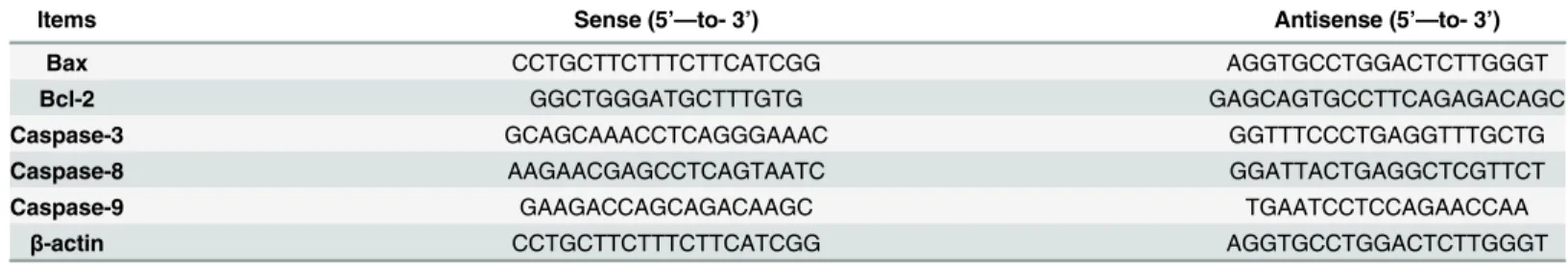
per-formed according to the“TUREscript 1st strand cDNA Synthesis Kit”from Aidlab (China), the mRNA was reverse transcribed into cDNA. The cDNA was used as a template for qRT-PCR analysis. Reaction conditions were set to 3 min at 95°C (first segment, one cycle), 10 s at 95°C and 30 s at Tm of a specific primer pair (second segment, 39 cycles) followed by 10 s at 95°C, and 72°C for 10 s (dissociation curve segment) using Thermal Cycler(C1000, BIO RAD, USA). Relative gene expression was defined as a ratio of target gene expression versusβ -actin gene expression [31]. Gene expression values of control group were used for gene expres-sion calibration, respectively. With 2-44Ctassay, the results were analyzed [32]. The following primers (Table 1) were designed and synthesized by Liuhe Beijing company (China).
TUNEL assay
Nephridial tissues were fixed in 4% paraformal-dehyde, embedded in paraffin and cut into 6μm sections. TdT-mediated dUTP nick end labelling (TUNEL) assay was conducted to study
DNA fragmentation using the in situ cell death detection kit (Vazyme, Piscataway, NJ, USA) according to the manufacturer’s instructions. After mounting the TUNEL positive cells, nuclei were counterstained with DAPI and the sections were observed at ×1000 magnification under a Nikon microscope (Nikon Inc., Japan).
Apoptosis assessment by DAPI and AO/EB staining
The kidneys were removed and minced using scissors to form a cell suspension that was filtered through a 300-mesh nylon screen. For DAPI staining, the renal cells were fixed with 80% etha-nol at room temperature for 30 min. The fixative was removed and the renal cells were washed with PBS for 3 times, and then incubated with DAPI (1μg/ml) for 45 min at room temperature
in the dark. For AO/EB staining, the cells without fixation were loaded with a 100μl
fresh-pre-pared AO/EB staining solution (100μg/ml), then immediately observed under a Nikon
fluores-cence microscope (Nikon Inc., Japan) in less than 20 min.
Caspase activity measurement
Caspases activities were measured by colorimetric assay kits (BioVision, Inc., Mountain View, California, US), according to the manufacture’s recommendations. Briefly, renal cells were har-vested and incubated in ice-cold cell lysis buffer for 30 min on ice. The supernatants were col-lected and protein concentrations were determined using BCA Protein Assay Reagent (Pierce,
Table 1. The primers used for qRT- PCR.
Items Sense (5’—to- 3’) Antisense (5’—to- 3’)
Bax CCTGCTTCTTTCTTCATCGG AGGTGCCTGGACTCTTGGGT
Bcl-2 GGCTGGGATGCTTTGTG GAGCAGTGCCTTCAGAGACAGC
Caspase-3 GCAGCAAACCTCAGGGAAAC GGTTTCCCTGAGGTTTGCTG
Caspase-8 AAGAACGAGCCTCAGTAATC GGATTACTGAGGCTCGTTCT
Caspase-9 GAAGACCAGCAGACAAGC TGAATCCTCCAGAACCAA
β-actin CCTGCTTCTTTCTTCATCGG AGGTGCCTGGACTCTTGGGT
Rockford, IL, US). Equivalent amount of proteins for each sample was incubated with inter-ested caspase substrate. After incubation at 37°C for 4h, the protease activity was determined at 405 nm with microplate spectrophotometer (Bio-Tek Instruments, Inc., Winooski, US)
Western blot analysis
Mouse monoclonal antibodies against caspase-9, -3, -8, Cytc, Bcl-2, Bax, Bid, Apaf-1, COX4, PARP, Fas, FasL, p53 andβ-actin were purchased from Santa Cruz Biotechnology, Inc. (Santa Cruz, CA, US). Horseradish peroxidase-conjugated secondary antibody was purchased from Wuhan Boster Bio-Engineering Co., Ltd. (Wuhan, China). Renal cells were harvested and washed with ice-cold PBS, then lysed with ice-cold RIPA lysis buffer (Beyotime Inst. Biotech, Beijing, China) with 1 mmol/L PMSF. Protein concentrations were calculated by BCA assay kits (Pierce). 20μg of total cellular protein was subjected to 12% SDS-PAGE and transferred to
PVDF membranes (Millipore, Atlanta, GA, US). The membranes were blocked with 5% defat-ted milk powder at room temperature for 1 hr and then immunoblotting was performed with primary antibodies at 4°C overnight, followed by HRP-conjugated secondary antibody at room temperature for 1 h. Following each step, the membranes were washed five times with PBS-T for 3 min. Finally, the blots were developed using the enhanced chemiluminescence (ECL) sys-tem (Pierce).
DNA fragmentation assay
Both control andE.adenophorum-administration Renal cells were collected and washed with PBS. DNA extraction was performed according to previous studies[33]. After dissolved in TE buffer, DNA was subjected to 2% agarose gel electrophoresis for DNA fragmentation analysis.
Mitochondrial membrane potential assay
The mitochondrial membrane potential was detected using 5,50,6,60-tetrachloro-1,10,3,30
tetra-ethylbenzimidazolcarbocyanine iodide (JC-1; Molecular Probes, Eugene, OR, USA). Renal cells were rinsed once with PBS and incubated with JC-1 for 30 min at 37°C, centrifuged, washed twice with cold PBS, transferred to a 96-well plate (105 cells/well), and assayed in a fluores-cence plate reader.
Electron microscopy observation
The ultrastructural morphology changes were observed under a transmission electron micro-scope. AfterE.adenophorumadministration, the cells were fixed with 3% glutaraldehyde, and post-fixed with 1% OsO4. Then samples were dehydrated in graded ethanol solutions, followed by embedment and section. Ultra-thin sections were stained with uranyl acetate and lead cit-rate, and then observed under a transmission electron microscope (JEM-1230, Tokyo, Japan) at 60 kV.
Statistical Analysis
Results
Cell Cycle of Renal Cells
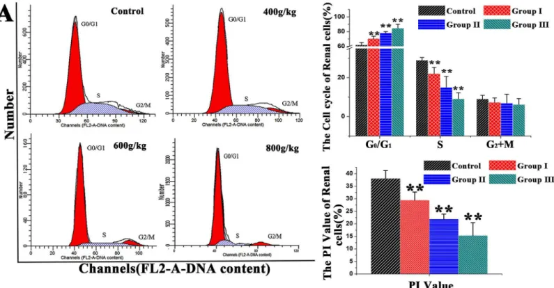
The distribution of renal cells in different phases of the cell cycle was analyzed by flow cytome-try (Fig 1). Following feeding saanen goats on different dose levels ofE.adenophorum, in the experimental groups, the proportion of renal cells in the G0/G1 phase was increased substan-tially (P<0.01) and the percentage of renal cells was decreased to different extent in the S (P<0.01, P<0.01 and P<0.01) and PI value (P<0.01). Besides, the percentages of renal cells in the G0/G1 were increased by 13.8%, 26.0%, 36.6% and decreased by 23.8%, 48.2%, 68.6% in S phase than control, respectively, but there is no significant difference in G2/M phase (P>0.05), which suggested G0/G1 phase arrest. Our data suggests thatE.adenophoruminhibits renal cell growth of saanen goats by blocking the G0/G1 to S phase transition in the cell cycle.
The detection of apoptosic renal cells
We further detected the apoptosis occurrence in renal cells. The renal cells apoptotic rate of saanen goats was assessed by using the Annexin V/PI staining assay, DAPI and AO/EB staining and TUNEL assay. By flow cytometry, the results indicated that the percentage of normal renal cells in the experimental groups was decreased markedly (P<0.05, P<0.01 and P<0.01). With the increase ofE.adenophorumadding proportion, the apoptosis ratio was also on the increase, the percentages of early and late apoptotic renal cells in groupⅡ,Ⅲwere significantly increased (P<0.01) (Fig 2A). The TUNEL assay is capable of detecting DNA strand breaks that occur
Fig 1. DNA histogram of renal cell cycle in control. (A)The Saanen goat was treated with different dose ofE.adenophorumfor 3 months, Then DNA histogram of renal cells cell cycle was analyzed by flow cytometry for PI staining. the G0/G1%,S% and G2+M% phases of the renal cells were analyzed using flow cytometry. Flow cytometric histograms are representative of 3 separate experiments. Proliferating index (PI) value = [S+(G2+M)]/[(G0/G1)+S+(G2+M)]×100%.
Data are presented with the means±SD and mean values of three independent experiments.*p<0.05 and**p<0.01, compared with the control group.
prior to the nucleus fragmenting[34]. We also evaluated cell apoptosis through TUNEL stain-ing. TUNEL-positive cells were quantified through manual countstain-ing. Our TUNEL staining results revealed that TUNEL-positive cells were not significantly observed in control group, whereas the proportion of renal cells undergoing apoptosis and showing signs of apoptosis increased significantly compared to the control group. Quantification indicated a 22.20% and
Fig 2.E.adenophorumadministration induces apoptosis in renal cells. (A)The scattergram of apoptotic renal cells. The renal cells were analyzed for apoptosis by flow cytometry for Annexin V and PI staining. The percentage number of renal cells apoptosis(%) were shown.E.adenophorumsignificantly induced apoptosis in renal cells.(B)The Detection of apoptotic renal cells by DAPI, AO/EB staining and TUNEL assay. Representative kidney sections from saanen goats were analyzed using TUNEL assay for apoptotic cell death. The number of TUNEL positive cells (indicated arrows) in kidney was counted from five random microscopic fields. Magnification, x400. Nuclear morphological changes in renal cells were observed under fluorescent microscope after DAPI staining (indicated arrows, 200×) and DAPI or AO/EB staining (400×).(C)Induction of DNA fragmentation. DNA isolated fromE.adenophorum
-administration renal cells was subjected to 2% agarose gel electrophoresis, followed by visualization of bands and photography. Data are presented with the means±SD and mean values of three independent experiments.*p<0.05 and**p<0.01, compared with the control group.
31.45% amount of TUNEL-positive cells at dose of 600, 800g/dE.adenophorumand both were significantly increased (P<0.01). The proportion of apoptosic renal cells positively correlated with the doses ofE.adenophorum. DAPI and AO/EB staining showed that the cell nuclei and cell membrane integrity of renal cells of control did not appear significant changes, however, the renal cells of experimental groups appeared different extent of chromatin condensation, nuclear fragmentation and destruction of cell membrane integrity (Fig 2B). Besides the mor-phological changes of apoptosis inE.adenophorum–administrated renal cells, DNA fragmen-tation assay also showed that characteristic ladder patterns appeared in renal cells. Also, DNA ladder started became more evident in renal cells when at the dose level of 600 and 800g/kg/d (Fig 2C). The proportion of apoptosic renal cells positively correlated with the doses ofE. ade-nophorum. These results demonstrated thatE.adenophoruminduced renal cells apoptosis in a dose-dependent manner.
The contributions of caspase-8, 9 and 3 in apoptosis
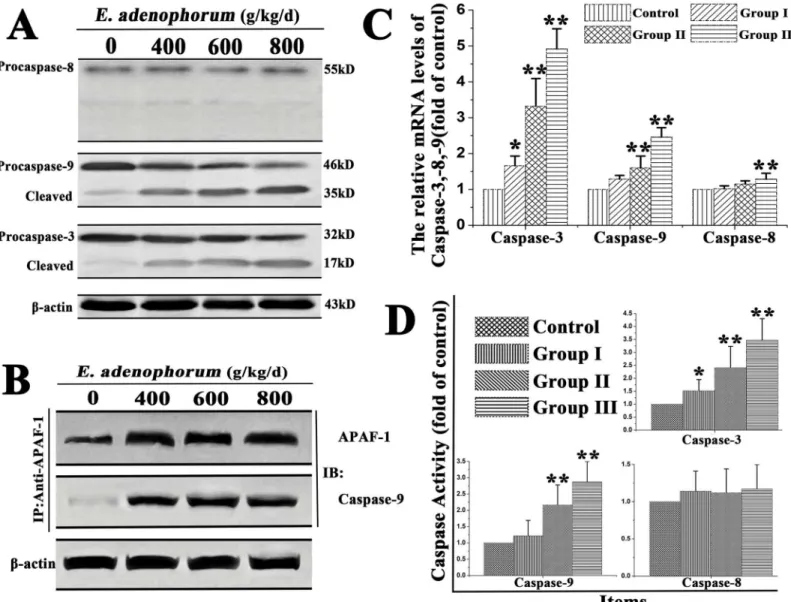
Caspases are the central components in the execution of apoptosis. Respectively, caspase-8 and -9 are both the initiator caspases in the death receptor pathway and the mitochondrial pathway. Caspase-3 is the key executioner caspase in apoptosis pathway and the downstream caspase of caspase-8 and -9. The contributions of caspase-8, -9 and -3 inE.adenophorum-induced apopto-sis were confirm by qRT-PCR caspase activity and Western blot. The results of Western-blot indicated that full-length procaspase-9 and procaspase-3 were decreased with the increased treated times, while their cleaved form were increased. However, the cleavage of procaspase-8 and the cleaved form of it did not show any difference (Fig 3A). In cytoplasm, released Cytc usu-ally combines with Apaf-1 and procaspase-9 to form the apoptosome in the presence of ATP, resulting in the activation of caspase-9. The cell lysates were immunoprecipitated with an anti-Apaf-1 antibody and subsequently subjected to Western blot with anti-Caspase-9 antibodies, which was conducted to detect whetherE.adenophorumpromotes the formation of apoptosome. The results showed that Apaf-1 was interacted with Caspase-9 (Fig 3B). Besides, qRT-PCR showed thatE.adenophorumevidently induced the activation of caspase-3 and caspase-9, but not induce the activation of caspase-8 in groupⅠ,Ⅱ(P>0.05) (Fig 3C). The caspase molecules involved inE.adenophorum-induced apoptosis was analysed to measure the activities of caspase-8, -9, and -3 using colorimetric assay kits.E.adenophorumsignificantly induced the activation of caspases-9 and -3, but not caspase-8 (Fig 3D).
E
.
adenophorum
treatment activates the mitochondrial apoptotic
pathway
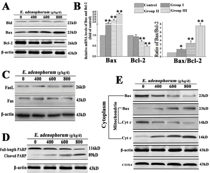
show significant variations, suggesting that the activation of mitochondrial pathway was inde-pendent of the activation of caspase-8 and Bid (Fig 4A). qRT-PCR indicated that the activation of Bax was increased and Bcl-2 was decreased, resulting in the ratio of Bax/Bcl-2, which incline to the activation of the mitochondrial pathway(Fig 4B). Western blot indicated thatE. adeno-phorumfailed to affect the levels of Fas and FasL, suggesting thatE.adenophorummight not activate Fas-mediated death receptor pathway in renal cells (Fig 4C). PARP is a downstream target for caspases during apoptosis. The western blot showed that PARP was shown to be cleaved from 116 to 85 kDa fragments obviously in the renal cells adminitrated withE. adeno-phorum(Fig 4D). The location of Bax and Cytcin the proteins extracts from both
Fig 3.E.adenophorum-induced apoptosis is mediated by activation of caspase-9, caspase-3. (A)The protein levels of procaspase-3, -8, -9 and the cleaved form of them. The expression of apoptosis-related proteins, including caspase-3, -9, -8 were shown withβ-actin as a control, were detected by Western-blot analysis.(B)E.adenophoruminduced apoptosome formation. Protein extractions from renal cells were collected and then used in
immunoprecipitation assays against Apaf-1. The level of caspase-9 was detected by western blot to indicate the formation of apoptosome complex.(C)The relative mRNA levels of caspase-3, -8 and -9. The Saanen goat was treated with different dose ofE.adenophorumfor 3 months and the mRNA of renal cells was extraced and used for qRT-PCR assay.E.adenophoruminduced the activiation of caspase-3, -9.(D)Caspase activities in renal cells. BCA assay was used to equal protein amounts and the enzymatic activities of caspases-8, -9, and -3 were measured using the colorimetric assay kits. Data are presented with the means±SD and mean values of three independent experiments.*p<0.05 and**p<0.01, compared with the control group.
mitochondrial and cytosolic fractions of renal cells was detected. A translocation of Bax from cytosol to mitochondria was observed. Consistent with this, a dose-dependent decrease in mitochondrial Cytcand a concomitant increase in the cytosolic fraction were also observed (Fig 4E). These results suggested thatE.adenophorum-induced apoptosis was mainly through the activation of mitochondrial pathway.
Mitochondrial localization of p53 promotes mitochondrial apoptosis
pathways
Previous reports confirmed that p53 promotes apoptosis through transcription-independent mechanisms, primarily signals through the mitochondrial pathway[37]. Previous studies dem-onstrated that a fraction of wild-type p53 protein rapidly migrates to the mitochondria early in the course of p53-dependent apoptosis[38]. Activation of p53-dependent apoptosis leads to
Fig 4. The renal cells apoptosis induced byE.adenophorumwas mediated by mitochondrial pathway. (A)E.adenophorumdid not promote the cleavage of Bid, but increased the protein levels of Cytcand Bax, and decrease the protein level of Bcl-2.(B)E.adenophorumdecreased ralative mRNA level of Bcl-2, but increased the level of Bax, resulting in the change of Bax/Bcl-2.(C)The protein levels of Fas or FasL measured by western blot did not reveal any changes.(D)The renal cells were subjected to western blot analysis to detectfull-length and cleaved PARP.(E)E.adenophoruminduced Bax translocation and Cytcrelease. The cytosolic and mitochondrial fraction proteins were collected and then detected by western blot. COX 4 andβ-actin were used as internal controls for the mitochondrial fractions and the cytosolic fraction, respectively.
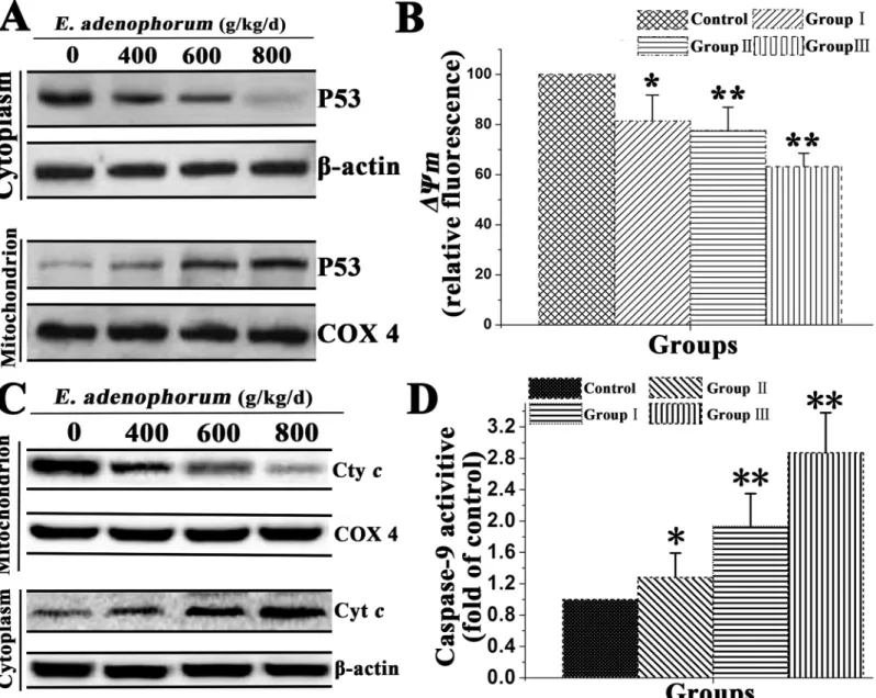
mitochondrial apoptotic changes via the intrinsic and extrinsic pathways triggering cell death execution most notably by release of Cytcand activation of the caspase cascade. Although it was previously believed that p53 induces apoptotic mitochondrial changes exclusively through transcription-dependent mechanisms, recent studies suggest that p53 also regulates apoptosis via a transcription-independent action at the mitochondria[39]. We conducted the study to verify that mitochondrial p53 accumulation is associated withE.adenophorum-induced apo-ptosis of renal cells. As shown inFig 5A,E.adenophoruminduced mitochondrial p53 translo-cation from cytosol to mitochondrion. To investigate the downstream of the mitochondrial p53 pathway, that Cytcrelease to the cytosol from mitochondrion, caspase-9 activation and
Fig 5. Mitochondrial p53 activates mitochondrial pathway in renal cells.To determine localization of p53, renal cells were incubated as indicated(A) Western blot analyzed the effect ofE.adenophorumon p53 mitochondrial translocation. COX 4, mitochondrial loading control.(B)Flow cytometry and JC-1 measure the effet ofE.adenophorumonΔΨm.(C)Western blot was conducted to analyze the release of Cytcin renal cells.(D)The effect ofE.
adenophorumon enzymatic activity of caspase-9 was measured and was expressed relative to the control. Date are presented with the means±SD and mean values of three independent experiments.*p<0.05 and**p<0.01 compared with the control group.
mitochondrial membrane potential(ΔCm) in renal cells were examined. The collapse ofΔCm was also observed in renal cells as compared to control (Fig 5B). A significant increase in cyto-solic Cytc(Fig 5C), and caspase-9 activation (Fig 5D) were observed. These results indicated that p53 molecules accumulated into the mitochondria and activated mitochondrial apoptosis pathway.
Ultrastructural morphology changes
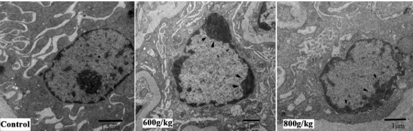
The apoptotic features of renal cells were further confirmed by electron microscopy. Transmis-sion electron microscope observation showed that renal cells displayed characteristically mor-phological changes afterE.adenophorum-administration. In contrast to necrotic cells, the plasma membrane of apoptotic cells remains intact, under high-power (×10000), renal cells had an intact plasma membrane and a markedly reduced cytoplasmic volume. Besides the high-power views showed condensation of nuclear chromatin, which were the features typical of apoptosis that does not occur in necrosis. Chromatin condenses against the nuclear mem-brane, producing the crescentic pattern in early apoptosis. Then chromatin condenses into solid, rounded masses that undergo fragmentation (arrowheads inFig 6). whereas the control cells did not appear significant changes in cell nuclei and cell membrane integrity.
Discussion
E.adenophorum, a worldwide noxious invasive weed, has become a major threat to economy and ecology in some regions of the world[40]. Regular ingestion ofE.adenophorumcaused chronic pulmonary disease in horses[1] and Verma’s studies reported that usingE. adeno-phorumgrowing in India as diet supplement caused cattle anorexia, suspension of rumination and photosensitization[41]. In mice, feeding containingE.adenophorumfreeze-dried leaf pow-der caused hepatotoxicity. However, no toxic effects were seen in goats when the comprising of E.adenophorumup to 67% of their intake[42]. The experiment period was probably the reason why our result was on the contrary. These studies provide rational for exploringE. adeno-phorumas a cause of nephrotoxicity and an inducer of apoptosis in renal cells of saanen goat.
Apoptosis, a highly regulated process, is used to eliminate dysplastic and damaged cells from multicellular organisms[43]. By means of flow cytometry[44], We found that the renal cells that stained positive for (FITC+/PI+) and (FITC+/PI-) were increased, this indicated that E.adenophorumreduced the survival and inhibited the growth of the renal cells through
Fig 6. Ultrastructural morphology changes of renal cells. (A)Saanen goats were administrated with various doses ofE.adenophorumfor 3 months, and the ranal cells of saanen goats were conducted with ultrastructural observation. After a series of washing, fixation, dehydration and stained, cells were visualized under transmission. Magnification, x10000.
induction of apoptosis and cell cycle arrest, suggesting that renal cells was sensitive toE. adeno-phorumin Saanen goat. In cell level, the study showed thatE.adenophoruminduced renal cells apoptosis with the typical morphological characteristics including cellular shrinkage, chroma-tin condensation, DNA fragmentation. Here we showed thatE.adenophorumdecreased the expression of Bcl-2 while increased that of Bax both in protein and mRNA levels, and pro-moted the translocation of Bax from cytoplasm to mitochondria and that of Cytcfrom mito-chondria to cytoplasm, indicating that mitomito-chondrial pathway was activated[45,46]. The mitochondrial pathway requires the release of mitochondrial Cytcand the formation of a large multiprotein complex comprising Cytc, Apaf-1 and procaspase-9. In renal cells, the Cytc released from mitochondria into cytosol to form apoptosomes together with Apaf-1 and pro-caspase-9, followed by the activation of caspse-9, -3 and the cleavage of PARP. Besides,E. ade-nophorumfailed to activate Fas, FasL, Bid and the death receptor-mediated caspase-8 pathway, suggesting that mitochondria-mediated apoptosis pathway is activated byE.adenophorum administration andE.adenophoruminduced apoptosis through the Cytc-mediated and cas-pase-dependent pathway. Furthermore, the dysregulation of mitochondria integrity associated molecules Bcl-2 and Bax inE.adenophorum-administration renal cells further suggest that the activation of mitochondria-mediated apoptosis pathway is the main events in the process of apoptosis occurrence. We found thatE.adenophorumcould induce p53's mitochondrial trans-location, followed by the release of Cytcand caspase-9 activation, as well as aggravatedΔCm decrease in renal cells. Thus, it is evident that the p53 in renal cells translocated to mitochon-dria where it triggered mitochonmitochon-drial apoptotic pathways. These results indicated thatE. ade-nophorumcan induce renal cells apoptosis and cause kidney impairment by induction of mitochondrial dysfunction, and further confirm that mitochondria as the center of cell metab-olism play an essential role in maintaining the normal physiological function of renal cells. The study indicated that mitochondrial dysfunction in renal cells induced byE.adenophorumwas considered to responsible for the apoptosis occurrence[47]. The cell cycle detection and PI value showedE.adenophorumintake could effectively inhibit the growth of renal cells by caus-ing cell cycle arrest.
The present study demonstrated thatE.adenophorumsignificantly inhibits the growth of renal cells by causing G0/G1 phase cell cycle arrest and the induction of apoptosis. TheE. ade-nophorum-induced apoptosis is dependent on the mitochondria-mediated caspase activation and involvement of the regulation of Bcl-2 and Bax, followed by the significant increases in activation of caspases-9 and -3, the release of Cytcto cytosol and the cleavage of PARP. This study provides us a new insight into understanding the mechanisms of saanen goat renal cells apoptosis caused byE.adenophorum.
Acknowledgments
This research was supported by Special Fund for Agroscientific Research in the Public Interest (Grant No. 201203062) and Chang-jiang Scholars and the Innovative Research Team in Uni-versity (Grant No. IRT0848).
Author Contributions
References
1. Oelrichs PB, Calanasan CA, Macleod JK, Seawright AA, Ng JC. Isolation of a compound from Eupator-ium adenophorum(Spreng.)[Ageratina adenophora(Spreng.)] causing hepatotoxicity in mice. Natural toxins. 1995; 3(5):350–4. PMID:8581319
2. Sharma OP, Dawra RK, Kurade NP, Sharma PD. A review of the toxicosis and biological properties of the genusEupatorium. Natural Toxins. 1998; 6(1):1–14. PMID:9851506
3. Guo S, Li W, Zhang L, Peng J, Xia H, Zhang S. Kinetics and equilibrium adsorption study of lead (II) onto the low cost adsorbent—Eupatorium adenophorumspreng. Process Safety and Environmental Protection. 2009; 87(5):343–51.
4. Nong X, Ren YJ, Wang JH, Xie Y, Fang CL, Yang DY. Clinical efficacy of botanical extracts from Eupa-torium adenophorumagainst theSarcoptes scabiei(Sarcoptidae:Sarcoptes) in Rabbits. Veterinary parasitology. 2013: doi:10.1016/j.vetpar.2013.02.020
5. Seddiek SA, Khater HF, El-Shorbagy MM, Ali AM. The acaricidal efficacy of aqueous neem extract and ivermectin againstSarcoptes scabieivar. cuniculi in experimentally infested rabbits. Parasitology Research. 2013; 112(6):2319–30. doi:10.1007/s00436-013-3395-2PMID:23572045
6. Liao F, Hu Y, Tan H, Wu L, Wang Y, Huang Y, et al. Acaricidal activity of 9-oxo-10, 11-dehydroagera-phorone extracted fromEupatorium adenophorumin vitro. Experimental parasitology. 2014; 140:8–11. doi:10.1016/j.exppara.2014.02.009PMID:24631419
7. Kundu A, Saha S, Walia S, Ahluwalia V, Kaur C. Antioxidant potential of essential oil and cadinene ses-quiterpenes ofEupatorium adenophorum. Toxicological & Environmental Chemistry. 2013; 95(1):127–
37.
8. Liao F, Hu Y, Wu L, Tan H, Mo Q, Luo B, et al. The Antitumor Activity in Vitro by 9-oxo-10, 11-dehydroa-geraphorone Extracted fromEupatorium adenophorum. Asian Journal of Chemistry. 2014; 26 (21):7321–3.
9. Chakravarty AK, Mazumder T, Chatterjee SN. Anti-inflammatory potential of ethanolic leaf extract of
Eupatorium adenophorumSpreng. Through Alteration in Production of TNF-α, ROS and expression of certain genes. Evidence-Based Complementary and Alternative Medicine. 2011; doi:10.1093/ecam/ neq033
10. Sani Y, Harper P, Cook R, Seawright A, Ng J, James L, et al., editors. The toxicity ofEupatorium adeno-phorumfor the liver of the mouse. Poisonous plants Proceedings of the Third International Symposium; 1992: Iowa State University Press.
11. Singh Y, Mukhopadhayay S. Ali M. Ayub, Tolenkhomba TC and Shah MA Ayub. Short-term toxicity studies ofEupatorium adenophorumin Swiss albino mice. Int J Res Phytochem Pharmacol. 2011; 1:165–71.
12. Katoch R, Sharma OP, Dawra RK, Kurade NP. Hepatotoxicity ofEupatorium adenophorumto rats. Toxicon. 2000; 38(2):309–14. PMID:10665812
13. Kaushal V, Dawra R, Sharma O, Kurade N. Hepatotoxicity in rat induced by partially purified toxins from
Eupatorium adenophorum(Ageratina adenophora). Toxicon. 2001; 39(5):615–9. PMID:11072039
14. Bhardwaj R, Singh A, Sharma OP, Dawra RK, Kurade NP, Mahato SB. Hepatotoxicity and cholestasis in rats induced by the sesquiterpene, 9-oxo-10, 11-dehydroageraphorone, isolated fromEupatorium adenophorum. Journal of biochemical and molecular toxicology. 2001; 15(5):279–86. PMID:11835625
15. Kantari C, Walczak H. Caspase-8 and bid: caught in the act between death receptors and mitochondria. Biochimica et Biophysica Acta (BBA)-Molecular Cell Research. 2011; 1813(4):558–63.
16. Fan T-J, Han L-H, Cong R-S, Liang J. Caspase family proteases and apoptosis. Acta biochimica et bio-physica Sinica. 2005; 37(11):719–27. PMID:16270150
17. Li J, Yuan J. Caspases in apoptosis and beyond. Oncogene. 2008; 27(48):6194–206. doi:10.1038/ onc.2008.297PMID:18931687
18. Schlegel J, Peters I, Orrenius S, Miller DK, Thornberry NA, Yamin T-T, et al. CPP32/apopain is a key interleukin 1 converting enzyme-like protease involved in Fas-mediated apoptosis. Journal of Biological Chemistry. 1996; 271(4):1841–4. PMID:8567626
19. Nicholson DW, Ali A, Thornberry NA, Vaillancourt JP, Ding CK, Gallant M, et al. Identification and inhibi-tion of the ICE/CED-3 protease necessary for mammalian apoptosis. 1995. PMID:7596430
20. Wang X, Zelenski NG, Yang J, Sakai J, Brown MS, Goldstein JL. Cleavage of sterol regulatory element binding proteins (SREBPs) by CPP32 during apoptosis. The EMBO journal. 1996; 15(5):1012. PMID:
8605870
22. Schuler M, Bossy-Wetzel E, Goldstein JC, Fitzgerald P, Green DR. p53 induces apoptosis by caspase activation through mitochondrial cytochrome c release. Journal of Biological Chemistry. 2000; 275 (10):7337–42. PMID:10702305
23. Salvesen GS, Dixit VM. Caspases: intracellular signaling by proteolysis. Cell. 1997; 91(4):443–6. PMID:9390553
24. Sawada M, Nakashima S, Banno Y, Yamakawa H, Hayashi K, Takenaka K, et al. Ordering of ceramide formation, caspase activation, and Bax/Bcl-2 expression during etoposide-induced apoptosis in C6 gli-oma cells. Cell death and differentiation. 2000; 7(9):761–72. PMID:11042671
25. Ye J, Wang S, Leonard SS, Sun Y, Butterworth L, Antonini J, et al. Role of reactive oxygen species and p53 in chromium (VI)-induced apoptosis. Journal of Biological Chemistry. 1999; 274(49):34974–80. PMID:10574974
26. Boulakia CA, Chen G, Ng F, Teodoro JG, Branton PE, Nicholson DW, et al. Bcl-2 and adenovirus E1B 19 kDA protein prevent E1A-induced processing of CPP32 and cleavage of poly (ADP-ribose) polymer-ase. Oncogene. 1996; 12(3):529–35. PMID:8637709
27. Liu X, Kim CN, Yang J, Jemmerson R, Wang X. Induction of apoptotic program in cell-free extracts: requirement for dATP and cytochromec. Cell. 1996; 86(1):147–57. PMID:8689682
28. Yang J, Liu X, Bhalla K, Kim CN, Ibrado AM, Cai J, et al. Prevention of apoptosis by Bcl-2: release of cytochromecfrom mitochondria blocked. Science. 1997; 275(5303):1129–32. PMID:9027314
29. Oltval ZN, Milliman CL, Korsmeyer SJ. Bcl-2 heterodimerizes in vivo with a conserved homolog, Bax, that accelerates programed cell death. Cell. 1993; 74(4):609–19. PMID:8358790
30. Sahoo A, Singh B, Sharma O. Evaluation of feeding value ofEupatorium adenophorumin combination with mulberry leaves. Livestock Science. 2011; 136(2):175–83.
31. Iqbal M, Philbin VJ, Smith AL. Expression patterns of chicken Toll-like receptor mRNA in tissues, immune cell subsets and cell lines. Veterinary immunology and immunopathology. 2005; 104(1):117–
27.
32. Livak KJ, Schmittgen TD. Analysis of Relative Gene Expression Data Using Real-Time Quantitative PCR and the 2−ΔΔCTMethod. methods. 2001; 25(4):402–8. PMID:11846609
33. Li Z, Xu X, Huang Y, Ding L, Wang Z, Yu G, et al. Swainsonine Activates Mitochondria-mediated Apo-ptotic Pathway in Human Lung Cancer A549 Cells and Retards the Growth of Lung Cancer Xenografts. International journal of biological sciences. 2012; 8(3):394–405. doi:10.7150/ijbs.3882PMID:
22393311
34. O'Brien IE, Reutelingsperger CP, Holdaway KM. Annexin-V and TUNEL use in monitoring the progres-sion of apoptosis in plants. Cytometry. 1997; 29(1):28–33. PMID:9298808
35. Xu Y, Ge R, Du J, Xin H, Yi T, Sheng J, et al. Corosolic acid induces apoptosis through mitochondrial pathway and caspases activation in human cervix adenocarcinoma HeLa cells. Cancer letters. 2009; 284(2):229–37. doi:10.1016/j.canlet.2009.04.028PMID:19457606
36. Tong X, Lin S, Fujii M, Hou D-X. Echinocystic acid induces apoptosis in HL-60 cells through mitochon-dria-mediated death pathway. Cancer letters. 2004; 212(1):21–32. PMID:15246558
37. Green DR, Kroemer G. Cytoplasmic functions of the tumour suppressor p53. Nature. 2009; 458 (7242):1127–30. doi:10.1038/nature07986PMID:19407794
38. Zhao Y, Chaiswing L, Velez JM, Batinic-Haberle I, Colburn NH, Oberley TD, et al. p53 translocation to mitochondria precedes its nuclear translocation and targets mitochondrial oxidative defense protein-manganese superoxide dismutase. Cancer research. 2005; 65(9):3745–50. PMID:15867370
39. Wang DB, Kinoshita C, Kinoshita Y, Morrison RS. p53 and mitochondrial function in neurons. Biochi-mica et Biophysica Acta (BBA)-Molecular Basis of Disease. 2014; 1842(8):1186–97.
40. Lu P, Sang W, Ma K. Progress and prospects in research of an exotic invasive species, {Eupatorium adenophorum}. Acta Phytoecological Sinica. 2004; 29(6):1029–37.
41. Verma A, Yadav B, Sampath K. Possible use of Spreng (Eupatorium adenophorum) in animal feeding. Indian journal of animal nutrition. 1987.
42. Neopane S. Performance of goats given different levels of Banmara (Eupatorium adenophorum) at Pakhribas Agricultural Centre: Pakhribas Agricultural Centre; 1992.
43. King KL, Cidlowski JA. Cell cycle and apoptosis: common pathways to life and death. Journal of cellular biochemistry. 1995; 58(2):175–80. PMID:7673325
45. Youle RJ, Strasser A. The BCL-2 protein family: opposing activities that mediate cell death. Nature reviews Molecular cell biology. 2008; 9(1):47–59. PMID:18097445
46. Finucane DM, Bossy-Wetzel E, Waterhouse NJ, Cotter TG, Green DR. Bax-induced caspase activation and apoptosis via cytochromecrelease from mitochondria is inhibitable by Bcl-xL. Journal of Biological Chemistry. 1999; 274(4):2225–33. PMID:9890985