Abieslactone Induces Cell Cycle Arrest and
Apoptosis in Human Hepatocellular
Carcinomas through the Mitochondrial
Pathway and the Generation of Reactive
Oxygen Species
Guo-Wei Wang1., Chao Lv2,3., Zhi-Ran Shi2, Ren-Tao Zeng2, Xue-Yun Dong3, Wei-Dong Zhang1,2, Run-Hui Liu2, Lei Shan2*, Yun-Heng Shen2*
1.School of Pharmacy, Shanghai Jiao Tong University, Shanghai 200240, PR China,2.School of Pharmacy, Second Military Medical University, Shanghai 200433, PR China,3.School of Pharmacy, Fujian University of Traditional Chinese Medicine, Fujian 350108, PR China
*shenyunheng@hotmail.com (YHS);shanleicn@126.com (LS)
.These authors contributed equally to this work.
Abstract
Abieslactone is a triterpenoid lactone isolated from Abiesplants. Previous studies have demonstrated that its derivative abiesenonic acid methyl ester possesses anti-tumor-promoting activity in vitroandin vivo. In the present study, cell viability assay demonstrated that abieslactone had selective cytotoxicity against human hepatoma cell lines. Immunostaining experiments revealed that abieslactone induced HepG2 and SMMC7721 cell apoptosis. Flow cytometry and western blot analysis showed that the apoptosis was associated with cell cycle arrest during the G1 phase, up-regulation of p53 and p21, and down-regulation of CDK2 and cyclin
D1. Furthermore, our results revealed that induction of apoptosis through a mitochondrial pathway led to upregulation of Bax, down-regulation of Bcl-2, mitochondrial release of cytochrome c, reduction of mitochondrial membrane potential (MMP), and activation of caspase cascades (Casp-9 and -3). Activation of caspase cascades also resulted in the cleavage of PARP fragment. Involvement of the caspase apoptosis pathway was confirmed using caspase inhibitor Z-VAD-FMK pretreatment. Recent studies have shown that ROS is upstream of Akt signal in mitochondria-mediated hepatoma cell apoptosis. Our results showed that the accumulation of ROS was detected in HepG2 cells when treated with abieslactone, and ROS scavenger partly blocked the effects of abieslactone-induced HepG2 cell death. In addition, inactivation of total and phosphorylated Akt activities was found to be involved in abieslactone-induced HepG2 cell apoptosis. Therefore, our OPEN ACCESS
Citation:Wang G-W, Lv C, Shi Z-R, Zeng R-T, Dong X-Y, et al. (2014) Abieslactone Induces Cell Cycle Arrest and Apoptosis in Human
Hepatocellular Carcinomas through the Mitochondrial Pathway and the Generation of Reactive Oxygen Species. PLoS ONE 9(12): e115151. doi:10.1371/journal.pone.0115151
Editor:Yi-Hsien Hsieh, Institute of Biochemistry and Biotechnology, Taiwan
Received:September 8, 2014
Accepted:November 19, 2014
Published:December 11, 2014
Copyright:ß2014 Wang et al. This is an
open-access article distributed under the terms of the
Creative Commons Attribution License, which permits unrestricted use, distribution, and repro-duction in any medium, provided the original author and source are credited.
Data Availability:The authors confirm that all data underlying the findings are fully available without restriction. All relevant data are within the paper.
Funding:The work was supported by program NCET Foundation, NSFC (81230090), Shanghai Leading Academic Discipline Project (B906), Key laboratory of drug research for special environ-ments, PLA, Shanghai Engineering Research Center for the Preparation of Bioactive Natural Products (10DZ2251300), the Scientific Foundation of Shanghai China (12401900801, 13401900101), National Major Project of China (2011ZX09307-002-03) and the National Key Technology R&D Program of China (2012BAI29B06). The funders had no role in study design, data collection and analysis, decision to publish, or preparation of the manuscript.
findings suggested that abieslactone induced G1cell cycle arrest and
caspase-dependent apoptosis via the mitochondrial pathway and the ROS/Akt pathway in HepG2 cells.
Introduction
Hepatocellular carcinoma, also called malignant hepatoma, is the fifth common cancer in human and the third leading cause of cancer death worldwide, responsible for over 600,000 deaths every year [1]. Clinically, the only medical treatments for hepatocellular carcinoma are surgical resection and liver
transplantation in patients [2]. Unfortunately, owing to high recurrence rate after resection, most patients are not eligible for surgery [3]. Conventional
chemotherapeutic and radiotherapeutic treatments have led to serious health problems, as they can kill healthy cells as well. Moreover, resistance to
chemotherapy is frequently observed. Therefore, developing novel efficient drugs with minimal side effect and understanding their molecular mechanisms are necessary for improving hepatocellular carcinoma therapy.
Apoptosis, a process of programed cell death, is the most common mechanism exploited by targeted chemotherapies that can induce death in cancer cells [4]. Apoptosis is characterized by distinct morphological changes, including
membrane blebbing, cell shrinkage, loss of mitochondrial membrane potential (MMP), chromatin condensation and DNA fragmentation [5]. There are two established pathways that result in apoptosis: the extrinsic cell death pathway (cell death receptor pathway) and the intrinsic cell death pathway (the mitochondria-initiated pathway) [6]. At the biochemical level, apoptosis is mediated by the activation of a class of cysteine proteases known as caspases [7]. Caspase
activation mainly occurs via death receptor pathway activation or mitochondrial membrane depolarization. Mitochondrial-dependent apoptosis is regulated principally by the Bcl-2 protein family. Bax is a cardinal proapoptotic member of Bcl-2 family proteins, which regulates the critical balance between cell survival and death [8]. In response to apoptotic signals, Bax transforms into a lethal
mitochondrial oligomer and becomes activated to cause mitochondrial damage, a key step for the intrinsic pathway to apoptosis [9,10].
have shown that ROS acts upstream of mitochondria-mediated apoptosis by promoting Bax translocation to mitochondria [16–18], activating JNK activity [19], or repressing Akt and NF-kB activity [20,21]. Therefore, ROS play a key role in mitochondria-mediated apoptosis.
Plants are considered to be one of the most important sources of anticancer agents. Plant-derived natural products (such as taxol [22], curcumin [23], and tetrandrine [21,24]), that can activate cell apoptosis, have great potential in cancer therapy. Abieslactone, previously reported from the bark and leaves of A. mariesi in 1965 [25], is a natural triterpenoid lactone that we recently isolated from the branches and leaves of A. faxoniana. It has been reported that its derivative abiesenonic acid methyl ester could suppress tumor promoter-induced
phenomena in vitro and in vivo[26]. In this study, we demonstrated that
abieslactone inhibited the growth and proliferation of three human hepatoma cell lines (HepG2, SMMC7721, and Huh7) but had low cytotoxicity to normal hepatic cells (QSG7701). HepG2 and SMMC7721 cells were more sensitive to abieslactone treatment than Huh7 cells. We further investigated its mechanism of action using HepG2 and SMMC7721 as representative cell line models. Although abieslactone could induce cell cycle arrest and apoptosis in liver cancer cells HepG2 and SMMC7721, the molecular mechanisms in two cell lines are not the same. Abieslactone induced cell cycle arrest at G1 phase and caspase-dependent
apoptosis via both mitochondrial pathway and the ROS/Akt pathway in HepG2
cells, but the ROS/Akt pathway was not involved in abieslactone-induced SMMC7721 cells apoptosis.
Materials and Methods
Drugs and antibodies
Abieslactone was isolated from the branches and leaves of A.faxoniana
(purity.98% as determined by analytical HPLC). Propidium iodide (PI),
Hoechst 33258, dimethylsulfoxide (DMSO), [3-(4,5-dimethylthiazol-2-yl)-2,5-diphenyltetrazolium bromide] (MTT), Z-VAD-FMK, N-acetyl-L-cysteine (NAC), doxorubicin (DOX), Dulbecco’s Modified Eagle’s Medium (DMEM), fetal bovine serum (FBS), phosphate buffered saline (PBS), RNase A, penicillin and
streptomycin were purchased from Sigma Chemical Co. (St. Louis, MO, USA). Rhodamine 123 and DCFH-DA were purchased from Eugene Co. (OR, USA). The annexin V-FITC apoptosis detection kit was purchased from Beyotime Institute of Biotechnology (Shanghai, China). Mouse polyclonal anti-human Bcl-2, rabbit polyclonal anti-human Bax, cytochrome c, p53, p21, cyclin D1, CDK2, caspase-3, caspase-9, PARP, p-Akt, Akt and NF-kB p65 antibodies were purchased from Cell
Signaling Technology (Beverly, MA, USA). Antibodies specific to b-actin and
Cell lines and cell culture
The human hepatomacell lines (HepG2, SMMC7721, and Huh7) as well as the normal cell lines (QSG7701) were obtained from Shanghai Institute of Materia Medica, Chinese Academy of Sciences. The cells were grown in plastic culture flasks under standard conditions (37
˚
C with 5% CO2in a completely humidifiedatmosphere) using DMEM medium supplemented with 10% heat-inactivated
FBS, 2 mM L-glutamine, 100 units/mL penicillin and 100 mg/mL streptomycin.
Cell viability assay
Cell viability was determined by the MTT assay. Briefly, cells were seeded in 96-well plates at 66103cells/well and were treated with abieslactone (0, 1, 5, 10, 25,
50 mM) for various time periods (24, 48, 72 h) [27]. Doxorubicin (0, 0.25, 0.5, 1, 2.5, 5, 10 mM) was used as a positive control in this experiment. Cultures were
also treated with (0.1%) DMSO as the untreated control. After treatment, 10 mL
of MTT solution (5 mg/mL) was added to each well and the plates were incubated for 2–4 h at 37
˚
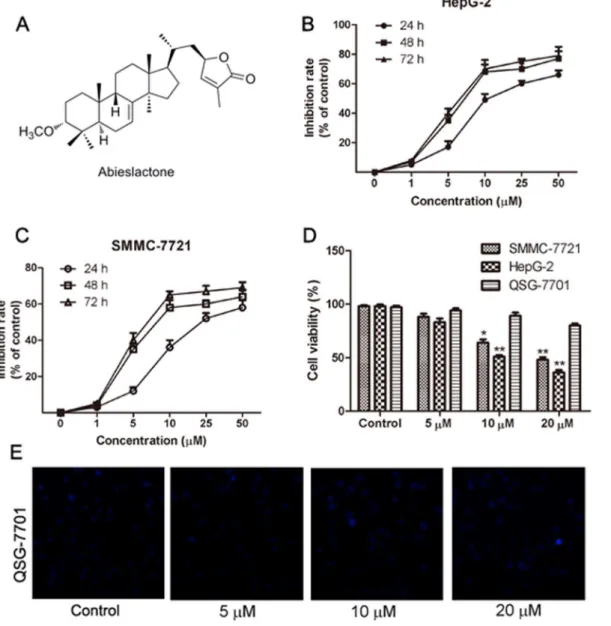
C. The supernatant was then removed from formazan crystals and 100 mL of DMSO was added to each well. The absorbance at 570 nm was read using an OPTImax microplate reader. The cell viability was calculated by dividing the mean optical density (OD) of compound-containing wells by that of DMSO-control wells. Three separate experiments were accomplished to determine the IC50 values. As shown in Fig. 1Band C, a clear dose-dependent cell death wasobserved after the cells were treated with abieslactone for 24 h. Thus, 24 hours was the preferred time period of choice for the rest of the experiments.
DNA fragmentation assay
Cells were treated with 5, 10 or 20 mM abieslactone for 24 h. DNA fragmentation was measured using Hoechst 33258 staining [28]. The cells were fixed with 4% paraformaldehyde for 15 min at room temperature. Then, washing with PBS, the cells were stained with Hoechst 33258 (50 mg/mL) at 37
˚
C in the dark for 30 min. The cells were washed and resuspended in PBS to assess nuclear morphology under fluorescence microscopy.Cell apoptosis assay
The apoptotic cells were quantified using the annexin V and PI double staining kit [28]. Briefly, cells were treated with abieslactone (5, 10 and 20 mM) for 24 h. After treatment, the cells were collected, washed with PBS, and resuspended in 200 mL binding buffer containing 5 mL annexin V (10 mg/mL) for 10 min in the dark. The
cells were then incubated with 10 mL PI (20 mg/mL), and the samples were
immediately analyzed using flow cytometry. For the caspase inhibitor analysis, the
cells were pretreated with 20 mM Z-VAD-FMK for 2 h and then incubated with
Cell cycle assay
The DNA content of cells in the G0/G1, S and G2/M phases were measured using
flow cytometry [27,28]. Cells were incubated with abieslactone (5, 10 and 20 mM) for 24 h. After treatment, the cells were collected, washed with PBS containing 2% FBS. 36105 cells/mL were fixed with cold absolute ethanol overnight at 4
˚
C in a15 mL polypropylene and V-bottomed tube. After washing with PBS twice, the cells were incubated with 1 mL PI staining solution (3.8 mM sodium citrate,
Fig. 1. The chemical structure of abieslactone and its growth-inhibiting effect on HepG2, SMMC7721 and QSG7701 cells.(A) The chemical structure of abieslactone. (B and C) Viability of HepG2 and SMMC7721 cells after exposure to 0.1% DMSO or various concentrations of abieslactone for 24, 48, and 72 h. The data are expressed as the means¡SEM of three independent experiments. (D) Cell viability after 5, 10, and 20mM abieslactone treatment in HepG2, SMMC7721 and QSG7701 cells for 24 h. The results are the mean¡SEM from three independent experiments. *P,0.05; **P,0.01vs. the untreated control. (E) The morphological nuclear changes in QSG7701 cells treated with abieslactone at different concentrations (5, 10, and 20mM). The cells were stained with Hoechst 33258 for 30 min in the dark to examine the cleaved nuclei, which is a sign of apoptosis.
50 mg/mL PI in PBS). Add 50 mL of RNase A solution (100 mg/mL RNase A) and incubated for 30 min at room temperature in the dark. The DNA content of cells and cell-cycle distribution were analyzed by flow cytometry.
Mitochondrial membrane potential measurement
The mitochondrial membrane potential (MMP) was measured by flow cytometry after staining liver cancer cells with Rhodamine 123 (Rh123), a cationic lipophilic fluorochrome [28]. The Rh123 uptake by the mitochondria is proportional to the MMP. Briefly, cells were treated with abieslactone (5, 10 and 20 mM) for 24 h and
were then incubated with Rh123 at a final concentration of 10 mM for 30 min at
room temperature in the dark. After being washed twice with PBS, cells were
resuspended in 1000 mL PBS and analyzed using flow cytometry with excitation
and emission wavelengths of 488 and 530 nm, respectively.
Intracellular ROS production measurement
ROS levels were detected using a flow cytometer and a microplate spectro-photometer (Molecular Devices, Sunnyvale, CA, USA) [20]. Cells pretreated with
10 mM ROS scavenger NAC for 1 h were incubated with 20 mM abieslactone for
24 h. Cellular viability was determined in the presence of 10 mM NAC for 1 h. After treatment, cells were harvested and washed with PBS and suspended in DMEM containing 10 mM 5(6)-carboxy-29,79-dichlorodihydrofluorescein diace-tate (carboxy-H2DCFDA; Invitrogen) at 37
˚
C for 20 min. The cells were thenwashed twice with PBS and subjected to flow cytometry analysis.
Western blot analysis
Cells were treated with abieslactone for 24 h, washed twice with PBS, and lysed for 30 min on ice using WIP cell lysis reagent (150 mM NaCl, 50 mM Tris, pH 8.0,
1% Triton X-100, 1 mM Na2EDTA, 1 mM EGTA, 2.5 mM sodium
pyropho-sphate, 1 mM b-glycerophosphate, 1 mM Na3VO4 and 1mg/mL leupeptin) [28].
The insoluble protein lysate was removed by centrifugation at 12000 rpm for 15 min at 4
˚
C [21]. The protein concentrations were determined using a NanoDrop 1000 spectrophotometer (Thermo Scientific, USA). Proteins were electrophoresed using 15% SDS-PAGE and transferred to a PVDF membrane. After blocking with 5% (w/v) non-fat milk and washing with Tris-bufferedsaline-Tween solution (TBST), the membranes were incubated overnight at 4
˚
C withStatistical analysis
Results were expressed as mean¡SEM. Data were analyzed by one-way analysis
of variance (ANOVA) followed by the Dunnett’s test. A value of P,0.05 was
considered significant.
Results
Abieslactone exhibits selective cytotoxicity toward tumor cells
The hepatoma cell lines (HepG2, SMMC7721, and Huh7) and normal hepatic cell line (QSG7701) were used to assess the cytotoxic effects of abieslactone (chemical structure shown in Fig. 1A). Doxorubicin was employed as a positive control in this experiment, with IC50values of 0.5, 0.3, 0.7 and 2.9 mM, respectively. In thethree hepatoma cell lines, HepG2 and SMMC7721 cells were more sensitive to abieslactone treatment than Huh7 cells; the 50% inhibitory concentrations of cell
viability (IC50) were determined to be 9.8 mM (HepG2), 14.3 mM (SMMC7721)
and 17.2 mM (Huh7). Interestingly, abieslactone demonstrated low toxicity to
normal hepatic cells (QSG7701, IC50.50 mM).
We also confirmed the inhibition effect of abieslactone against hepatoma cell growth by analyzing the percentages of living and dead cells. The number of living and dead cells was measured using a cell viability analyzer. The MTT assay showed that abieslactone inhibited HepG2 and SMMC7721 cells growth in a dose and time dependent manner (Fig. 1Band C). Fig. 1Dshowed the survival ratio of hepatoma cells (HepG2 and SMMC7721) and normal hepatic cells (QSG7701)
treated with 5, 10 or 20 mM abieslactone for 24 h. Compared to the untreated
control, the number of living cells in abieslactone-treated groups was significantly decreased in a dose-dependent manner. The normal hepatic cells QSG7701 were less sensitive to the inhibitory effects of abieslactone than those of HepG2 and SMMC7721 cells, suggesting that abieslactone has tumor cell selectivity. We also tested morphological nuclear changes in QSG7701 cells using Hoechst 33258 staining (Fig. 1E). QSG7701 cells showed less nuclear change after being treated with abieslactone at different concentrations.
Abieslactone induces cell apoptosis
First, we determined whether abieslactone-induced cell death was caused by apoptosis. Cell apoptosis was revealed by Annexin V-FITC/PI staining in HepG2 and SMMC7721 cells treated with abieslactone at different concentrations, though SMMC7721 cells were more resistant to abieslactone treatment (Fig. 2). 24 h after 20 mM abieslactone treatment, most HepG2 cells were undergoing apoptosis,
whereas SMMC7721 cells showed less apoptosis after being treated with 20 mM
abieslactone for 24 h. Furthermore, no significant early or late apoptosis was observed in HepG2 and SMMC7721 cells pretreated with Z-VAD-FMK, an extensive caspase inhibitor (Fig. 2). These results suggested that abieslactone
To further verify abieslactone-induced apoptosis in HepG2 and SMMC7721 cells, we analyzed morphological nuclear changes using Hoechst 33258 staining. DNA fragmentation and loss of plasma membrane asymmetry are the most typical characteristics of apoptotic cell death. Fig. 3showed increased nuclear shrinkage, condensation and DNA fragmentation in abieslactone-treated cells compared to the untreated control. Cells pretreated with Z-VAD-FMK had no significant nuclear change after abieslactone treatment at a dose of 20 mM (Fig. 3). The data indicated that the caspase pathway was involved in abieslactone-induced HepG2 and SMMC7721 cell apoptosis.
Fig. 2. Abieslactone-induced apoptosis in HepG2 and SMMC7721 cells.(A) Apoptosis was evaluated using an annexin V-FITC apoptosis detection kit and flow cytometry. The X- and Y-axes represent annexin V-FITC staining and PI, respectively. The representative pictures are from HepG2 and SMMC7721 cells incubated with different concentrations of abieslactone or caspase inhibitor (Z-VAD-FMK 20mM). (B) Abieslactone induced apoptosis in HepG2 and SMMC7721 cells in a dose dependent manner. Z-VAD-FMK markedly reduced apoptosis in HepG2 and SMMC7721 cells treated with high-dose abieslactone. The data are expressed as the means¡SEM of three independent experiments with the similar results. *P,0.05; **P,0.01vs. the untreated control.
Abieslactone induces cell cycle arrest in G
1phase
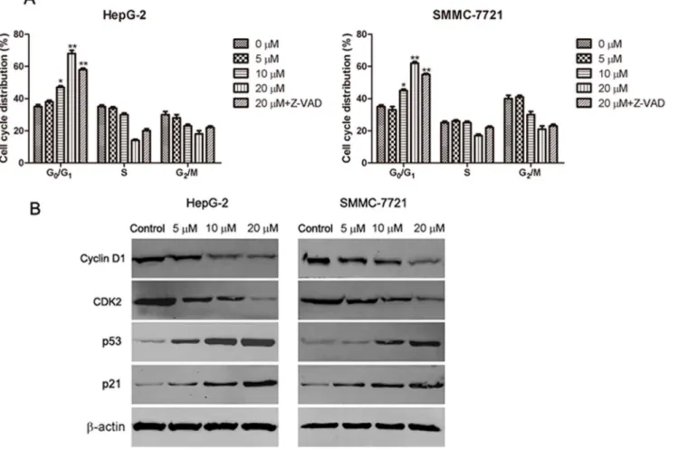
Cell cycle arrest is one of the major causes of cell death. To explore whether abieslactone-induced apoptosis was associated with cell cycle arrest, we examined cell cycle distribution in HepG2 and SMMC7721 cells using flow cytometry to analyze the DNA content in each cell cycle phase. As shown in Fig. 4A,
abieslactone treatment induced a dose-dependent increase in the proportion of cells in the G1phase and decrease in cells in the S and G2phases compared to the
untreated control. Furthermore, we used a general caspase inhibitor Z-VAD-FMK for the cell cycle analyses in HepG2 and SMMC7721 cells. Flow cytometric analysis showed that Z-VAD-FMK treatment did not prevent cell cycle arrest after high-dose abieslactone treatment (Fig. 4A), although abieslactone induced HepG2 and SMMC7721 cell apoptosis. The data indicate that abieslactone
induced HepG2 and SMMC7721 cell death through cell cycle arrest in G1 phase
and by the induction of apoptosis.
Abieslactone induces the expression of cell cycle regulators
Having established that abieslactone induce cell-cycle arrest, we attempted to characterize, at the molecular level, the mechanisms by which this effect is achieved. The tumor suppressor protein, p53, regulates the cell cycle, and its target gene, p21, directly inhibits cyclin D1 and CDK2. This pathway results in cell cycle arrest in the G1phase. To investigate the mechanism of abieslactone-induced cellcycle arrest in HepG2 and SMMC7721 cells, we analyzed p53, p21 cyclin D1 and CDK2 expression by western blotting. As illustrated in Fig. 4B, the expression levels of p53 and p21 were markedly increased, whereas cyclin D1 and CDK2 expression were significantly decreased in a dose dependent manner. These results revealed that abieslactone induced cell death in HepG2 and SMMC7721 cells through p53 activation, leading to cell cycle arrest in G1 phase.
Fig. 3. Nuclear morphology of HepG2 and SMMC7721 cells treated with 5, 10, and 20mM abieslactone
or 20mM Z-VAD-FMK for 24 h was determined by staining with Hoechst 33258.
Abieslactone induces apoptosis in the mitochondria
Another characteristic feature of apoptosis is depolarization of the mitochondrial membrane potential (MMP). To investigate whether abieslactone-induced cell apoptosis was associated with mitochondrial dysfunction, we analyzed MMP changes in HepG2 and SMMC7721 cells by staining with Rh123, a mitochondria-sensitive dye, and analyzing the cells by flow cytometry. Our results show that the MMP of HepG2 and SMMC7721 cells decreased significantly after treatment with abieslactone in a dose dependent manner (Fig. 5), suggesting that abieslactone induced cell apoptosis through the intrinsic pathway.
Fig. 4. The effect of abieslactone on the cell cycle and the expression of cell cycle regulators in HepG2 and SMMC7721 cells.(A) Abieslactone treatment induced a dose dependent increase in the proportion of cells in the G1phase and a decrease in cells in the S and G2phases compared to the untreated control. Z-VAD-FMK treatment did not prevent cell cycle arrest following high-dose abieslactone treatment. The results are represented as the mean¡SEM for three independent experiments with similar results. *P,0.05; **P,0.01vs. the untreated control. (B) Representative pictures for p53, p21 CDK2 and cyclin D1 protein expression by western blot analysis.b-actin was used as a control.
Abieslactone induces cell apoptosis through a mitochondrial
pathway
Mitochondrial damage facilitates cytochrome c release from mitochondria into the cytoplasm and activates apoptotic factors (Bcl-2 family proteins), which leads to activation of the caspase cascade (apoptotic markers) and mitochondria-mediated apoptosis [28]. Activation of caspase cascade leading to PARP cleavage is regarded as a major pathway in apoptosis induction. To test whether
abieslactone induces apoptosis through this mechanism in HepG2 and
Fig. 5. The effect of abieslactone on the MMP in HepG2 and SMMC7721 cells.(A) The MMP of HepG2 and SMMC7721 cells treated with abieslactone at different concentrations was analyzed by flow cytometry. (B) The loss of the MMP in HepG2 and SMMC7721 cells following abieslactone treatment in a dose dependent manner. The data are expressed as the means¡SEM for three independent experiments with similar results. *P,0.05vs. the untreated control.
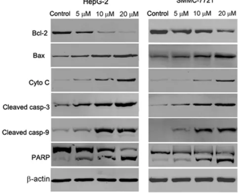
SMMC7721 cells, we examined the expression of cytochrome c, the pro-apoptotic protein Bax, the anti-apoptotic protein Bcl-2, caspase 3, caspase 9 and PARP by western blot analysis. In a dose dependent manner, abieslactone increased cytosolic cytochrome c, Bax, cleaved caspase 3 and cleaved caspase 9 expressions with a concomitant decrease in Bcl-2 expression compared to the untreated
control (Fig. 6). Meanwhile, exposure of HepG2 and SMMC7721 cells to
abieslactone also resulted in the cleavage of PARP fragment (Fig. 6), which is an endogenous substrate of activated caspase-3 and its cleavage is considered to be a hallmark of cell apoptosis. These results suggested that the mitochondria and Bcl-2 family members are involved in abieslactone-mediated cell apoptosis in HepGBcl-2 and SMMC7721 cells.
ROS/Akt pathway is involved in the abieslactone-induced cell
apoptosis in HepG2 cells but not in SMMC7721 cells
As ROS generation is important in mitochondria-mediated apoptosis, the effect of abieslactone on the generation of ROS was investigated. HepG2 and SMMC7721 cells were exposed to abieslactone at different concentrations for 24 h and analyzed for the accumulation of ROS by fluorescence microscopy following
staining with DCFH-DA. As shown in Fig. 7A, treatment with abieslactone
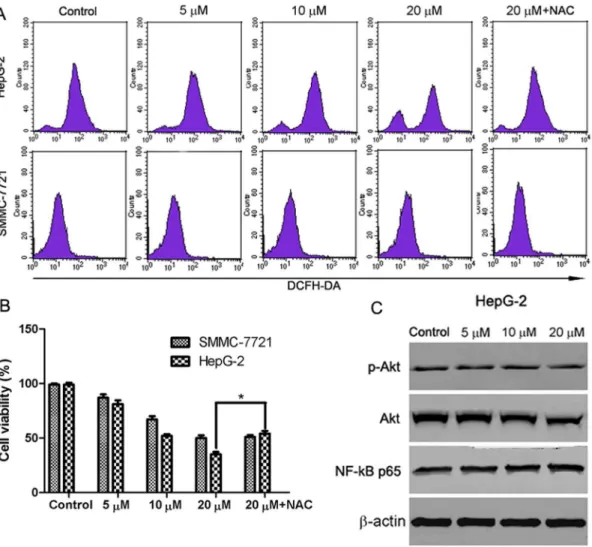
resulted in a significant increase in intracellular ROS in HepG2 cells. However, no significant increase in ROS levels was observed in SMMC7721 cells after 24 h of abieslactone treatment. Furthermore, we pretreated HepG2 cells with the ROS
scavenger NAC at 10 mM for 1 h followed by abieslactone (20 mM) treatment for
additional 24 h. The viability of HepG2 cells was partly rescued by ROS scavenger NAC (Fig. 7B). These results indicate that abieslactone may induce cell apoptosis by the generation of ROS in HepG2 cells.
Recent studies have demonstrated that downregulation of Akt and NF-kB activity is involved in mitochondria-mediated apoptosis and Akt and NF-kB signals are the downstream event of ROS generation in mitochondria-dependent apoptosis [20,21]. We further investigated the effects of abieslactone on the expression of Akt and the p65/RelA subunit of NF-kB in HepG2 cells, which is necessary for the transactivation activity of NF-kB. Our results showed that abieslactone inhibited Akt activity in HepG2 cells; not only phosphorylated levels of Akt were reduced, but total Akt levels were decreased after abieslactone treatment for 24 h (Fig. 7C). However, abieslactone applied to HepG2 cells for 24 h did not change the expression of p65/RelA protein levels (Fig. 7C). Based on the results described above, abieslactone may induce HepG2 cell apoptosis viathe ROS/Akt pathway.
Discussion
clinical improvement is marginal [27]. In addition, severe toxicities and drug resistance often occur, hindering the effective application of these agents. Considerable attention has been focused on identifying naturally occurring bioactive compounds and their derivatives capable of inhibiting or reversing the development of liver cancer. In the liver, the elimination of transformed cells via apoptosis induction is considered to be a crucial step for the treatment of liver cancer. We have been interested in discoverying new, effective, and safe drugs from natural products for cancer therapy. Previous studies have indicated that the
Abiesplants have ideal natural compounds for targeted treatment of many cancer cell lines [29–32]. Although some studies have demonstrated in vitro antitumor activity of triterpenoid lactones from Abies plants in various human tumor cell lines, their underlying mechanisms of action remain to be elucidated.
Abieslactone is a triterpenoid lactone isolated fromA. faxoniana, a folk medicine used to treat bronchitis, digestive disorders, and inflammation. Our data
demonstrated that abieslactone inhibited HepG2 and SMMC7721 cell growth in a dose dependent manner. Interestingly, we found that the growth-inhibiting effect of abieslactone was tumor cell selective, as normal hepatic cell line (QSG7701) did not display a significant toxic effect in vitro.
Cell cycle and apoptosis are considered to be two major regulatory mechanisms for cell growth. When specific checkpoints during the cell cycle are arrested,
Fig. 6. The effect of abieslactone on the expression of caspase-dependent mitochondrial apoptosis pathway proteins in HepG2 and SMMC7721 cells.Representative images of cytochrome c, Bax, Bcl-2, PARP, cleaved caspase 9 and cleaved caspase 3 protein expression detected by western blot.b-actin was used as a control.
apoptotic cell death occurs [33]. Moreover, many chemotherapeutic agents cause cell cycle arrest through microtubule damage and have been proven to be clinically effective for treating cancer [34]. Flow cytometric studies showed that the growth inhibition induced by abieslactone occurs through the cell cycle arrest of HepG2 and SMMC7721 cells in the G1phase. The G1to S cell cycle progression
is controlled by several cyclin-dependent kinase (CDK) complexes, the activities of which are dependent on the balance of cyclins and cyclin-dependent kinase inhibitors (CKIs). p53, the most extensively studied tumor suppressor, mediates a variety of anti-proliferative processes through cell cycle checkpoints, DNA repair and apoptosis [35]. Previous reports have found that p21, the target of p53, is one of the major CKIs, which directly inhibit the activity of CDKs, thereby leading to cell cycle arrest in the G phase [36–38]. Upregulation of p21 and p53 expression
Fig. 7. The involvement of ROS/Akt pathway in abieslactone-induced HepG2 cell apoptosis.(A) HepG2 and SMMC7721 cells were treated with different concentrations of abieslactone or ROS scavenger (NAC 10 mM), and then ROS was measured by DCF fluorescence analysis. The increased fluorescence of DCF was determined as the increased intracellular ROS accumulation. (B) HepG2 and SMMC7721 cells pretreated with 10 mM NAC for 1 h were incubated with 20mM abieslactone for 24 h, and cell viability was determined by MTT assay. *P,0.05vs. the untreated control. (C) The effect of abieslactone on the expression of p-Akt, Akt and NF-kB p65 proteins in HepG2 cells.b-actin was used as a control.
may inhibit cyclin/CDK complexes, thus leading to cell G1 cycle arrest. To gain
insight into the molecular mechanisms of abieslactone-induced G1 arrest in
HepG2 and SMMC7721 cells, we examined p53, p21 cyclin D and CDK2 expression. Western blot analysis revealed that abieslactone treatment down-regulated CDK2 and cyclin D expression and reversed the reduction in p53 and p21 expression, which enhances the formation of heterotrimeric complexes with the CDKs and cyclins, thereby leading to cell cycle arrest in the G1 phase. The
results indicated that one of the mechanisms of abieslactone in the suppression of
HepG2 and SMMC7721 cell growth may be the inhibition of cell G1 cycle
progression through a p53-dependent pathway.
G1phase arrest of cell cycle regulation provides an opportunity for cells to
follow the apoptotic pathway. Although it was shown that abieslactone suppresses
HepG2 and SMMC7721 cell growth through the induction of G1phase arrest, we
cannot be sure that they are the only factors reducing cell growth. To determine whether abieslactone-induced inhibition of HepG2 and SMMC7721 cell growth is dependent on apoptosis, we performed flow cytometric analysis of apoptosis after treating with abieslactone. The data showed that abieslactone induced HepG2 and SMMC7721 cell apoptosis in a dose dependent manner. Furthermore, after preincubation with Z-VAD-FMK, apoptosis was greatly attenuated, indicating that apoptosis occurs via a caspase-dependent pathway. Moreover, we also found that inhibition of caspase activation did not prevent cell cycle arrest, suggesting that abieslactone inhibited HepG2 and SMMC7721 cell growth through either cell cycle arrest or apoptosis induction.
involved in abieslactone-induced apoptosis. Therefore, we concluded that abieslactone induced caspase-dependent apoptosis via the mitochondrial pathway.
ROS generation is also an important mediator of many anti-cancer agents. Previous studies have shown that ROS produced by chemotherapy are essential for inducing apoptosis in some kinds of cancer [40]. ROS, which were
predominantly produced in the mitochondria, if excessive, may lead to the free radical attack of membrane phospholipids and loss of MMP, which causes the release of apoptosis-inducing factors that activate caspase cascades and cause nuclear condensation [41]. ROS generation is viewed as one of the main
mechanisms of mitochondria-dependent apoptosis [42]. The results showed that the accumulation of ROS was detected in HepG2 cells when treated with
abieslactone. However, no significant increase in ROS levels was observed in SMMC7721 cells after abieslactone treatment. NAC is a potent antioxidant that may inhibit oxidative stress by directly scavenging ROS. To identify the role of ROS in abieslactone-induced apoptosis in HepG2 cells, cell death was measured following treatment of abieslactone with NAC. Abieslactone (20 mM) significantly increased HepG2 cell death, whereas removing intracellular ROS by NAC partly inhibited induced cell death. These results revealed that abieslactone-induced ROS accumulation was involved in HepG2 cell apoptosis.
Akt is a critical kinase involved in cell signal transduction cascades promoting cell survival by inhibiting apoptosis through its ability to phosphorylate and inactivate several proapoptotic proteins [43]. It has been reported that some clinical chemotherapeutic agents could induce cancer cell apoptosis by regulating Akt signal pathway [44]. NF-kB, a pro-inflammatory nuclear transcription factor, regulates genes important for tumor invasion, metastasis, angiogenesis, and chemoresistance [45]. Exposure of cancer cells to anticancer drugs can induce the activation of the NF-kB pathway, leading to the expression of anti-apoptotic genesand resistance to apoptosis. Recent studies have shown that downregulation of Akt and NF-kB activity is involved in mitochondria-mediated apoptosis, and Akt and NF-kB signals appear to be a downstream regulator of ROS generation. To determine if abieslactone-induced apoptosis was mediated by regulating Akt and NF-kB activity, we treated HepG2 cells with increasing concentrations of abieslactone. Our results show that abieslactone inhibited total and phosphory-lated Akt activities. However, abieslactone applied to HepG2 cells did not change p65/RelA protein levels. Thus, we concluded that abieslactone induced HepG2 cell apoptosis via the ROS/Akt pathway.
In conclusion, our present studies show that abieslactone exerted an inhibitory effect on hepatoma cells by arresting the cell cycle in the G1phase and inducing
apoptosis (Fig. 8). G1 phase arrest was found to be associated with the
Author Contributions
Conceived and designed the experiments: GWW CL YHS LS WDZ RHL. Performed the experiments: GWW CL ZRS RTZ XYD. Analyzed the data: GWW CL. Contributed reagents/materials/analysis tools: GWW CL. Wrote the paper: GWW CL.
References
1. Llovet JM, Burroughs A, Bruix J(2003) Hepatocellular carcinoma. Lancet 362: 1907–1917.
2. Breitenstein S, Apestegui C, Petrowsky H, Clavien PA(2009) ‘‘State of the art’’ in liver resection and living donor liver transplantation: a worldwide survey of 100 liver centers. World J Surg 33: 797–803.
3. Zender L, Spector MS, Xue W, Flemming P, Cordon-Cardo C, et al. (2006) Identification and validation of oncogenes in liver cancer using an integrative oncogenomic approach. Cell 125: 1253– 1267.
4. Ghobrial IM, Witzig TE, Adjei AA (2005) Targeting apoptosis pathways in cancer therapy. CA Cancer J Clin 55: 178–194.
5. Reed JC(2001) Apoptosis-regulating proteins as targets for drug discovery. Trends Mol Med 7: 314– 319.
6. Budihardjo I, Oliver H, Lutter M, Luo X, Wang X(1999) Biochemical pathways of caspase activation during apoptosis. Annu Rev Cell Dev Biol 15: 269–290.
7. Zhang Y, Bao YL, Wu Y, Yu CL, Huang YX, et al.(2013) Alantolactone induces apoptosis in RKO cells through the generation of reactive oxygen species and the mitochondrial pathway. Mol Med Rep 8: 967– 972.
8. Walensky LD, Gavathiotis E (2011) BAX unleashed: the biochemical transformation of an inactive cytosolic monomer into a toxic mitochondrial pore. Trends Biochem Sci 36: 642–652.
9. Czabotar PE, Colman PM, Huang DC(2009) Bax activation by Bim? Cell Death Differ 16: 1187–1191. Fig. 8. Schematic form of the proposed mechanisms for abieslactone-induced G1arrest and apoptosis in HepG2 cells.
10. Dejean LM, Martinez-Caballero S, Guo L, Hughes C, Teijido O, et al.(2005) Oligomeric Bax is a component of the putative cytochrome c release channel MAC, mitochondrial apoptosis-induced channel. Mol Biol Cell 16: 2424–2432.
11. Chatterjee S, Kundu S, Bhattacharyya A(2008) Mechanism of cadmium induced apoptosis in the immunocyte. Toxicol Lett 177: 83–89.
12. Pathak N, Khandelwal S (2007) Role of oxidative stress and apoptosis in cadmium induced thymic atrophy and splenomegaly in mice. Toxicol Lett 169: 95–108.
13. Trachootham D, Alexandre J, Huang P(2009) Targeting cancer cells by ROS mediated mechanisms: a radical therapeutic approach? Nat Rev Drug Discov 8: 579–591.
14. Deavall DG, Martin EA, Horner JM, Roberts R (2012) Drug-induced oxidative stress and toxicity. J Toxicol 2012: 645460.
15. Martindale JL, Holbrook NJ (2002) Cellular response to oxidative stress: signaling for suicide and survival. J Cell Physiol 192: 1–15.
16. D’Alessio M, De Nicola M, Coppola S, Gualandi G, Pugliese L, et al. (2005) Oxidative Bax dimerization promotes its translocation to mitochondria independently of apoptosis. FASEB J 19: 1504– 1506.
17. Oh SH, Lee BH, Lim SC (2004) Cadmium induces apoptotic cell death in WI 38 cells via caspase-dependent Bid cleavage and calpain-mediated mitochondrial Bax cleavage by Bcl-2-incaspase-dependent pathway. Biochem Pharmacol 68: 1845–1855.
18. Jungas T, Motta I, Duffieux F, Fanen P, Stoven V, et al.(2002) Glutathione levels and BAX activation during apoptosis due to oxidative stress in cells expressing wild-type and mutant cystic fibrosis transmembrane conductance regulator. J Biol Chem 277: 27912–27918.
19. Hanawa N, Shinohara M, Saberi B, Gaarde WA, Han D, et al.(2008) Role of JNK translocation to mitochondria leading to inhibition of mitochondria bioenergetics in acetaminophen-induced liver injury. J Biol Chem 283: 13565–13577.
20. Gong K, Li W (2011) Shikonin, a Chinese plant-derived naphthoquinone, induces apoptosis in hepatocellular carcinoma cells through reactive oxygen species: A potential new treatment for hepatocellular carcinoma. Free Radic Biol Med 51: 2259–2271.
21. Liu C, Gong K, Mao X, Li W(2011) Tetrandrine induces apoptosis by activating reactive oxygen species and repressing Akt activity in human hepatocellular carcinoma. Int J Cancer 129: 1519–1531.
22. Holmes FA, Walters RS, Theriault RL, Forman AD, Newton LK, et al.(1991) Phase II trial of taxol, an active drug in the treatment of metastatic breast cancer. J Natl Cancer Inst 83: 1797–1805.
23. Kawamori T, Lubet R, Steele VE, Kelloff GJ, Kaskey RB, et al.(1999) Chemopreventive effect of curcumin, a naturally occurring anti-inflammatory agent, during the promotion/progression stages of colon cancer. Cancer Res 59: 597–601.
24. Dong Y, Yang MM, Kwan CY, Dong Y, Yang MM, et al.(1997) In vitro inhibition of proliferation of HL-60 cells by tetrandrine and Coriolus versicolor peptide derived from Chinese medicinal herbs. Life Sci 60: 135–140.
25. Matsunaga S, Okada J, Uyeo S(1965) The structure of abieslactone, a methoxy-tetracyclic triterpene lactone. Chem Commun 21: 525–527.
26. Takayasu J, Tanaka R, Matsunaga S, Ueyama H, Tokuda H, et al.(1990) Anti-tumor-promoting activity of derivatives of abieslactone, a natural triterpenoid isolated from severalAbiesgenus. Cancer Lett 53: 141–144.
27. Li X, Yang X, Liu Y, Gong N, Yao W, et al.(2013) Japonicone A suppresses growth of burkitt lymphoma cells through its effect on NF-kB. Clin Cancer Res 19: 2917–2928.
28. Wu M, Zhang H, Hu J, Weng Z, Li C, et al.(2013) Isoalantolactone inhibits UM-SCC-10A cell growth via cell cycle arrest and apoptosis induction. Plos One 8: e76000.
29. Li YL, Gao YX, Yang XW, Jin HZ, Ye J, et al. (2012) Cytotoxic triterpenoids fromAbies recurvata. Phytochemistry 81: 159–164.
31. Lavoie S, Legault J, Gauthier C, Mshvildadze V, Mercier S, et al.(2012) Abibalsamins A and B, two new tetraterpenoids fromAbies balsamea. Org Lett 14: 1504–1507.
32. Li YL, Zhang SD, Jin HZ, Tian JM, Shen YH, et al. (2012) Abiestetranes A and B, two unique tetraterpenes fromAbies fabri. Tetrahedron 68: 7763–7767.
33. Orren DK, Petersen LN, Bohr VA (1997) Persistent DNA damage inhibits S phase and G 2 progression, and results in apoptosis. Mol Biol Cell 8: 1129–1142.
34. Shapiro GI, Harper JW(1999) Anticancer drug targets: cell cycle and checkpoint control. J Clin Invest 104: 1645–1653.
35. Fridman JS, Lowe SW(2003) Control of apoptosis by p53. Oncogene 22: 9030–9040.
36. Zhang Z, He H, Chen F, Huang C, Shi X(2002) MAPKs mediate S phase arrest induced by vanadate through a p53-dependent pathway in mouse epidermal C141 cells. Chem Res Toxicol 15: 950–956.
37. Gartel AL, Tyner AL(2002) The role of the cyclin-dependent kinase inhibitor p21 in apoptosis. Mol Cancer Ther 8: 639–649.
38. Ogryzko VV, Wong P, Howard BH(1997) WAF1 retards S-phase progression primarily by inhibition of cyclin-dependent kinases. Mol Cell Biol 17: 4877–4882.
39. Yin XM(2000) Signal transduction mediated by Bid, a pro-death Bcl-2 family proteins, connects the death receptor and mitochondria apoptosis pathways. Cell Res 10: 161–167.
40. Simon HU, Haj-Yehia A, Levi-Schaffer F(2000) Role of reactive oxygen species (ROS) in apoptosis induction. Apoptosis 5: 415–418.
41. Zamzami N, Marchetti P, Castedo M, Decaudin D, Macho A, et al.(1995) Sequential reduction of mitochondrial transmembrane potential and generation of reactive oxygen species in early programmed cell death. J Exp Med 182: 367–377.
42. Zafarullah M, Li WQ, Sylvester J, Ahmad M(2003) Molecular mechanisms of N-acetylcysteine actions. Cell Mol Life Sci 60: 6–20.
43. Sun ZJ, Chen G, Hu X, Zhang W, Liu Y, et al.(2010) Activation of PI3K/Akt/IKK-alpha/NF-kappaB signaling pathway is required for the apoptosis-evasion in human salivary adenoid cystic carcinoma: its inhibition by quercetin. Apoptosis 15: 850–863.
44. Vivanco I, Sawyers CL(2002) The phosphatidylinositol 3-kinase AKT pathway in human cancer. Nat Rev Cancer 2: 489–501.


