America,5Department of Marine Biotechnology and Resources, National Sun Yat-Sen University, Kaohsiung, Taiwan
Abstract
Lipids are a source of metabolic energy, as well as essential components of cellular membranes. Although they have been shown to be key players in the regulation of cell proliferation in various eukaryotes, including microalgae, their role in the cell cycle of cnidarian-dinoflagellate (genusSymbiodinium) endosymbioses remains to be elucidated. The present study examined the effects of a lipid synthesis inhibitor, cerulenin, on the cell cycle of both culturedSymbiodinium(clade B) and those engaged in an endosymbiotic association with the sea anemoneAiptasia pulchella. In the former, cerulenin exposure was found to inhibit free fatty acid (FFA) synthesis, as it does in other organisms. Additionally, while it also significantly inhibited the synthesis of phosphatidylethanolamine (PE), it did not affect the production of sterol ester (SE) or phosphatidylcholine (PC). Interestingly, cerulenin also significantly retarded cell division by arresting the cell cycles at the G0/G1phase. Cerulenin-treatedSymbiodiniumwere found to be taken up by anemone hosts at a significantly depressed quantity in comparison with controlSymbiodinium. Furthermore, the uptake of cerulenin-treated Symbiodinium in host tentacles occurred much more slowly than in untreated controls. These results indicate that FFA and PE may play critical roles in the recognition, proliferation, and ultimately the success of endosymbiosis with anemones.
Citation:Wang L-H, Lee H-H, Fang L-S, Mayfield AB, Chen C-S (2013) Fatty Acid and Phospholipid Syntheses Are Prerequisites for the Cell Cycle ofSymbiodinium
and Their Endosymbiosis within Sea Anemones. PLoS ONE 8(8): e72486. doi:10.1371/journal.pone.0072486
Editor:Eric Cascales, Centre National de la Recherche Scientifique, Aix-Marseille Universite´, France ReceivedMarch 14, 2013;AcceptedJuly 10, 2013;PublishedAugust 29, 2013
Copyright:ß2013 Wang et al. This is an open-access article distributed under the terms of the Creative Commons Attribution License, which permits unrestricted use, distribution, and reproduction in any medium, provided the original author and source are credited.
Funding:This study was supported by grants from the National Museum of Marine Biology and Aquarium (981001092 and 99200312) and National Science Council (NSC 101-2311-B-291-002-MY3). ABM was supported by a postdoctoral research fellowship from the Living Oceans Foundation. The funders had no role in study design, data collection and analysis, decision to publish, or preparation of the manuscript.
Competing Interests:The authors have declared that no competing interests exist.
* E-mail: cchen@nmmba.gov.tw
Introduction
Lipids are important components of all living organisms, as they are a source of metabolic energy and serve as essential components of cellular membranes. They are also involved in processes such as cell proliferation, cell differentiation, and organ morphogenesis, which are all intimately associated with the progression of the cell cycle [1]. For instance, the concentration of phospholipids in the photosynthetic bacterium Rhodopseudomonas sphaeroidsdoubles be-fore cell division [2]. Polar glycerolipids (glycolipid, phosolipid, and ether lipid) are synthesized sequentially during the cell cycle of
Chlamydomonas reinhardtii [3]. In the heterotropic dinoflagellate,
Crypthecodinium cohnii, cells exhibit a stepwise increase in polar lipids and a continuous increase in neutral lipids over the course of the cell cycle [4]. The same study showed that inhibiting lipid synthesis caused cell cycle arrest at the early G1phase, not the G2/ M phase, demonstrating the essential role of lipid synthesis in regulating cell cycle progression.
The Cnidaria-Symbiodinium association is an endosymbiosis in which the dinoflagellate symbionts reside within the anthozoan host’s gastrodermal cells and contribute to the latter’s nutrition by translocating photosynthetically fixed carbon and other metabo-lites into the host cytoplasm [5]. Among the photosynthates produced by the symbionts, lipids and their roles in regulating the
association with hosts are being subjects for intensive studies [6– 11]. Whether all lipid synthates of symbionts would be transferred to the host remains to be determined, as a recent study of lipidomic examination has shown that the free fatty acids of symbionts did not translocate to hosts [10]. Nonetheless, elevated temperature-induced bleaching events in which Symbiodinium
photosynthesis was impaired, led to reduced productions of neutral lipids such as triacylglycerols (TAGs) and wax ester (WE) in bothPorites compressaand Montipora verrucosa [7]. Furthermore, the formation of lipid bodies (LBs) in the gastrodermal cells of
Euphyllia glabrescenshas been shown to depend upon the presence of symbionts [12]. Although the cellular mechanism of LB formation remains to be elucidated, concentrations of three major lipids of LBs (TAGs, WE and sterol) are regulated by the diel cycle and photosynthesis [13]. The depletion or alter of these lipid energy reserves also increase susceptibility to diseases and mortality, possibly through an increase in microbes in the damaged tissue [14,15]. It is thus conceivable that the role of lipid synthesis of symbionts during the symbiosis could be pivotal and remains to be elucidated.
phase. Blue light (450610 nm) alone mimics regular white light, while red (660610 nm) and infrared (735610 nm) lights have little or no effect on the cell cycle [16]. In hospite, the picture becomes more complicated, as it has been suggested that
Symbiodiniumcell proliferation may be under the regulation of host cells. For example, the frequency of Symbiodinium division in fed anemones is two times higher than in starved specimens [17]. Moreover, Symbiodinium densities vary little in healthy symbioses [18,19], possibly because newly generated Symbiodinium cells are released into the host gastrovascular cavity [20].
However, lipids may also play a role in regulation of the cell cycle in stable endosymbiotic associations. In order to gain more insight into this notion, cultured Symbiodinium (clade B) were treated with 2,3-epoxy-4-oxo-7,10-dodecadienamide (cerulenin), a fungal metabolite [21,22] that specifically inhibits three types of fatty acid synthases in various eukaryotes [23]. It was hypothesized that treatment with cerulenin might alter the lipid synthesis and cell cycle progression of culturedSymbiodinium. Our current study also focused on the ability of cerulenin-treated Symbiodinium to infect sea anemones, aiming to elucidate the role of lipid synthesis in the establishment of anemone-dinoflagellate endosymbiosis.
Materials and Methods
Symbiodiniumculture and identification
Symbiodiniumcells isolated fromA. pulchellawere cultured at 25uC at 40mmol m22s21over a 12 h light/12 h dark (12L/12D) cycle, and f/2 medium (Sigma, USA) containing antibiotics (10mg ml21 streptomycin and 10 units ml21penicillin; Invitrogen, USA) was replenished every 5 days. Restriction fragment length polymor-phism (RFLP) analysis of theSymbiodinium18S rDNA was used to identify the clade of the cultured Symbiodinium following a published procedure [24]. The RFLP genotype pattern 885/505 bp forTaq I (New England Biolabs, USA) and 765/500 bp for
Sau3AI (New England Biolabs, USA) confirmed that the cultured
Symbiodiniumwere of clade B.
Flow cytometry analyses of cell cycle progression The cell cycle progression was determined by flow cytometry according to a published procedure [16]. Briefly, three cultures maintained at the exponential growth stage (56104 cells ml21) were sampled for analyses. 106cells were collected at the times indicated in Fig. 1. Cells were harvested by centrifugation at 686g for 5 min and fixed in ice-cold 70% ethanol for 1 h at 18mmol m22s21to extract the chlorophyll. Cells were then incubated in phosphate-buffered saline (PBS) containing Triton-X 100 (0.1%, Pharmacia Biotech, Sweden), RNase (10mg ml21, Sigma, USA), and propidium iodide (PI; 30mg ml21, Invitrogen, USA) at 4uC overnight in the dark. The DNA content per cell calculated from the DNA–PI complex fluorescence under 488/610 nm (Excita-tion/Emission) with an EPICS ALTRATM flow cytometer (Beckman Coulter, Inc., USA). Histograms of relative DNA content were analyzed using MultiCycle AV for Windows V5.0 (Phoenix Flow Systems, CA) in order to quantify the percentage of cells at each stage: G1, S, and G2/M.
Cerulenin treatments
Cerulenin (Sigma, USA), a fatty acid synthesis inhibitor, was used to modulate the lipid synthesis in culturedSymbiodinium, in order to examine the role of lipid synthesis in cell cycle progression. To determine the optimal concentration required to elicit a biological response, cerulenin (in DMSO) was added to
Symbiodiniumcultures to final concentrations of 0, 1027 , 1026
, or 1025
M (final DMSO concentration: 0.02%) at T00 (time zero before the light on, see Fig. 1). The effect of cerulenin on cell cycle propagation of treatedSymbiodinium during a 12L/12D photope-riod was then analyzed by flow cytometry as described in the preceding section, with three biological replicates sampled at T00, as well as after 5 (T05), 11, (T11), 17 (T17), 23 (T23), and 29 (T29) h. Lipid analyses were assessed in a separate aliquot from the same cultures of each of the four concentrations of cerulenin at T00, T05, T11, and T17.
Figure 1. Sampling time forSymbiodiniumsp. during a light–dark photoperiod. doi:10.1371/journal.pone.0072486.g001
Table 1.Gradient elution program for HPLC separation of lipids.
Time (min) Flow rate
Solvents (ml/min)
A (%) B (%) C (%)
0 100 0 0 1.0
4 100 0 0 1.0
5 85 15 0 1.0
10 80 20 0 1.0
12 75 25 0 1.0
15 50 50 0 1.0
18 30 50 20 1.0
20 30 40 30 1.0
25 25 30 45 1.0
30 30 70 0 1.0
40 100 0 0 1.0
solvent A: isohexane.
solvent B: propan-2-ol: acetonitrile:butan-2-one (butan-2-one or 2-butanone) (7:2:1, v/v/v).
solvent C: propan-2-ol:acetonitrile: butan-2-one: methanol:water:N -ethylmorpholine: acetic acid = 56:14:7.2:14:8.4:0.42:0.15 v/v/v/v/v/v/v. doi:10.1371/journal.pone.0072486.t001
Analysis of lipids and starch
Cells (107) from each treatment were collected at specific times after centrifugation (686g for 5 min), and the pellets were stored at 280uC. Before assays, cells were re-suspended in PBS and homogenized using a ball mill (Retsch MM301, USA) with glass beads (425–600mm, Sigma, USA). Each cell homogenate was quantified for total protein using Pierce BCA Protein Assay Kit (Thermo Scientific, USA) and then cell homogenate containing exactly twenty-five microgram total protein was used to assay the lipid profiles and starch contents.
Lipids were extracted by methyl tert-butyl ether (MTBE; Merck, Germany) according to Matyash et al. [25]. Methanol (1.5 ml) was added to a 200ml sample aliquot, and the tube was vortexed. Then, 5 ml of MTBE was added and the mixture was incubated for 1 h at room temperature in a shaker. The phase separation was induced by adding 1.25 ml of deionized water. Upon 10 min of incubation at room temperature, the sample was then centrifuged at 1,0006g for 10 min. The upper organic phase was collected; and the lower phase was re-extracted with 2 ml of MTBE/methanol/water (10:3:2.5, v/v/v) to collect the upper Figure 2. HPLC profiles of standard lipids and extracted lipids from control and cerulenin-treatedSymbiodiniumat T11.WE, wax ester; SE, sterol ester; TAG, triacylglyceride; FFA, free fatty acid; PE, phophatidylethanolamine; PC, phosphatidylcholine; X1, unknown peak 1; X2, unknown peak 2.
organic phase. The collections of organic phases containing extracted lipids were combined and then dried by Rotary Evaporator (Panchum Scientific Corp, Taiwan).
The extracted lipids were analyzed by a high-performance liquid chromatography equipped with evaporative light scattering detector (HPLC-ELSD) [26]. A Hitachi Model L7100 HPLC pump, equipped with an auto-sampler (L7200, Hitachi, Japan), Figure 3. Dose-dependent response of cerulenin on theSymbiodiniumcell cycle.Cell cycle analyses ofSymbiodiniumsp. were performed across a 12L/12D treatment in the absence (0 M) or presence of cerulenin (1027M, 1026M, and 1025M). (panel a). The percentage of cells at each
stage of the cell cycle. (panel b). Values were expressed as mean6SEM. Letters (a–b and a’ –b’) denote the statistical significance of different cerulenin concentrations according to one-way ANOVA followed by Duncan’s multiple-range procedure (p,0.05).
doi:10.1371/journal.pone.0072486.g003
was used with a Sedex 80 evaporative light-scattering detector (Sedere, France). The drift-tube temperature was maintained at 55uC, and the flow-rate of the nebulizer gas (nitrogen) was 2.5 kg/
cm2. The detector response was quantified by electronic integra-tion. Solvents were de-aerated with nitrogen gas. A column of YMC-PVA-SIL (10063 mm i.d.; 5 mm particles) was obtained Figure 4. Effect of cerulenin on different stages of theSymbiodiniumcell cycle.Cell cycle analyses ofSymbiodiniumsp. were performed across a 12L/12D photoperiod with cerulenin (1026M) added at different times (panel a). The percentage of cells at each stage of the cell cycle are
shown with bar graphs. (panel b). The percentage of cells at the G2/M phase at the five treatments across five sampling times. Values were expressed
as mean6SEM. Letters (a–c and a’–c’) denote the statistical significance of different time points according to one-way ANOVA followed by Duncan’s multiple-range procedure (p,0.05). F = 1.889,p= 0.187 at T05; F = 3.444,p= 0.051 at T11; F = 182.065,p,0.001 at T17; F = 192.214,p,0.001 at T23; F = 57.748,p,0.001 at T29).
from Hichrom Ltd. (Reading, UK). This experiment required a ternary gradient elution scheme consisting of isohexane (Merck, Germany) (solvent A), propan-2-ol (Merck, Germany): acetonitrile (Merck, Germany): butan-2-one (butan-2-one or 2-butanone) (Merck, Germany) (7:2:1, v/v/v; solvent B), and propan-2-ol:acetonitrile: butan-2-one: methanol (Merck, Germany): water: N-ethylmorpholine (Sigma, USA): acetic acid (Merck, Germany) (56:14:7.2:14:8.4:0.42:0.15 v/v; solvent C), with the gradient elution program described as in Table 1.
As shown in the upper panel of Fig. 2, six major lipid standards (from Sigma, USA and Lipid Products, England) could be clearly separated and analyzed by the current HPLC analysis, including wax ester (WE: arachidyl dodecanoate; RT = 0.9 min), sterol ester (SE: cholosteryl oleate; RT = 1.4 min), triacylglycerol (TAG: trilaurin; RT = 2.9 min), free fatty acid (FFA: linoleic acid; RT = 8.9 min), phophatidylethanolamine (PE; RT = 22.1 min), and phosphatidylcholine (PC: lecithin; RT = 26.8 min). In comparison with the lipid standards, HPLC analyses of lipid extracts from both Figure 5. Comparison of lipid and starch content in control and cerulenin-treatedSymbiodiniumat different cell cycle stages.(a) SE; (b) FFA; (c) PE; (d) PC; (e) starch. Values were expressed as mean6SEM. Letters (a–c and a’–c’) denote the statistical significance of different time points according to one-way ANOVA followed by Duncan’s multiple-range tests (p,0.05). * denotes the statistical significance of cerulenin treatment comparing to control at the same time point according student’s t-test (p,0.05).
doi:10.1371/journal.pone.0072486.g005
control (0.02% DMSO, middle panel of Fig. 2) and cerulenin-treated (the lower panel of Fig. 2) Symbiodinium were lack of significant amount of WE and TAG. On the other hand, they contained two unidentified lipid species (X1 and X2 with RT at 7.8 and 9.3 min, respectively). It was confirmed that they are not wax esters, triacylglycerides, monogalactosyldiacylglycerols, diga-lactosyldiacylglycerols, nor phosphatidylserines, and thus were not analyzed further in the present study. As a consequence, the integrated areas in the HPLC profile of four different concentra-tions of lipid standards (i.e. SE, FFA, PE and PC) were used to create the linear regression equation (Y=aX+b,Y= lipid concen-tration and X = integrated area). The lipid concentration of
Symbiodiniumextracts was then calculated by interpolating into the lipid standard equation. Final lipid concentrations were then acquired by the normalization based on protein concentration of the extracted sample.
The starch content of each sample was assayed using a Starch Assay Kit (Sigma, USA), and normalized to the total protein concentration.
Preparation of bleachedAiptasia pulchellaspecimens Endosymbiotic sea anemones,Aiptasia pulchella, were cultured in a circulating seawater tank with temperature maintained at 25uC. Small endosymbiotic anemones (oral disc diameters,1 to 2 mm) were treated with cold seawater (4uC) for 5 h. Afterwards, sea anemones were immediately returned to 25uC seawater, resulting in the release of Symbiodinium from these samples. Bleached sea anemones were fed withArtemia sp.every five days and maintained in the dark for at least 6 months. They were transferred to new containers and reared in filtered (0.22mm) seawater to prevent
Symbiodiniumre-infection. The small (oral disc diameter,1 mm), bleached sea anemones were first examined using a fluorescence stereomicroscope (Discovery V8, Zeiss, Germany) and subse-quently cultured at 40mmol m22s21) over a 12L/12D cycle for 10 d before being re-examined with a fluorescence stereomicroscope. To ensure that there were no residualSymbiodiniumliving within tissues, only completely bleached anemones were used for the infection experiment.
Infection of Aiptasia pulchella with Symbiodinium To prepareSymbiodiniumfor reinfection of bleachedA. pulchella, cultured Symbiodinium at T00 were treate ml d with or without 1026
M cerulenin for 11 h under light irradiations. These cerulenin-treatedSymbiodiniumwere then harvested at T11 (686g
control (DMSO alone)
1963 1963 060 101610 4268 5866 341630.2 79610 262622 1072664 8167 991664
1026M
cerulenin
361* 361* 060 1464* 1264* 161* 2465* 963* 1564* 1964* 562* 1463*
t value 4.91 4.91 0 7.90 3.24 9.37 9.88 6.55 10.51 16.05 10.22 15.19
pvalue ,0.001 0.002 1.00 ,0.001 0.002 ,0.001 ,0.001 ,0.001 ,0.001 ,0.001 ,0.001 ,0.001
aThe replication of each group at each time point was 31 (N = 31). bData represented as mean
6SEM.
*denotes the statistical significance of cerulenin treatment comparing to control at the same time point according student’s t-test (p,0.05). doi:10.1371/journal.pone.0072486.t002
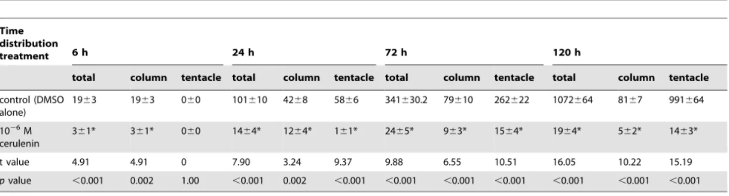
Figure 6. Effect of cerulenin on Symbiodinium distribution inside the host. (a) The schematic demonstration of dynamic Symbiodiniumdistribution from ‘‘column’’ to ‘‘tentacle’’ fractions during the infection process. (b) The effect of cerulenin treatment on the dynamic change ofSymbiodiniumpopulation in tentacle fraction were examined. Percentage of tentacle fraction was calculated by dividing Symbiodiniumnumber in tentacles with total number (i.e. numbers in tentacle plus column fractions, see also Table 2). All data are presented as mean6SEM,N= 31. * denote the statistical significance of cerulenin treatment comparing to control at the same time point according Mann-Whitney Rank Sum Test (p,0.05).
for 5 min), and re-suspended in seawater for infection experi-ments.
After being washed twice with filtered seawater (FSW), the bleached sea anemones were incubated with 10421of the control or cerulenin-treated Symbiodinium cells for 1 h. The infected sea anemones were then washed twice with FSW, moved to a new container with FSW, and cultured in across a 12L/12D cycle. The numbers ofSymbiodiniuminside sea anemones (The number of sea anemones investigated at each time points is 31, N = 31) were then examined under an epifluorescence microscope (Axiovision, Zeiss, Germany) after 6, 24, 72, and 120 h of culture.
In order to identify the intracellular distribution ofSymbiodinium
in host tissue, some anemones were fixed in 3.6% paraformalde-hyde (two hours at room temperature) for histological examina-tion. Anemones were then washed with 200 mM phosphate buffer twice, dehydrated by a series of increasing ethanol concentrations (50%, 75%, 95%, 95%, 100% and 100%, for 30 minutes each). They were infiltrated with JB-4 catalyzed solution A (Electron Microscopy Sciences, USA) for overnight at 4uC, followed by the embedding with embedding medium (2 ml JB-4 catalyzed solution A plus with JB-4 solution B). After the sample is solidified, a microtome (Leica, Germany) was used to section tissue at 5mm thickness. Each section was then stained with hematoxylin (Merck, Germany) and trichrome (Sigma, USA) to examine the distribu-tion of ingestedSymbiodiniumby the microscope (Axiovision, Zeiss, Germany).
Statistical analysis
In order to determine the statistical significance of the treatments, student’s t-tests or one-way analysis of variance (ANOVA) followed by Duncan’s multiple-range procedure (p,0.05) were used. Non-parametric Mann-Whitney Rank Sum Tests were performed on non-normal data.
Results
Inhibitory effects of cerulenin on theSymbiodiniumcell cycle
The cell cycle propagation of controlSymbiodinium(clade B) from the growing/DNA synthesis stage (i.e., G1-S-G2/M) to cytokinesis (i.e., G2/M-G1) was entrained by the 12L/12D cycle (Fig. 3a). Cells progressed from G1(T05) to S (T11), and then to the G2/M phase during the first 12 h of light stimulation. During darkness (T17), DNA synthesis was greatly decreased, and more cells entered the G2/M phase followed by cytokinesis to generate G1 cells for the next cycle (T29 of Fig. 3b). Approximately 50–60% of the G1cells progressed through the entire cell cycle for each 12L/ 12D cycle.
To determine the optimal concentration of cerulenin required to interfere with the cell cycle progression, cerulenin was added at T00 in three different concentrations: 1027
, 1026
, or 1025 M (Fig. 3a). At both 1026
and 1025
M, cerulenin effectively arrested the cell cycle at the G1 phase and greatly decreased the progression toward G2/M. On the other hand, cerulenin was not able to alter the cell cycle progression at 1027
M (Fig. 3b; F = 16.374, p,0.001 at T11; F = 351.483, p,0.001 at T17; F = 358.025,p,0.001 at T23). Consequently, cerulenin at a 1026 M concentration was used in the following experiments.
To further examine the effect of cerulenin on specific cell cycle phases, Symbiodinium were incubated with 1026
M cerulenin at different times (T00, T05, T11, or T17) during the light-dark cycle (Fig. 4). As shown in Fig. 4a, when cerulenin was added at T00 or T05 when most cells were in the G1 phase, treated cells were unable to progress throughout the remainder of their cell cycle
over the duration of the experiment and remained at G1phase. Furthermore, when added at T11, at which point the majority of cells were either at the G1or S phase (Fig. 4a), cerulenin treatment significantly decreased the percentage of cells transitioned from S to G2/M between T17 and T23 (see Fig. 4b; F = 182.065,
p,0.001 at T17; F = 192.214,p,0.001 at T23 in comparison to control), which further resulted in a delay of mitotic division at T29 (Fig. 4b, F = 57.748,p,0.001). This indicates that cerulenin was also able to decrease the transition from S to G2/M. Cerulenin was also able to inhibit the mitotic division of G2/M cells when it was added at T17 (Fig. 4a–b).
Effect of cerulenin on lipid contents of cells at different phases of the cell cycle
The lipid contents of Symbiodinium populations 5 or 6 h after cerulenin was added at T00, T05, T11 and T17 (i.e. T05, T11, T17 and T23, respectively) were analyzed. First, as shown in Fig. 5, there were dynamic concentration changes of SE, FFA, and PE, but not PC, in untreatedSymbiodiniumpopulation. Although 0.02% DMSO was used as carrier in untreated Symbiodinium (see the ‘‘Materials and methods’’ section), the lipid concentration changes were intrinsic nature of the microalgal population, but not artifacts induced by DMSO (see Table S1). Both concentrations of SE (Fig. 5a) and FFA (Fig. 5b) increased significantly to the maximum level at T11 (0.10860.021 and 1.12660.138mg/25mg protein, respectively) and then decreased during T17 and T23 (SE: F = 4.460,p= 0.025; FFA: F = 9.400,p= 0.002). The concentra-tion of PE gradually decreased over the 12L:12D cycle and reached the minimum level (0.15860.015mg/25mg protein) at T23 (Fig. 5c; F = 10.765, p= 0.001). On the other hand, the concentration of PC remained unchanged throughout the 12L:12D cycle (Fig. 5d; F = 3.527,p= 0.052).
The SE concentration was not significantly changed after cerulenin treatment at T05, T11, T17, or T23 (Fig. 5a; T5: t = 1.938, p= 0.125; T11: t = 1.161, p= 0.290; T17: t = 1.239,
p= 0.270; T23: t = 0.953, p= 0.378). Nevertheless, FFA concen-trations at T11 and T17 significantly decreased (i.e. 0.42260.065 and 0.42660.020mg/25mg protein, respectively) when cerulenin was added at T05 and T11 for 6 h, compared to the control treatment (Fig. 5b) (T11: t = 4.481, p= 0.007; T17: t = 5.431,
p= 0.003). Moreover, cerulenin treatment significantly decreased PE concentrations at T05 (0.14660.002mg/25mg protein), T11 (0.14860.014mg/25mg protein), T17 (0.15560.004mg/25mg protein), and T23 (0.14060.004mg/25mg protein) (Fig. 5c; t = 11.234, p,0.001; t = 3.195, p= 0.024; t = 2.594, p= 0.049; t = 6.631, and p,0.001, respectively). However, PC concentra-tions remained almost unchanged relative to the controls after treatments at T05, T11, T17, or T23 (Fig. 5d; t = 2.624,p= 0.059; t = 2.449, p= 0.058; t = 0.955, p= 0.3842; t = 1.166, p= 0.288, respectively). The result demonstrated that syntheses of specific lipid species, i.e. PE and FFA, were inhibited by cerulenin at different cell phases over the cell cycle.
Effect of cerulenin on the starch content of cells of different phases of the cell cycle
The starch concentration of untreatedSymbiodiniumincreased to the maximum level at T11 and then decreased to the basal level at T23 (Fig. 5e; F = 110.46, p,0.001). Starch concentrations also remained unchanged when Symbiodinium were treated with cerulenin at T05, T11, T17, and T23 for 6 h (t = 1.068,
p= 0.310; t = 1.068, p= 0.311; t = 0.311, p= 0.762; t = 1.000,
p= 0.341, respectively).
affect the symbiotic association between Symbiodinium and anem-ones, Symbiodinium at T11, resulting from 11 h treatments of 0 (control) and 1026
M cerulenin were collected to infect bleached anemones for 1 h in FSW. The number of ingestedSymbiodinium
and their distribution inside the host animal were then examined after 6, 24, 72, and 120 h after infection. Once the control
Symbiodiniumwere ingested by the anemones, they first aggregated in the mesenteries of the host body column, and then later appeared in the tentacles (Fig. 6a). The cerulenin-treated
Symbiodinium not only exhibited reduced uptake but also an abnormal distribution inside the anemone host, in comparison with controlSymbiodinium(see Table 2 and Fig. 6b). First, the initial uptake of control Symbiodinium after 6 h of incubation was significantly higher than that of cerulenin-treated Symbiodinium (1963vs.361 per anemone; p,0.001, see Table 2). During the following incubation, number of control Symbiodinium gradually increased and reached 1072664 per anemone at 120 h, indicating rapid proliferation of the ingested microalgae inside the host. On the other hand, number of cerulenin-treated Symbiodinium prolif-erated slowly and only reached 1964 per anemone after 120 h incubation. Secondly, there was a significant retardation of
Symbiodinium population translocation from the mesenteries (i.e. the ‘‘column’’ fraction) toward the tentacle (Fig. 6b). After the first 6 h of incubation, all Symbiodinium distributed in the ‘‘column’’ fraction and noSymbiodiniumcould be identified in tentacle of the host (Fig. 6b; also see Table 2). After 24 h of incubation, 64.764.7% of the control Symbiodinium population redistributed from the mesenteries to the tentacles. On the other hand, most cerulenin-treated Symbiodinium still remained in the mesenteries, and only 21.066.2% of them had redistributed to the tentacles. The percentage of the cerulenin-treated Symbiodinium in the tentacles gradually increased to 77.165.6% after 72 h incubation, reaching a similar percentage as the controls.
Discussion
The cell cycle of free-living clade BSymbiodiniumhas been shown to be entrained by a 12L/12D photoperiod [16]. The light irradiation initiates the cell propagation from G1to S, while dark treatment drives cells to progress toward G2/M and then cytokinesis. Furthermore, the cell motility is dynamic and increased during the first 7–8 h of light irradiation (G1 phase) relative to darkness [16]. In the heterotropic dinoflagellate,
Crypthecodinium cohnii, cellular lipid profile varies with their cell cycle [4]. The present study shows that abnormal lipid synthesis not only affect the cell cycle ofSymbiodinium, but also their ingestion and eventual symbiotic association with the sea anemone host.
Although starch content exhibited a diurnal pattern that coincided with cell cycle progression, starch content was not altered by cerulenin (Fig. 5e). The fact that cerulenin blocks cell cycle progression without altering starch content implies that newly synthesized lipids are more critical than starch during the cell cycle progression, especially at the G1/S and G2/M transitions. Four major lipids, including FFA, PE, PC and SE, were identified inSymbiodinium, with dynamic expression during the cell cycle. Among these lipids, cerulenin significantly inhibited synthesis of PE and FFA.Symbiodinium exhibited the highest PE
exposure of PE on the yeast plasma membrane involved in the polarized organization of the actin cytoskeleton and membrane curvature changes at the bud cortex of late mitotic cell, indicating the involvement of PE in cytokinesis [28]. As a consequence, inhibition of PE synthesis by cerulenin alters the cell cycle, as observed in the present study (Figs. 3–4).
Besides the PE synthesis, the biosynthesis of FFA turns out to be another critical regulation for cell cycle ofSymbiodinium. Cerulenin deactivates three major types of fatty acid synthases [29]. Inhibition of FFA by cerulenin treatment is concurrent with G1 arrest or a transition delay from S to G2/M and G2/M to G1, depending on the time of cerulenin application (Figs. 3, 4, 5). Furthermore, in addition to the fatty acid synthases, the synthesis of FFA could be regulated by the acetyl-CoA carboxylase whose activity is protein kinase A (i.e. cAMP dependent protein kinase or PKA)-dependent [30]. As a consequence, the decrease of FFA synthesis by inhibiting PKA could alter the cell cycle. This was confirmed by a previous study showing that the inhibition of adenylyl cyclase (AC) resulted in the cell cycle arrest of clade B
Symbiodiniumat the G1/S transition [16].
The present study has also attempted to examine how alteration of lipid synthesis might affectSymbiodiniumingestion and symbiotic distribution in the hostA. pulchella. After the initial ingestion, the
Symbiodinium-gastroderm recognition in tentacles of the host is the next important task to establish highly specific mutualistic associations [31]. Although the cellular mechanisms underlying the ingestion/recognition between free-living Symbiodinium and cnidarian hosts remain unclear, the decreased ingestion of
Symbiodinum induced by cerulenin treatment (see Table 2) was not due to different cellular motility upon the infection (see Figure S1). Wood-Charlson and colleagues have demonstrated that the glycan-lectin interaction may play a critical role during the recognition between the Symbiodinium and Fungia scutaria larvae [32]. The inhibition of Symbiodinium surface glycans, such as a -mannose/ a-glucose and a-galactose, by lectins greatly lowered their ingestions into the larvae [32]. Moreover, by binding surface galactose and glucosamine residues ofSymbiodinium with a lectin analogue concanavalin A, the ingestion rate ofSymbiodiniumby the
Aiptasia pulchellaalso significantly reduced [33]. These observations have collectively suggested that glycan ligands, such as a -mannose/ a-glucose and a-galactose, locating on the surface of
Supporting Information
Figure S1 Effect of cerulenin (1026
M) onSymbiodinium
motility.Cells were treated with or without cerulenin (1026 M) at T00 (see Fig. 1). The percentage of motile cells was counted at different time using an epifluorescence microscope (Axiovision, Zeiss, Germany). Values were expressed as mean 6 SEM.
N= 5. (EPS)
Table S1
(DOC)
Author Contributions
Conceived and designed the experiments: LHW CSC LSF. Performed the experiments: LHW CSC HHL. Analyzed the data: LHW CSC. Contributed reagents/materials/analysis tools: LHW CSC. Wrote the paper: LHW CSC ABM.
References
1. Donnelly PM, Bonetta D, Tsukaya H, Dengler RE, Dengler NG (1999) Cell Cycling and Cell Enlargement in Developing Leaves ofArabidopsis. Deve Biol 215:407–419.
2. Knacker T, Harwood JL, Hunter CN, Russell NJ (1985) Lipid biosynthesis in synchronized cultures of the photosynthetic bacterium Rhodopseudomonas sphaeroides. Biochem J 229: 701–710.
3. Janero DR, Barrnett R (1981) Thylakoid membrane biogenesis inChlamydomonas reinhardi137+: Cell-cycle variations in the synthesis of polar glycerolipid. J Cell Biol 91:126–134.
4. Kwok ACM, Wong JTY (2005) Lipid Biosynthesis and its Coordination with Cell Cycle Progression. Plant Cell Physiol 46:1973–1986.
5. Whitehead LF, Douglas AE (2003) Metabolite comparisons and the identity of nutrients translocated from symbiotic algae to an animal host. J Exp Biol 206:3149–3157.
6. Papina M, Meziane T, van Woesik R (2003) Symbiotic zooxanthellae provide the host-calralMontipora digitatawith polyunsaturated fatty acids. Comp Biochem Physiol B 135: 533–537.
7. Grottoli AG, Rodrigues LJ, Juarez C (2004) Lipids and stable carbon isotopes in two species of Hawaiian corals,Porites compressaandMontipora verrucosa, following a bleaching event. Mar Biol 145:621–631.
8. Yamashiro H, Oku H, Onaga K (2005) Effect of bleaching on lipid content and composition of Okinawan corals. Fish Sci 75:448–453.
9. Zhukova NV, Titlyanov EA (2006) Effect of light intensity on the fatty acid composition of dinoflagellates symbiotics with hermatyptic corals. Botanica Marina 49:339–346.
10. Dunn SR, Thomas MC, Nette GW, Dove SG (2012) A lipidomics approach to understanding free fatty acid lipogenesis derived from dissolved inorganic carbon within cnidarian-dinoflagellate symposis. PLoS ONE 7:e46801.
11. Garrett TA, Schmeitzel JL, Klein JA, Hwang JJ, Schwarz JA (2013) Comparative lipid profiling of the cnidarianAiptasia pallidaand its dinoflagellate symbiont. PLoS ONE 8:e57975.
12. Luo YJ, Wang LH, Chen WNU, Peng SE, Tzen JTC, et al. (2009) Ratiometric image of gastrodermal lipid bodies in coral-dinoflagellate endosymbiosis. Coral Reefs 28:289–301.
13. Chen WNU, Kang HJ, Weis VM, Mayfield AB, Jiang PL, et al. (2012) Diel rhythmicity of lipid-body formation in a coral-Symbiodiniumendosymbiosis. Coral Reefs 31: 521–534.
14. Meesters EH, Bak RPM (1993) Effects of coral bleaching on tissue regeneration potential and colony survival. Mar Ecol Prog Ser 96:189–198.
15. Mascarrelli PE, Bunkley-Williams L (1999) An experimental field evaluation of healing in damaged, unbleached and artificially bleached star coral,Montastraea annularis. Bull Mar Sci 65:577–586.
16. Wang LH, Liu YH, Ju YM, Hsiao YY, Fang LS, et al. (2008) Cell cycle propagation is driven by light–dark stimulation in a cultured symbiotic dinoflagellate isolated from corals. Coral Reefs 27:823–835.
17. Smith GJ, Muscatine L (1999) Cell cycle of symbiotic dinoflagellates: variation in G1 phase-duration with anemone nutritional status and macronutrient supply in theAiptasia pulchella-Symbiodinium pulchrorumsymbiosis. Mar Biol 134:405–418.
18. Reimer AA (1971) Observations on the relationships between several species of tropical Zoanthids (Zoanthidae, Coelenterata) and their zooxanthellae. J Exp Mar Biol Ecol 7:207–214.
19. Falkowski PG, Dubinsky Z, Muscatine L, McCloskey L (1993) Population control in symbiotic corals. Bioscience 43:606–611.
20. Hoegh-Guldberg O, McCloskey LR, Muscatine L (1987) Expulsion of zooxanthellae by symbiotic cnidarians from the Red Sea. Coral Reefs 5:201– 204.
21. Hata T, Sano Y, Matsumae A, Kamio Y, Nomura S, et al. (1960) Study of new antifungal antibiotic. Jpn J Bacteriol 15:1075–1077.
22. Fishman Y, Rotham S, Citri N (1978) Evidence linking penicillinase formation and secretion to lipid metabolism inBacillus lichenijormis. J Bacteriol 134:434–439. 23. Omura S (1976) The antibiotic cerulenin, a novel tool for biochemistry as an
inhibitor of fatty acid synthesis. Bacteriol Rev. 40: 681–697.
24. Rowan R, Powers DA (1991) Molecular genetic identification of symbiotic dinoflagellates (zooxanthellae). Mar Ecol Prog Ser 71:65–73.
25. Matyash V, Liebisch G, Kurzchalia TV, Shevchenko A, Schwudke D (2008) Lipid extraction by methyl-tert-butyl ether for high-throughput lipidomics. J Lipid Res 49:1137–1146.
26. Christie WW, Gill S, Nordba¨ck J, Itabashi Y, Sanda S (1998) New procedures for rapid screening of leaf lipid components fromArabidopsis. Phytochem Anal 9:53–57.
27. Janero DR, Barrnett R (1982) Membrane biogenesis inChlamydomonas reinhardi
137+: Cell-cycle variations in the synthesis of phospholipids of non-photosyn-thetic membranes. Exp Cell Res 138:451–454.
28. Iwamto K, Kobayashi S, Fukuda R, Umeda M, Kobayashi T, et al. (2004) Local exposure of phosphatidylethamine on the yeast plasma membrane is implicated in cell polarity. Genes to Cells 9:891–903.
29. Vance D, Goldberg I, Mitsuhashi O, Bloch K (1972) Inhibition of fatty acid synthetases by the antibiotic cerulenin. Biochem Biophys Res Commu 48:649– 656.
30. Kim KH, Lopez-Casillas F, Bai DH, Luo X, Pape ME (1989) Role of reversible phosphorylation of acetyl-Co A carboxylase in long-chain fatty acid synthesis. FASEB J 3:2250–2256.
31. Peng SE, Wang YB, Wang LH, Chen WNU, Lu CY, et al. (2010) Proteomics anaylsis of symbiosome membranes in Cnidaria-dinoflagellate endosymbiosis. Proteomics 10:1002–1016.
32. Wood-Charleson EM, Hollingsworth LH, Krupp DA, Weis VM (2006) Lectin/ glycan interactions play a role in recognition in a coral/dinoflagellate symbiosis. Cell Microbiol 8:1985–1994.
33. Lin KL, Wang JT, Fang LS (2000) Participation of glycoproteins on zooxanthellal cell walls in the establishment of a symbiotic relationship with sea anemone,Aiptasia pulchella. Zool Studies 39:172–178.
34. Fraysse N, Couderc F, Poinsot V (2003) Surface polysaccharide involvement in establishing the rhizobium–legume symbiosis. Eur J Biochem 270:1365–1380. 35. Petrovics G, Putnoky P, Reuhs B, Kim J, Thorp TA, et al. (1993) The presence
of a novel type of surface polysaccharide in Rhizobium meliloti requires a new fatty acid synthase-like gene cluster involved in symbiotic nodule development. Mol Microbiol 8:1083–1094.