BGD
10, 8851–8886, 2013DMS and C cycling in dynamic light fields
M. Galí et al.
Title Page
Abstract Introduction
Conclusions References
Tables Figures
◭ ◮
◭ ◮
Back Close
Full Screen / Esc
Printer-friendly Version Interactive Discussion
Discussion
P
a
per
|
Dis
cussion
P
a
per
|
Discussion
P
a
per
|
Discussio
n
P
a
per
Biogeosciences Discuss., 10, 8851–8886, 2013 www.biogeosciences-discuss.net/10/8851/2013/ doi:10.5194/bgd-10-8851-2013
© Author(s) 2013. CC Attribution 3.0 License.
Open Access
Biogeosciences
Discussions
Geoscientiic Geoscientiic
Geoscientiic Geoscientiic
This discussion paper is/has been under review for the journal Biogeosciences (BG). Please refer to the corresponding final paper in BG if available.
Di
ff
erential response of planktonic
primary, bacterial, and dimethylsulfide
production rates to vertically-moving and
static incubations in upper mixed-layer
summer sea waters
M. Galí1, R. Simó1, G. L. Pérez2, C. Ruiz-González1,*, H. Sarmento1,**, S.-J. Royer1, A. Fuentes-Lema3, and J. M. Gasol1
1
Institut de Ciències del Mar (CSIC), Passeig Marítim de la Barceloneta, 37–49, 08003 Barcelona, Catalonia, Spain
2
Laboratorio de Ecología y Fotobiología Acuática (IIB-INTECH), Chascomús, Argentina
3
Departamento de Ecoloxía e Bioloxía Animal, Facultade de Ciencias do Mar, Vigo (Pontevedra), Spain
*
now at: Département des Sciences Biologiques, Université du Québec à Montréal, Québec, Canada
**
BGD
10, 8851–8886, 2013DMS and C cycling in dynamic light fields
M. Galí et al.
Title Page
Abstract Introduction
Conclusions References
Tables Figures
◭ ◮
◭ ◮
Back Close
Full Screen / Esc
Printer-friendly Version Interactive Discussion
Discussion
P
a
per
|
Dis
cussion
P
a
per
|
Discussion
P
a
per
|
Discussio
n
P
a
per
Received: 6 May 2013 – Accepted: 11 May 2013 – Published: 29 May 2013
Correspondence to: M. Galí ([email protected])
BGD
10, 8851–8886, 2013DMS and C cycling in dynamic light fields
M. Galí et al.
Title Page
Abstract Introduction
Conclusions References
Tables Figures
◭ ◮
◭ ◮
Back Close
Full Screen / Esc
Printer-friendly Version Interactive Discussion
Discussion
P
a
per
|
Dis
cussion
P
a
per
|
Discussion
P
a
per
|
Discussio
n
P
a
per
Abstract
Microbial plankton experience fluctuations in total solar irradiance and in its spectral composition as they are vertically moved by turbulence in the oceanic upper mixed layer (UML). The fact that the light exposure is not static but dynamic may have im-portant consequences for biogeochemical processes and ocean-atmosphere fluxes.
5
However, most biogeochemical processes other than primary production, like bacte-rial production or dimethylsulfide (DMS) production, are seldom measured in sunlight and even less often in dynamic light fields. We conducted four experiments in olig-otrophic summer stratified Mediterranean waters, where a sample from the UML was incubated in ultraviolet (UV)-transparent bottles at three fixed depths within the UML
10
and on a vertically-moving basket across the same depth range. We assessed the response of the phyto- and bacterioplankton community with physiological indicators based on flow cytometry singe-cell measurements, Fast Repetition Rate fluorometry (FRRf), phytoplankton pigment concentrations and particulate light absorption. Dy-namic light exposure caused a disruption of the photoinhibition and photoacclimation
15
processes associated to ultraviolet radiation (UVR), which slightly alleviated bacterial
photoinhibition but did not favor primary production. Gross DMS production (GPDMS)
decreased sharply with depth in parallel to shortwave UVR, and displayed a dose-dependent response that mixing did not significantly disrupt. To our knowledge, we
provide the first measurements of GPDMSunder in situ UV-inclusive optical conditions.
20
1 Introduction
The characteristic response times of microbial plankton match the natural variability
of light exposure, which changes at different temporal scales with solar elevation, the
passage of clouds, vertical mixing and even wave focusing (Gallegos and Platt, 1985). In transparent oceanic waters, exposure to high irradiance (photosynthetically available
25
BGD
10, 8851–8886, 2013DMS and C cycling in dynamic light fields
M. Galí et al.
Title Page
Abstract Introduction
Conclusions References
Tables Figures
◭ ◮
◭ ◮
Back Close
Full Screen / Esc
Printer-friendly Version Interactive Discussion
Discussion
P
a
per
|
Dis
cussion
P
a
per
|
Discussion
P
a
per
|
Discussio
n
P
a
per
of the water column (Vincent and Neale, 2000). Short-term irradiance fluctuations elicit fast and reversible responses (Roy, 2000), whereas continued exposure to high PAR and UVR may elicit photoacclimation (MacIntyre et al., 2002) or permanent physiolog-ical changes, i.e., irreversible damage (Buma et al., 2001).
Vertical mixing can have either a positive, neutral or negative effect on water
column-5
integrated processes depending on the interplay between mixing rates, damage and repair kinetics, and underwater attenuation of PAR and UVR (Neale et al., 2003). In the absence of repair mechanisms, damage will be proportional to cumulative exposure (i.e., it will be dose-dependent). If moderate repair exists, mixing will allow the cells to recover in the UVR shaded portion of the UML (Fig. 1a), so that damage will no longer
10
be dose-dependent, and a steady state will be achieved provided that the cells spend
sufficient time under constant exposure conditions. In the idealized situation where
damage is completely counteracted by repair in a timescale much shorter than the
mixing time, or in the absence of repair, vertical mixing will have neutral effects. These
responses can change with exposure time.
15
The effects of dynamic light exposure have concerned the aquatic photosynthesis
research community for almost 40 yr (see Gallegos and Platt, 1985 and references therein), and apparently contradictory findings have often been reached using either experimental or modeling approaches (Ross et al., 2011a,b). It appears that the ability to take advantage of dynamic light exposure may depend on the taxonomic
compo-20
sition and size structure of the phytoplankton community, their light history, and their nutritional status (Barbieri et al., 2002; Brunet and Lavaud, 2010; Helbling and Vil-lafañe, 2013). Knowledge on the photoresponse of (bacterial) heterotrophic activity is much more limited, but a number of studies suggest that significant PAR-driven stim-ulation frequently occurs (Moran et al., 2001; Church et al., 2004), as does inhibition
25
BGD
10, 8851–8886, 2013DMS and C cycling in dynamic light fields
M. Galí et al.
Title Page
Abstract Introduction
Conclusions References
Tables Figures
◭ ◮
◭ ◮
Back Close
Full Screen / Esc
Printer-friendly Version Interactive Discussion
Discussion
P
a
per
|
Dis
cussion
P
a
per
|
Discussion
P
a
per
|
Discussio
n
P
a
per
in the ocean (Kolber et al., 2000; Béjà et al., 2000; Kirchman and Hanson, 2012) or to their interaction with other light-driven processes (see references in Ruiz-González et al., 2013).
Besides carbon and nutrient cycling, solar radiation modulates the biogeochemical
cycles of other elements. It has recently been shown that sunlight stimulates GPDMS
5
(Galí et al., 2011) in an irradiance- and spectrum-dependent manner (Galí et al., 2013). The volatile DMS is produced mainly by the enzymatic cleavage of the phytoplankton osmolyte dimethylsulfoniopropionate (DMSP) as a result of microbial food web interac-tions (Simó, 2004). Marine DMS emission represents the main natural source of sulfur to the atmosphere (Lana et al., 2011) and has potential climate implications (Charlson
10
et al., 1987) which depend on its response to solar radiation (Vallina and Simó, 2007).
However, the climatic effects of DMS emission remain controversial (Quinn and Bates,
2011).
We designed an experiment where a single surface seawater sample was incubated in UVR-transparent bottles at three fixed optical depths, approximately corresponding
15
to the water sub-surface, the optical middle, and the bottom of the UML. An addi-tional set of bottles was regularly moved up and down across the same depth range and radiation gradient (Fig. 1; Table 1). Rather than simulating actual turbulent mixing
experimentally (which is extremely difficult), vertical motion was applied as a
pertur-bation of the photoinhibiton and photoacclimation processes occurring in upper mixing
20
waters, with fixed-depth incubations being the static end of such perturbation. The ex-perimental design was aimed at answering two questions: (1) photobiological: should the mixing bottles display the same response that the ones incubated at the middle optical depth considering that both treatments received a similar cumulative dose? If the response was the same this would imply that the measured processes were
dose-25
BGD
10, 8851–8886, 2013DMS and C cycling in dynamic light fields
M. Galí et al.
Title Page
Abstract Introduction
Conclusions References
Tables Figures
◭ ◮
◭ ◮
Back Close
Full Screen / Esc
Printer-friendly Version Interactive Discussion
Discussion
P
a
per
|
Dis
cussion
P
a
per
|
Discussion
P
a
per
|
Discussio
n
P
a
per
2 Methods
2.1 Experimental setting and irradiance calculations
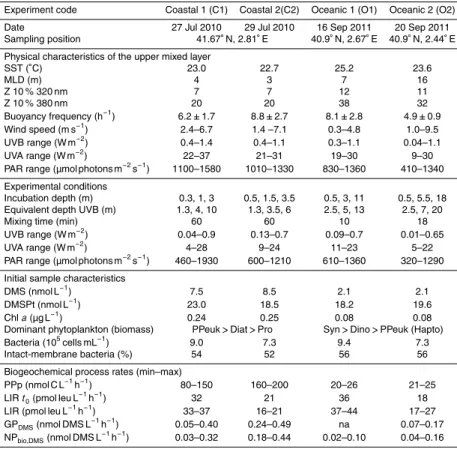
Surface (0.2 to 3 m deep) seawater samples were taken pre-dawn in 20–30 L poly-carbonate carboys (dimmed with a black plastic bag). In the coastal experiments (C1 and C2) the samples were taken from a boat at the Blanes Bay Microbial Observatory
5
coastal site (BBMO; 0.5 miles offshore over a water column depth of 20 m), brought
to the lab, and incubated at the pier of the Barcelona Olympic Harbor during 4 h cen-tered on the solar noon. The oceanic experiments (O1 and O2) were done in the open Mediterranean during a Lagrangian cruise over a water column depth of ca. 2000 m (R/V García del Cid). In these experiments the samples were incubated in situ,
be-10
ginning 4 h before solar noon and ending 2 h after solar noon (with an intermediate sample taken after the first 2 h). In C1 and C2 mixing was applied by moving the bottle basket (Fig. 1a) manually every 15 min, completing a mixing cycle every 60 min. In the ship-based experiments the mixing bottles were continuously moved using the winch of
the ship at the smallest possible vertical speed (3–4 cm s−1), completing a cycle in 10–
15
18 min. Since the waters were less transparent in the harbor than at the BBMO, in C1 and C2 the bottles were incubated at shallower depths to approximate the equivalent in situ optical depths (Table 1). Mixing layer depths (MLD) were estimated from CTD
tem-perature profiles, and defined by a>0.1◦C deviation with respect to 1 m depth. The
buoyancy or Brunt–Väisälä frequency was calculated in 1 m bins (Fig. 2), and used as
20
an additional criterion to distinguish the weakly-stratified UML from the more stratified waters below.
The irradiance just below the water surface (sub-surface irradiance) during the in-cubations was recorded with a PUV-2500 (Biospherical) multichannel filter radiometer, which was also used to measure underwater irradiance profiles in C1 and C2. In O1 and
25
O2, the vertical profiles were measured with a PRR-800 (Biospherical). Diffuse
attenu-ation coefficients of downward irradiance (Kd) were calculated as the linear regression
BGD
10, 8851–8886, 2013DMS and C cycling in dynamic light fields
M. Galí et al.
Title Page
Abstract Introduction
Conclusions References
Tables Figures
◭ ◮
◭ ◮
Back Close
Full Screen / Esc
Printer-friendly Version Interactive Discussion
Discussion
P
a
per
|
Dis
cussion
P
a
per
|
Discussion
P
a
per
|
Discussio
n
P
a
per
surface layer where the incubations were done. The time-series of sub-surface irradi-ance were converted to the irradiirradi-ance seen by each water sample by applying the
attenuation due to seawater (e−Kd·z) and the attenuation due to the incubation bottles.
We used polytetrafluoroethylene (Teflon, Nalgene) bottles, which according to our mea-surements transmit 65 %, 77 % and 100 % of spectral irradiance in the UVB, UVA and
5
PAR bands, respectively (Galí et al., 2013). The bottles were placed in a metallic bas-ket which caused a minimal alteration of the tridimensional light field. For the mixing bottles, the calculation was made using a time-varying depth that corresponded to the vertical displacement of the basket. In each incubation, the mean UVB (300–320 nm) and UVA (320–400 nm) irradiance was calculated by integrating over the spectrum the
10
mean spectral irradiance in the 6 bands measured by the PUV-2500 (centered at 305, 313, 320, 340, 380 and 395 nm) as described by Galí et al. (2013). PAR was measured in a single integrated band (400–700 nm) so that no spectral integration was required. The irradiance dose was calculated by multiplying the mean irradiance by the total incubation time.
15
2.2 Process measurements and analysis techniques
Primary production was measured as the 14C incorporated into particles in duplicate
40 mL Teflon bottles inoculated with NaH14CO3 (Morán et al., 1999) and incubated in
situ (including dark controls). Bacterial heterotrophic production rates were measured
as3H-leucine incorporation rates (LIR; Kirchman et al., 1985; Smith and Azam, 1992)
20
in the initial samples and on sub-samples taken from the larger (2.3 L) Teflon incubation bottles after in situ light exposure. Triplicate sub-samples plus one killed control from each Teflon bottle were further incubated for 2 h in the dark at in situ temperature in 1.5 mL eppendorf vials. In O1 and O2, incubation-averaged leucine incorporation rates (LIR) were calculated as the time-weighted average of intermediate (2 h) and final time
25
(6 h) time incubations. In C1 and C2 leucine incorporation was also measured during
BGD
10, 8851–8886, 2013DMS and C cycling in dynamic light fields
M. Galí et al.
Title Page
Abstract Introduction
Conclusions References
Tables Figures
◭ ◮
◭ ◮
Back Close
Full Screen / Esc
Printer-friendly Version Interactive Discussion
Discussion
P
a
per
|
Dis
cussion
P
a
per
|
Discussion
P
a
per
|
Discussio
n
P
a
per
Samples for pigment analysis were obtained by filtering 1–2 L seawater onto GF/F filters at the beginning and the end of the incubations (O1 and O2 only). Following filtra-tion, the filters were immediately stored in liquid nitrogen. Pigments were extracted and analysed by HPLC following Zapata et al. (2000) on a SpectraSYSTEM (Thermo)
us-ing a Waters Symmetry C8 column (150×4.6 mm, 3.5 µ particle size, 10 nm pore size).
5
Calibration was made using commercial external pigment standards (DHI, Denmark), and the pigments were identified according to their elution time.
The absorption spectra of total particulate matterapwere determined by the
quanti-tative filter technique, using the simple transmittance method in a Lambda 800 (Perkin– Elmer) spectrophotometer. Water samples (2 L) were filtered on-board using 25 mm
di-10
ameter GF/F filters. Immediately after filtration absorbance scans were measured from 350 to 750 nm at 1 nm intervals. The quantitative filter technique was applied
accord-ing to NASA’s optics protocols for absorption coefficient measurements (Mitchell et al.,
2000). In order to minimize light scattering, the wet filters were placed as close to the spectrophotometer detector as possible and measured against a blank clean filter
wet-15
ted with filtered (0.2 µm) seawater. Absorption coefficients were estimated according to
the relationshipap(λ)=
2.303Afilter(λ)s
Vfiltβ(λ) whereAfilteris the measured absorbance, s is the
clearance area of the filter,Vfiltis the volume of filtered water, andβ(λ) is the
amplifica-tion factor vector (Mitchell and Kiefer, 1984).
The maximum quantum yield of photosystem II photochemistry (Fv/Fm), an indicator
20
of phytoplankton photosynthetic performance and photoinhibtion, was measured by Fast Repetition Rate fluorometry (FastTracka I, Chelsea), as detailed by Galí et al. (2013).
A FACSCalibur (Becton & Dickinson) flow cytometer equipped with a 15 mW Argon-ion laser (488 nm emissArgon-ion) was used to enumerate picophyto- and bacterioplankton
25
populations and to measure their performance at the single-cell level. The cell-specific
fluorescence of each different picophytoplankton population (normalized to their side
BGD
10, 8851–8886, 2013DMS and C cycling in dynamic light fields
M. Galí et al.
Title Page
Abstract Introduction
Conclusions References
Tables Figures
◭ ◮
◭ ◮
Back Close
Full Screen / Esc
Printer-friendly Version Interactive Discussion
Discussion
P
a
per
|
Dis
cussion
P
a
per
|
Discussion
P
a
per
|
Discussio
n
P
a
per
beads (1 µm, Fluoresbrite carboxylate microspheres, Polysciences Inc., Warrington, PA) were added at a known density as internal standards. Two subpopulations of heterotrophic bacterioplankton were distinguished based on the Nucleic-Acid-Double-Staining (NADS) viability protocol: intact-membrane (or “live”) bacteria and membrane-compromised (or “dead”) bacteria (Grégori et al., 2001), which uses a combination of
5
the cell-permanent nucleic acid strain SybrGreen I (Molecular Probes, Eugene, OR) and the cell-impermeant propidium iodine (PI, Sigma Chemical Co.) fluorescent probe.
We used a 1 : 10 SG1 and 10 µg mL−1PI concentrations that were added to live
sam-ples less than 2 h after sampling. After simultaneous addition of each stain, the samsam-ples were incubated for 20 min in the dark at room temperature and then analyzed.
10
DMS and DMSP were measured by purge and trap gas chromatography (Shi-madzu GC14A) coupled to flame photometric detection. Net biological DMS production (NPbio,DMS) was obtained by incubating whole water samples in 2.3 L Teflon bottles and
correcting afterward for photochemical DMS loss, as described by Galí et al. (2013). Gross DMS production was measured in the same way in additional bottles amended
15
with 200 nmol L−1dimethyldisulfide (Galí et al., 2011), an effective inhibitor of bacterial
DMS consumption (Wolfe and Kiene, 1993; Simó et al., 2000).
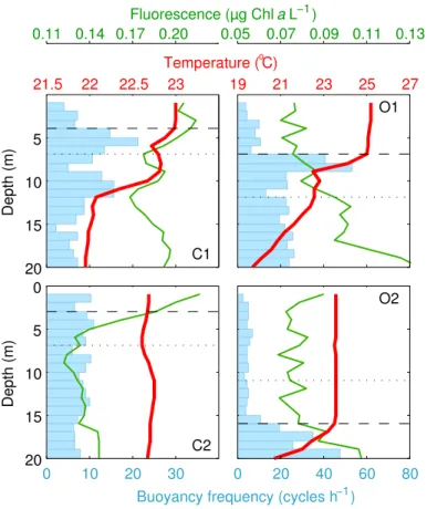
DMS photolysis was measured in 0.2 µm filtered-water incubations in 40 mL Teflon bottles or 50 mL quartz flasks. As expected, DMS photolysis was linearly related to the photochemically-weighted irradiance dose (Fig. 3). Since we observed distinct DMS
20
photolysis yields in coastal (C1–C2) versus oceanic (O1–O2) experiments, a photolysis
rate constant (k∗
photo) was used at each experimental location to correct the biological
rates for photochemical DMS loss.
All the variables were measured in duplicate incubation bottles except DMS produc-tion rates, which require large incubaproduc-tion volumes to properly account for food-web
25
BGD
10, 8851–8886, 2013DMS and C cycling in dynamic light fields
M. Galí et al.
Title Page
Abstract Introduction
Conclusions References
Tables Figures
◭ ◮
◭ ◮
Back Close
Full Screen / Esc
Printer-friendly Version Interactive Discussion
Discussion
P
a
per
|
Dis
cussion
P
a
per
|
Discussion
P
a
per
|
Discussio
n
P
a
per
2.3 Statistical analyses
Each variable was normalized within each experiment to the vertical integral of the fixed incubations. After pooling the four experiments together we checked for
signifi-cant differences among treatments. If the Bartlett’s equal variance test was
success-fully passed (p >0.05) a parametric one-way ANOVA was used. Otherwise, a
non-5
parametric Kruskal–Wallis ANOVA was performed. After a significant ANOVA (p <
0.05) multiple comparisons were done with the Tukey–Kramer test.
3 Results and discussion
3.1 Oceanographic settings
The sampled upper mixed layer was in all cases exposed to high proportions of UVR,
10
i.e.>10 % of the sub-surface UVA and UVB levels. Only in C2 the deeper portion of the
UML was exposed to<10 % of sub-surface UVB (Fig. 2; Table 1). The phytoplankton
community was typical of oligotrophic conditions, with low biomass and large
contribu-tions of the pico-sized fraction (Prochlorococcus, Synechococcusand picoeukaryotes)
though in different proportions (Table 1). The picoeukaryote fraction was likely
domi-15
nated by haptophytes (prymnesiophytes) and pelagophytes in O1 and O2 according to HPLC pigment data (Pérez et al., 2013). Diatoms in C1 and C2 and small
dinoflagel-lates (<10 µm) in O1 and O2 also made significant contributions to total phytoplankton
biomass. The presence of strong DMSP producers, such as dinoflagellates and
hap-tophytes, may explain the elevated DMSPt : Chlaratios of 196–315 µmol g−1 found in
20
the initial O1–O2 samples, compared to the 77–92 (already elevated) found in C1–C2. The mixing layer was very shallow at the coastal site (MLD of 3–4 m). In the oceanic setting, the UML deepened from 7 m (O1) to 16 m (O2) due to the passage of a storm (Fig. 2). The fact that all experiments took place in soft wind conditions, and the rela-tively high values of the buoyancy (Brunt–Väisälä) frequency within the UML suggest
BGD
10, 8851–8886, 2013DMS and C cycling in dynamic light fields
M. Galí et al.
Title Page
Abstract Introduction
Conclusions References
Tables Figures
◭ ◮
◭ ◮
Back Close
Full Screen / Esc
Printer-friendly Version Interactive Discussion
Discussion
P
a
per
|
Dis
cussion
P
a
per
|
Discussion
P
a
per
|
Discussio
n
P
a
per
that it was not mixing actively at the time of the CTD casts (Table 1). If we assume that
vertical diffusivity (Kz) in the UML interior was in the range 10−2–10−4m2s−1(Denman
and Gargett, 1983; Ross et al., 2011b), it would take between ca. 0.25 to 100 h for
a population of particles released at a single depth to diffuse across one optical depth
in the UML depending on the wavelengths and MLD considered (Gallegos and Platt,
5
1985). A similar range is obtained by calculating the mixing timescale as MLD2/Kz as
suggested by Ross et al. (2011a,b). The highestKz might be representative of
night-time convective overturning, while the lowestKz might be more representative of the
daytime, when mixing was likely inhibited by solar heating (Brainerd and Gregg, 1995). From these calculations we conclude that the simulated mixing times were
consid-10
erably faster than the actual mixing times. Although we tried to simulate the optical gradient experienced by the organisms and solutes within the UML, in practice the in-cubations spanned a larger optical gradient once the attenuation due to seawater and the incubation bottles was taken into account (Table 1).
Indeed, some of the differences between experiments and particularly between O1
15
and O2 may arise from slight differences in experimental exposure and prior light
his-tory of the plankton. Yet, our discussion will focus on the general trends rather than the
differences among individual experiments.
3.2 Phytoplankton photosynthetic performance and photoacclimation
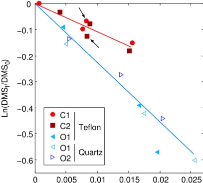
Particulate primary production (PPp) was moderately inhibited at the surface, optimal
20
at the middle depth, and slightly lower at the bottom, with the exception of C1 (Fig. 4a). PPp in mixing bottles resembled that in surface bottles and was 18 % lower than in
middle bottles except in C1 (p <0.01). As a result, vertically integrated PPp from fixed
bottles generally exceeded that in mixing bottles by 10–17 % (C1 excluded). This re-sult contrasts with that obtained by Bertoni et al. (2011), who observed a neutral to
25
positive effect of dynamic light exposure in coastal Mediterranean waters in late spring.
BGD
10, 8851–8886, 2013DMS and C cycling in dynamic light fields
M. Galí et al.
Title Page
Abstract Introduction
Conclusions References
Tables Figures
◭ ◮
◭ ◮
Back Close
Full Screen / Esc
Printer-friendly Version Interactive Discussion
Discussion
P
a
per
|
Dis
cussion
P
a
per
|
Discussion
P
a
per
|
Discussio
n
P
a
per
photoprotection and damage and repair processes that will be explored in the para-graphs below.
At the end of the incubations, the average fluorescence of Synechococcusand
pi-coeukaryote cell populations was generally lowest at the surface and increased with depth (Fig. 4b, c). Fluorescence was generally lower than average in mixing bottles
(al-5
though different patterns were observed for picoeukaryotes in O2). Similar responses
were observed for nanoeukaryotes in C2 and for Prochlorococcus in O1 (data not
shown), accompanied by marked decreases in Prochlorococcus cell counts, as
pre-viously shown by Sommaruga et al. (2005). In concordance with the response of
pop-ulations analysed with single-cell techniques, bulk phytoplankton Fv/Fm tended to
in-10
crease with incubation depth (Fig. 4d).Fv/Fmin mixing bottles was (again) lower than
the vertical integral of fixed bottles in C1 and O1, but not in O2, potentially due to the high fluorescence yields of the picoeukaryote population (Fig. 4c). The decrease in
fluorescence yields may simultaneously result from a decrease in chlorophylla(Chla)
content per cell (MacIntyre et al., 2002), an increase in excess energy dissipation as
15
heat by photoprotective carotenoids (non-photochemical quenching), photodamage of photosystem II, and pigment bleaching (Vincent and Neale, 2000).
Chla concentrations generally increased (by 10–30 %) during the experiments
ex-cept in O1, where a ca. 20 % decrease was found. In O1 and O2, the ratio of
photosyn-thetic carotenoids to Chla(PC/Chla) increased with depth, from ca. 0.48 at the surface
20
to ca. 0.56 in bottom bottles. PC/Chlain mixing bottles was close to the vertical integral
of fixed bottles (Fig. 4e). This suggests that phytoplankton photoacclimated during the
time frame of the experiment (6 h) by adjusting PC/Chla to the average spectral
irra-diance they were exposed to, likely seeking to optimize photosynthesis. Another phys-iological indicator that is worth analyzing is the ratio of photosynthetic carotenoids to
25
BGD
10, 8851–8886, 2013DMS and C cycling in dynamic light fields
M. Galí et al.
Title Page
Abstract Introduction
Conclusions References
Tables Figures
◭ ◮
◭ ◮
Back Close
Full Screen / Esc
Printer-friendly Version Interactive Discussion
Discussion
P
a
per
|
Dis
cussion
P
a
per
|
Discussion
P
a
per
|
Discussio
n
P
a
per
investment in photoprotection through non-photochemical quenching at higher spectral irradiance. This is consistent with the decrease in photosystem II fluorescence yields (Fig. 4d), since NPC compete for excitation energy with the other energy dissipation pathways: photochemistry and fluorescence emission. Surprisingly, mixing bottles dis-played the highest values of PC/NPC due to higher-than-average PC concentrations,
5
a response that remains difficult to interpret.
The xanthophyll cycle pigments diadinoxanthin (Dd) and diatoxanthin (Dt) were up-regulated by about 35 % (up to 75 %) during the exposure relative to their initial
con-centration. Likewise, (Dd+Dt) concentrations relative to Chl a increased by 50 % in
the ensemble of all treatments in O1 and O2. (Dd+Dt)/Chla generally increased
to-10
wards the surface, and showed intermediate values in mixing bottles (Fig. 4g). These xanthophylls constitute a photoprotective mechanism in haptophytes, dinoflagellates and diatoms (van de Poll and Buma, 2009) by which the epoxidated form (Dd) is enzymatically de-epoxidated to Dt, and vice-versa, depending on the cells’ need for photoprotection. Dd assists in light harvesting and Dt is thought to thermally dissipate
15
excess energy, so that a high value of the de-epoxidation state index Dt/(Dd+Dt)
indi-cates a stronger need for photoprotection. The highest Dt/(Dd+Dt) were observed in
surface bottles (and in the mixing bottle only in O1), with values between 0.50–0.70. However, this index must be viewed with caution because Dd to Dt interconversion can respond in a matter of minutes (i.e., faster than the filtration time after the experiment),
20
and because UV-driven de novo Dd synthesis may also lower the Dt/(Dd+Dt) index
(van de Poll and Buma, 2009). We also tried to calculate the de-epoxidation state of the violaxanthin-antheroxanthin-zeaxanthin (VAZ) cycle (a photoprotection system that
operates in chlorophytes and prasinophytes) as (V+0.5A)/(V+A+Z) following Sobrino
et al. (2005). However, we did not find meaningful trends in this index, perhaps
be-25
cause an active xanthophyll cycle is not found in cyanobacteria (Synechococcus) and
BGD
10, 8851–8886, 2013DMS and C cycling in dynamic light fields
M. Galí et al.
Title Page
Abstract Introduction
Conclusions References
Tables Figures
◭ ◮
◭ ◮
Back Close
Full Screen / Esc
Printer-friendly Version Interactive Discussion
Discussion
P
a
per
|
Dis
cussion
P
a
per
|
Discussion
P
a
per
|
Discussio
n
P
a
per
UV-absorbing (sunscreen) compounds, possibly mycosporine-like aminoacids (Shick and Dunlap, 2002), were observed in particulate absorption spectra in O1 and O2 (Fig. 4i). The ratio of particulate light absorption at 340 nm relative to that at the blue
peak of Chlaat 440 nm,ap,340/ap,440 was highest (1–1.5) in surface bottles and lower
(0.7–0.8) in middle and bottom bottles. Mixing bottles showed an ambiguous response,
5
with lowap,340/ap,440 in O1 and slightly higherap,340/ap,440 in O2.
The several photoresponse indicators we have explored indicate that, although
phy-toplankton deployed different photoprotection mechanisms, these were not enough to
counteract high PAR and UV-driven photoinhibition in surface bottles. Seen another way, the investment in photoprotection might have decreased the allocation of
re-10
sources to carbon fixation. In middle bottles, conversely, the combination of high PAR and longwave UVA (which can also be used for photosynthesis, Helbling et al., 2003) and a lower investment in photoprotection due to lower proportions of UVR resulted
in optimal PPp. It is also important to bear in mind that different phytoplankton groups
likely preferred different photoprotection mechanisms within those cited.
15
The response of mixing bottles is more difficult to interpret. The reduced
photosyn-thetic performance in C2, O1 and O2 might indicate that the short surface exposure received by mixing bottles was enough to cause some irreversible inhibition, and that phytoplankton repair capacity was limited. However, this is not clearly supported by
radiative stress indicators. In addition, repair is thought to be more efficient at elevated
20
temperatures like those encountered in our study (Campbell et al., 1998; van de Poll and Buma, 2009). The fact that surface inhibition was only moderate and that high-est PPp occurred in the middle bottle sugghigh-ests that the photosynthetic machinery of phytoplankton was well adapted to a stratified system and thus not geared to take ad-vantage of fast changes in spectral irradiance. This contrasts with what has been found
25
BGD
10, 8851–8886, 2013DMS and C cycling in dynamic light fields
M. Galí et al.
Title Page
Abstract Introduction
Conclusions References
Tables Figures
◭ ◮
◭ ◮
Back Close
Full Screen / Esc
Printer-friendly Version Interactive Discussion
Discussion
P
a
per
|
Dis
cussion
P
a
per
|
Discussion
P
a
per
|
Discussio
n
P
a
per
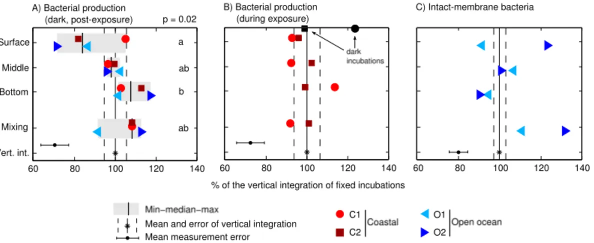
3.3 Response of bacterial heterotrophic production
In fixed bottle incubations, LIR were significantly inhibited at the surface by 14–28 % with respect to the vertical integral (except in C1), and increased with depth to find their optimum at the bottom of the mixed layer (Fig. 5a). LIR in mixing bottles re-sembled those of bottom bottles in 3 out of 4 experiments, and were higher (though
5
not significantly) than those in middle bottles and the vertical integral. This suggests that fast mixing favored recovery and photorepair over photodamage. It is well known that photolyase enzymes use UVA and blue light to repair damaged DNA. According to Kaiser and Herndl (1997), optimal photoreactivation occurs in a certain window of UVA/UVB that, in our experiments, would roughly correspond to the bottom half of
10
the UML (Fig. 1b). This interpretation is supported by the higher proportions of intact-membrane bacteria found in mixing bottles at the end of the incubations with respect to the surface bottles (O1 and O2 only; Fig. 5c). Yet, the vertical trend shown by this cytometric indicator in fixed bottles contradicts this view, especially in O2, where the proportion of intact-membrane bacteria decreased with depth.
15
In addition to the post-exposure dark incubations, in C1 and C2 we measured
LIR during the sunlit incubations, i.e., with the3H-leucine added into exposed bottles
(Fig. 5b). In these in situ incubations, surface and mixing bottles displayed more similar degrees of inhibition, and the trends of bacterial production with depth did not match those found in post-exposure dark incubations. We also measured LIR in aluminum
20
foil-darkened bottles placed in the in situ incubation basket. Dark LIR was 22 % higher
than the vertical integral of sunlit bottles in C1, but no differences were observed in C2
(Fig. 5b). The discrepancies between in situ and post-exposure leucine incorporation may be due to distinct photoinhibition and photorepair dynamics during sunlit and dark incubations, i.e., we do not know how much of the substrate was taken up before the
25
BGD
10, 8851–8886, 2013DMS and C cycling in dynamic light fields
M. Galí et al.
Title Page
Abstract Introduction
Conclusions References
Tables Figures
◭ ◮
◭ ◮
Back Close
Full Screen / Esc
Printer-friendly Version Interactive Discussion
Discussion
P
a
per
|
Dis
cussion
P
a
per
|
Discussion
P
a
per
|
Discussio
n
P
a
per
of bacterial heterotrophic production, which is particularly challenging in oligotrophic waters with low activity.
Different explanations have been invoked to explain the changes in bacterial
ac-tivity under sunlight. For instance, the occurrence of photoheterotrophic metabolisms in some bacterial groups, or the exudation of labile organic matter by phytoplankton at
5
high irradiance (see review by Ruiz-González et al., 2013). Unfortunately, we did not in-vestigate the phylogenetic composition of the bacterial communities in our experiments. No obvious patterns linking the response of LIR and PPp were found, perhaps because phytoplankton-bacteria interactions through the dissolved carbon pool are complex and group-specific (Sarmento and Gasol, 2012). Despite the numerous uncertainties, our
10
study adds valuable information to the only previous study of bacterial production un-der dynamic light exposure (Bertoni et al., 2011), and agrees with that work in that the
effect of mixing was neutral to positive compared to fixed incubations.
3.4 Response of gross DMS production
Gross DMS production showed the strongest vertical gradient among the three
pro-15
cesses, and increased significantly by about three-fold between the bottom and the surface of the UML in fixed incubations (Fig. 6a). Gross and DMS production in mixing
bottles was not significantly different from that in middle bottles, and not either from the
vertical integral, although a slight trend towards lower GP in mixing bottles occurred in C1 and C2.
20
Gross DMS production results from the addition and interaction of several processes, namely: exudation of DMS by phytoplankton, bacterial degradation of DMSP released by phytoplankton as a result of grazing, viral infection, or cell death, and even the re-duction of dimethylsulfoxide (Spiese et al., 2009; Asher et al., 2011). Galí et al. (2013)
showed that UVR stimulates GPDMS in a spectral irradiance-dependent manner, a
re-25
sult that is confirmed by our present study. They also demonstrated that the stimulation
is more effective at shorter and more energetic UVR wavelengths, with a spectral peak
BGD
10, 8851–8886, 2013DMS and C cycling in dynamic light fields
M. Galí et al.
Title Page
Abstract Introduction
Conclusions References
Tables Figures
◭ ◮
◭ ◮
Back Close
Full Screen / Esc
Printer-friendly Version Interactive Discussion
Discussion
P
a
per
|
Dis
cussion
P
a
per
|
Discussion
P
a
per
|
Discussio
n
P
a
per
caused by the additive effects of excess PAR (Stefels, 2000) and UVR stress (Sunda
et al., 2002). Furthermore, it was suggested that lethal UVR exposure could promote DMS production as a result of phytoplankton cell lysis and subsequent DMSP release. This mechanism would make more DMSP available to bacteria and to algal DMSP cleavage enzymes (“lyases”) released along with algal DMSP.
5
In the ensemble of all the experiments, experiment-normalized PPp and GPDMS
were weakly but significantly correlated (Pearson’sr =−0.58;p=0.018; Spearman’s
ρ=−0.51; p=0.044). Moreover, the response of GPDMS to radiative stress was
gen-erally consistent with the patterns of photoinhibition and photoprotection. Whether or not this response was the result of active physiological regulation of phytoplankton
10
cells remains to be elucidated. Clearly, better methods are needed to study the
rel-ative weight of different DMS production processes and their modulation by spectral
irradiance (Galí et al., 2013). Sunda et al. (2002) suggested that intracellular DMSP cleavage to DMS and further oxidation products might help phytoplankton cells
cop-ing with oxidative stress. Our data suggest that, even if GPDMS arose completely from
15
intracellular DMSP cleavage (which is very unlikely), this potential antioxidant mecha-nism would not be enough to counteract photoinhibition and ameliorate photosynthetic performance, even if working in tandem with other photoprotection mechanisms.
DMSP concentrations displayed only moderate changes (<5 % variation in 13 out of
16 incubations) and no clear trends were found across treatments (data not shown).
20
A strong DMSP depletion in surface bottles was only found in O2 (21 %). The stability of the DMSP concentration across spectral irradiance treatments is notable, given that (1) a lower amount of fixed carbon was available for DMSP synthesis in surface and mixing samples, and (2) higher amounts of DMSP were lost as DMS (and perhaps as
DMSP) at higher irradiance. The quotient of GPDMSto DMSPt was 0.42 d−
1
, 0.29 d−1,
25
0.18 d−1
, and 0.21 d−1
BGD
10, 8851–8886, 2013DMS and C cycling in dynamic light fields
M. Galí et al.
Title Page
Abstract Introduction
Conclusions References
Tables Figures
◭ ◮
◭ ◮
Back Close
Full Screen / Esc
Printer-friendly Version Interactive Discussion
Discussion
P
a
per
|
Dis
cussion
P
a
per
|
Discussion
P
a
per
|
Discussio
n
P
a
per
experiment-normalized net DMSP synthesis rates and PPp were correlated
(Pear-son’s r=0.50; p=0.048; Spearman’s ρ=0.65; p=0.006). Recent results suggest
that DMS can be produced intracellularly in phytoplankton through DMSP cleavage by OH radicals, without the need of DMSP cleavage enzymes (D. J. Kieber, personal
communication, 2012). In this case, intracellular DMSP would be a more effective
radi-5
cal scavenger than previously thought (Sunda et al., 2002), and this could explain why such an important proportion of the intracellular DMSP pool escapes as DMS without reacting with intracellular oxidants.
Net biological DMS production (NPbio,DMS) showed a pattern similar to that of GPDMS
(Fig. 6b). This variable is interesting in that it tells the net effect of sunlight on
biolog-10
ical DMS cycling, that is, on the difference between GPDMS and bacterial DMS
con-sumption. Bacterial DMS consumption rates, calculated by subtracting NPbio,DMS from
GPDMS, consumed on average 11 %, 31 %, 43 % and 14 % of GPDMS in surface,
mid-dle, bottom and mixing bottles, respectively. Thus, the imbalance between GPDMSand
bacterial DMS consumption increased with spectral irradiance due to UV and/or PAR
15
inhibition of bacterial DMS consumption and stimulation of GPDMS, making the vertical
gradient of NPbio,DMSeven larger than that of GPDMS(Fig. 6b). The net stimulating effect
of sunlight on biological DMS production was largely compensated by DMS photolysis, so that net overall DMS concentration changes were close to zero in all treatments, as already observed by Galí et al. (2013) with other experimental settings.
20
Bacterial DMS consumption, expressed as the % of vertically-integrated rates, was 49, 79, 125 and 78 in surface, middle, bottom and mixing bottles, respectively. Although
these results suffer from a large uncertainty due to error propagation, they suggest that
bacterial DMS consumption was more strongly inhibited than bulk LIR, and that it was photoinhibited in a dose-dependent manner. Severe photoinhibition was already
ob-25
served by Toole et al. (2006), who observed a similar response of bacterial DMS con-sumption and LIR. Since only a portion of the bacterial community is able to consume DMS through oxidation, it is likely that the photoresponse of bacterial DMS consumers
BGD
10, 8851–8886, 2013DMS and C cycling in dynamic light fields
M. Galí et al.
Title Page
Abstract Introduction
Conclusions References
Tables Figures
◭ ◮
◭ ◮
Back Close
Full Screen / Esc
Printer-friendly Version Interactive Discussion
Discussion
P
a
per
|
Dis
cussion
P
a
per
|
Discussion
P
a
per
|
Discussio
n
P
a
per
or even that the photoresponse of different metabolic activities differs in a given cell or
strain. Clearly, these issues deserve further investigation.
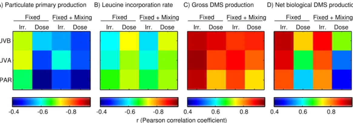
3.5 Differential irradiance- and dose-response among biogeochemical processes
The experiment-normalized PPp, LIR and community DMS production rates were
plot-5
ted against the mean (UVB, UVA, PAR) incubation irradiance in the ensemble of all the experiments, and the points corresponding to fixed bottles were fitted with a linear
regression (Fig. 7). We also calculated the Pearson correlation coefficient between the
experiment-normalized process rates and (1) mean irradiance and (2) total irradiance dose for each radiation band (Fig. 8). The aim of this exercise was to identify whether
10
a process was more dose-dependent or irradiance (“dosage-rate”)-dependent, follow-ing the rationale exposed in the Introduction. Note that in our experimental settfollow-ing it
is hard to discriminate between the effects of each band of the spectrum, since the
proportion of shortwave UV decreases along with total (or PAR) irradiance as we move deeper in the water column.
15
PPp showed a slight negative trend with respect to irradiance in fixed bottles in the three radiation bands, which was mainly driven by photoinhibition in surface bottles.
In fact, the response was rather flat below an irradiance threshold of ca. 0.4 W m−2
UVB, 16 W−2UVA and 1000 µmol photons m−2s−1. The correlation with irradiance was
higher than that with dose (Fig. 8a), suggesting that some balance between inhibition
20
and protection/repair could be attained in the different exposure regimes. The
high-est linear correlation was found with UVA irradiance, perhaps indicating that this band drives photoinhibition in UV-transparent waters. In concordance with this suggestion, some studies have shown that the spectral peak of UV photoinhibition occurs in the UVA, due to the combination of increasing irradiance and decreasing UV efectiveness
25
as we move towards longer wavelengths (Neale and Kieber, 2000).
BGD
10, 8851–8886, 2013DMS and C cycling in dynamic light fields
M. Galí et al.
Title Page
Abstract Introduction
Conclusions References
Tables Figures
◭ ◮
◭ ◮
Back Close
Full Screen / Esc
Printer-friendly Version Interactive Discussion
Discussion
P
a
per
|
Dis
cussion
P
a
per
|
Discussion
P
a
per
|
Discussio
n
P
a
per
strongly correlated to the dose than to irradiance (Fig. 8b), particularly in the UVB band, suggesting that cumulative UVB-induced DNA damage occurred in bacterial cells in fixed incubations (Buma et al., 2001). This fits with the general idea that the radiation bands causing damage (UVB) elicit more dose-dependent responses than the radiation bands that are used by the cells to conduct physiological processes (PAR and longwave
5
UVA).
Community DMS production rates showed a strong response to variations in spectral
irradiance, with a steeper slope observed for NPbio,DMSthan for GPDMS(Fig. 7c, d). The
strongest correlations were found between GPDMSand irradiance in the three bands,
particularly in the UVA. This agrees with previous studies that suggested, using distinct
10
approaches, that the spectral peak of sunlight-induced DMS production occurs in the 330–340 nm region in surface UV-transparent waters (Toole et al., 2008; Levine et al., 2012; Galí et al., 2013).
Finally, note that among all process and radiation combinations the correlation was stronger when mixing bottles were excluded. This illustrates in a loose way that mixing
15
disrupted the photoacclimation and photodamage processes, as thoroughly discussed in Sects. 3.2–3.4.
4 Conclusions
The photoresponse of phytoplankton, bacterioplankton, and community DMS produc-tion displayed clear trends in bottles incubated at fixed depths in the UML (Fig. 7)
de-20
spite the relatively small gradient in spectral irradiance. The irradiance dose-response in mixing bottles was distinct (though subtle) in each of the processes measured, as
well as for different physiological indicators. In the oligotrophic waters investigated,
dy-namic light exposuregenerally caused, compared to the middle bottles receiving the
same cumulative exposure (1) an adverse though non significant effect on particulate
25
primary production, concomitant with reduced cell-specific fluorescence in most
BGD
10, 8851–8886, 2013DMS and C cycling in dynamic light fields
M. Galí et al.
Title Page
Abstract Introduction
Conclusions References
Tables Figures
◭ ◮
◭ ◮
Back Close
Full Screen / Esc
Printer-friendly Version Interactive Discussion
Discussion
P
a
per
|
Dis
cussion
P
a
per
|
Discussion
P
a
per
|
Discussio
n
P
a
per
photoinhibition, related to an increase in the proportion of intact-membrane or “live”
heterotrophic bacteria in two of the experiments; and (3) a neutral effect or slight
re-duction in gross DMS prore-duction. These responses translated, in some experiments, into measurable deviations with respect to the vertically-integrated rates in the water
column; in others, the effects were close to neutral or too small to be reliably detected.
5
Incubating the samples at a fixed intermediate optical depth appears as a reasonable
and convenient solution for measuring GPDMS and leucine incorporation, at least in
UVR-transparent stratified UML waters. However, this solution might not be optimal for measuring UML-integrated primary production. Our results call for a more systematic assessment of the consequences of dynamic light exposure of microbial plankton in
10
different oceanic regimes. This way, the photobiological processes governing, among
other important processes, the ocean-atmosphere exchange of long-lived (CO2) and
short-lived (DMS) gases of climatic relevance will be better understood.
Acknowledgements. We thank the staffat Port Olímpic de Barcelona for their collaboration, and the crew and scientists aboard R/V García del Cid for their invaluable help in setting up
15
the experiments during the SUMMER-I cruise. We also thank David J. Kieber for his insightful comments on an earlier version of the manuscript. M. G. acknowledges the receipt of a CSIC JAE scholarship. This work was supported by the (former) Spanish Ministry of Science and Innovation through the project SUMMER (CTM2008-03309/MAR). This is a contribution of the Research Groups on Marine Biogeochemistry and Global Change and on Aquatic Microbial
20
Food Webs, supported by the Generalitat de Catalunya.
References
Aas, P., Lyons, M., Pledger, R., Mitchell, D., and Jeffrey, W.: Inhibition of bacterial activities by solar radiation in nearshore waters and the Gulf of Mexico, Aquat. Microb. Ecol., 11, 229– 238, doi:10.3354/ame011229, 1996. 8854
25
BGD
10, 8851–8886, 2013DMS and C cycling in dynamic light fields
M. Galí et al.
Title Page
Abstract Introduction
Conclusions References
Tables Figures
◭ ◮
◭ ◮
Back Close
Full Screen / Esc
Printer-friendly Version Interactive Discussion
Discussion
P
a
per
|
Dis
cussion
P
a
per
|
Discussion
P
a
per
|
Discussio
n
P
a
per
Alonso-Sáez, L., Gasol, J. M., Lefort, T., Hofer, J., and Sommaruga, R.: Effect of natural sunlight on bacterial activity and differential sensitivity of natural bacterioplankton groups in northwestern Mediterranean coastal waters, Appl. Environ. Microb., 72, 5806–5813, doi:10.1128/AEM.00597-06, 2006. 8854
Asher, E. C., Dacey, J. W. H., Mills, M. M., Arrigo, K. R., and Tortell, P. D.: High concentrations
5
and turnover rates of DMS, DMSP and DMSO in Antarctic sea ice, Geophys. Res. Lett., 38, 1–5, doi:10.1029/2011GL049712, 2011. 8866
Barbieri, E. S., Villafañe, V. E., Helbling, E. W., and Nov, N.: Experimental Assessment of UV Effects on Temperate Marine Phytoplankton When Exposed to Variable Radiation Regimes Experimental assessment of UV effects on temperate marine phytoplankton when exposed
10
to variable radiation regimes, Limnol. Oceanogr., 47, 1648–1655, 2002. 8854
Béjà, O., Aravind, L., Koonin, E. V., Suzuki, M. T., Hadd, A., Nguyen, L. P., Jovanovich, S. B., Gates, C. M., Feldman, R. A., Spudich, J. L., Spudich, E. N., and DeLong, E. F.: Bacterial Rhodopsin: evidence for a new type of phototrophy in the sea, Science, 289, 1902–1906, doi:10.1126/science.289.5486.1902, 2000. 8855
15
Bertoni, R., Jeffrey, W. H., Pujo-Pay, M., Oriol, L., Conan, P., and Joux, F.: Influence of wa-ter mixing on the inhibitory effect of UV radiation on primary and bacterial production in Mediterranean coastal water, Aquat. Sci., 73, 377–387, doi:10.1007/s00027-011-0185-8, 2011. 8861, 8864, 8866
Brainerd, K. and Gregg, M.: Surface mixed and mixing layer depths, Deep-Sea Res. Pt. I, 42,
20
1521–1543, 1995. 8861
Bricaud, A., Babin, M., Morel, A., and Claustre, H.: Variability in the chlorophyll-specific ab-sorption coefficients of natural phytoplankton: analysis and parameterization, J. Geophys. Res.-Oceans, 100, 13321–13332, doi:10.1029/95JC00463, 1995. 8862
Brunet, C. and Lavaud, J.: Can the xanthophyll cycle help extract the essence of the
microal-25
gal functional response to a variable light environment?, J. Plankton Res., 32, 1609–1617, doi:10.1093/plankt/fbq104, 2010. 8854
Buma, A. G., Helbling, E. W., de Boer, M. K., and Villafañe, V. E.: Patterns of DNA damage and photoinhibition in temperate South-Atlantic picophytoplankton exposed to solar ultraviolet radiation, J. Photoch. Photobio. B, 62, 9–18, 2001. 8854, 8870
30
BGD
10, 8851–8886, 2013DMS and C cycling in dynamic light fields
M. Galí et al.
Title Page
Abstract Introduction
Conclusions References
Tables Figures
◭ ◮
◭ ◮
Back Close
Full Screen / Esc
Printer-friendly Version Interactive Discussion
Discussion
P
a
per
|
Dis
cussion
P
a
per
|
Discussion
P
a
per
|
Discussio
n
P
a
per
Charlson, R., Lovelock, J., Andreae, M., and Warren, S.: Oceanic phytoplankton, atmospheric sulphur, cloud albedo and climate, Cah. Rev. The., 326, 655–661, 1987. 8855
Church, M. J., Ducklow, H. W., and Karl, D. M.: Light dependence of [3H]leucine incor-poration in the oligotrophic North Pacific ocean, Appl. Environ. Microb., 70, 4079–4087, doi:10.1128/AEM.70.7.4079-4087.2004, 2004. 8854
5
Denman, K. L. and Gargett, A. E.: Time and space scales of vertical mixing in the upper ocean, Limnol. Oceanogr., 28, 801–815, 1983. 8861
Galí, M. and Simó, R.: Occurrence and cycling of dimethylated sulfur compounds in the Arctic during summer receding of the ice edge, Mar. Chem., 122, 105–117, doi:10.1016/j.marchem.2010.07.003, 2010. 8868
10
Galí, M., Saló, V., Almeda, R., Calbet, A., and Simó, R.: Stimulation of gross dimethylsulfide (DMS) production by solar radiation, Geophys. Res. Lett., 38, 1–5, doi:10.1029/2011GL048051, 2011. 8855, 8859
Galí, M., Ruiz-González, C., Lefort, T., Gasol, J. M., Cardelús, C., Romera-Castillo, C., and Simó, R.: Spectral dependence of sunlight effects on plankton dimethylsulfide production,
15
Limnol. Oceanogr., 58, 489–504, 2013. 8855, 8857, 8858, 8859, 8866, 8867, 8868, 8870, 8881
Gallegos, C. L. and Platt, T.: Vertical advection of phytoplankton and productivity estimates: a dimensional analysis, Mar. Ecol.-Prog. Ser., 26, 125–134, 1985. 8853, 8854, 8861
Grégori, G., Citterio, S., Ghiani, A., Labra, M., Sgorbati, S., Brown, S., and Denis, M.: Resolution
20
of viable and membrane-compromised bacteria in freshwater and marine waters based on analytical flow cytometry and nucleic acid double staining, Appl. Environ. Microb., 67, 4662– 4670, 2001. 8859
Helbling, E. W., Gao, K., Gonçalves, R., Wu, H., and Villafañe, V. E.: Utilization of solar UV radiation by coastal phytoplankton assemblages offSE China when exposed to fast mixing,
25
Mar. Ecol.-Prog. Ser., 259, 59–66, doi:10.3354/meps259059, 2003. 8864
Helbling, E. W., Carrillo, P., Medina-Sánchez, J. M., Durán, C., Herrera, G., Villar-Argaiz, M., and Villafañe, V. E.: Interactive effects of vertical mixing, nutrients and ultraviolet radiation: in situ photosynthetic responses of phytoplankton from high mountain lakes in Southern Europe, Biogeosciences, 10, 1037–1050, doi:10.5194/bg-10-1037-2013, 2013. 8854
30
BGD
10, 8851–8886, 2013DMS and C cycling in dynamic light fields
M. Galí et al.
Title Page
Abstract Introduction
Conclusions References
Tables Figures
◭ ◮
◭ ◮
Back Close
Full Screen / Esc
Printer-friendly Version Interactive Discussion
Discussion
P
a
per
|
Dis
cussion
P
a
per
|
Discussion
P
a
per
|
Discussio
n
P
a
per
Kirchman, D. L. and Hanson, T. E.: Bioenergetics of photoheterotrophic bacteria in the oceans, Environ. Microbiol. Rep., 5, 188–199, doi:10.1111/j.1758-2229.2012.00367.x, 2012. 8855 Kirchman, D., K’nees, E., and Hodson, R.: Leucine incorporation and its potential as a measure
of protein synthesis by bacteria in natural aquatic systems, Appl. Environ. Microb., 49, 599– 607, 1985. 8857
5
Kolber, Z., Van Dover, C., Niederman, R., and Falkowski, P.: Bacterial photosynthesis in surface waters of the open ocean, Cah. Rev. The., 407, 177–179, 2000. 8855
Lana, A., Bell, T. G., Simó, R., Vallina, S. M., Ballabrera-Poy, J., Kettle, A. J., Dachs, J., Bopp, L., Saltzman, E. S., Stefels, J., Johnson, J. E., and Liss, P. S.: An updated climatology of surface dimethlysulfide concentrations and emission fluxes in the global ocean, Global Biogeochem.
10
Cy., 25, 1–17, doi:10.1029/2010GB003850, 2011. 8855
Levine, N. M., Varaljay, V. A., Toole, D. A., Dacey, J. W. H., Doney, S. C., and Moran, M. A.: Envi-ronmental, biochemical and genetic drivers of DMSP degradation and DMS production in the Sargasso Sea, Environ. Microbiol., 14, 1210–1223, doi:10.1111/j.1462-2920.2012.02700.x, 2012. 8870
15
MacIntyre, H. L., Kana, T. M., Anning, T., and Geider, R. J.: Photoacclimation of photosyn-thesis irradiance response curves and photosynthetic pigmentspigments in microalgae and cyanobacteria, J. Phycol., 38, 17–38, 2002. 8854, 8862
Marie, D. and Partensky, F.: Analyse de micro-organismes marins, in: La Cytométrie en Flux, edited by: Ronot, X., Grunwald, D., Mayol, J. F., and Boutonnat, J., Lavoisier, 211–233, 2006.
20
8858
Mitchell, B. G. and Kiefer, D. A.: Determination of absorption and fluorescence excitation spec-tra for phytoplankton, in: Marine Phytoplankton and Productivity, edited by: Holm-Hansen, O., Bolis, L., and Gilles, R., Springer, 157–169, 1984. 8858
Mitchell, B. G., Bricaud, A., Carder, K., Cleveland, J., Ferrari, G., Gould, R., Kahru, M., Kishino,
25
M., Maske, H., Moisan, T., Moore, L., Nelson, N., Phinney, D., Reynolds, R., Sosik, H., Stram-ski, D., Tassan, S., Trees, C., Weidemann, A., Weiland, J., and Vodacek, A.: Determination of spectral absorption coefficients of particles, dissolved material and phytoplankton for dis-crete water samples, in: Ocean Optics Protocols for Satellite Ocean Color Sensor Validation, Revision 2, edited by: Fargion, G., Mueller, J., and McClain, C., NASA, 125–153, 2000. 8858
30
BGD
10, 8851–8886, 2013DMS and C cycling in dynamic light fields
M. Galí et al.
Title Page
Abstract Introduction
Conclusions References
Tables Figures
◭ ◮
◭ ◮
Back Close
Full Screen / Esc
Printer-friendly Version Interactive Discussion
Discussion
P
a
per
|
Dis
cussion
P
a
per
|
Discussion
P
a
per
|
Discussio
n
P
a
per
Moran, X. A. G., Massana, R., and Gasol, J. M.: Light conditions affect the measurement of oceanic bacterial production via leucine uptake, Appl. Environ. Microb., 67, 3795–3801, doi:10.1128/AEM.67.9.3795-3801.2001, 2001. 8854
Neale, P. J. and Kieber, D. J.: Assessing biological and chemical effects of UV in the marine environment: spectral weighting functions, Issues in Environmental Science and Technology,
5
2000. 8869
Neale, P. J., Helbling, E. W., and Zagarese, H. E.: Modulation of UVR exposure and effects by vertical mixing and advection, in: UV Effects in Aquatic Organisms and Ecosystems, chap. 4, Royal Society of Chemistry, 107–134, 2003. 8854
Quinn, P. K. and Bates, T. S.: The case against climate regulation via oceanic phytoplankton
10
sulphur emissions, Cah. Rev. The., 480, 51–6, doi:10.1038/nature10580, 2011. 8855 Ross, O. N., Geider, R. J., Berdalet, E., Artigas, M. L., and Piera, J.: Modelling the effect of
vertical mixing on bottle incubations for determining in situ phytoplankton dynamics. I. Growth rates, Mar. Ecol.-Prog. Ser., 435, 13–31, doi:10.3354/meps09193, 2011a. 8854, 8861 Ross, O. N., Geider, R. J., and Piera, J.: Modelling the effect of vertical mixing on bottle
incuba-15
tions for determining in situ phytoplankton dynamics. II. Primary production, Mar. Ecol.-Prog. Ser., 435, 33–45, doi:10.3354/meps09194, 2011b. 8854, 8861
Roy, S.: The strategies for minimization of UV damage, in: The Effects of UV Radiation in the Marine Environment, chap. 3, edited by: de Mora, S. J., Demers, S., and Vernet, M., Cambridge University Press, Cambridge, 177–205, 2000. 8854
20
Ruiz-González, C., Galí, M., Lefort, T., Cardelús, C., Simó, R., and Gasol, J. M.: Annual vari-ability in light modulation of bacterial heterotrophic activity in surface northwestern Mediter-ranean waters, Limnol. Oceanogr., 57, 1376–1388, doi:10.4319/lo.2012.57.5.1376, 2012. 8854
Ruiz-González, C., Simó, R., Sommaruga, R., and Gasol, J. M.: Away from darkness: a review
25
on the effects of solar radiation on heterotrophic bacterioplankton activity, Front. Microbiol., 4, 131, doi:10.3389/fmicb.2013.00131, 2013. 8855, 8866
Saló, V., Simó, R., and Calbet, A.: Revisiting the dilution technique to quantify the role of micro-zooplankton in DMS(P) cycling: laboratory and field tests, J. Plankton Res., 32, 1255–1267, doi:10.1093/plankt/fbq041, 2010. 8859
30
BGD
10, 8851–8886, 2013DMS and C cycling in dynamic light fields
M. Galí et al.
Title Page
Abstract Introduction
Conclusions References
Tables Figures
◭ ◮
◭ ◮
Back Close
Full Screen / Esc
Printer-friendly Version Interactive Discussion
Discussion
P
a
per
|
Dis
cussion
P
a
per
|
Discussion
P
a
per
|
Discussio
n
P
a
per
Shick, J. M. and Dunlap, W. C.: Mycosporine-like amino acids and related Gadusols: biosyn-thesis, acumulation, and UV-protective functions in aquatic organisms, Annu. Rev. Physiol., 64, 223–62, doi:10.1146/annurev.physiol.64.081501.155802, 2002. 8864
Simó, R.: From cells to globe: approaching the dynamics of DMS(P) in the ocean at multiple scales, Can. J. Fish. Aquat. Sci., 61, 673–684, doi:10.1139/F04-030, 2004. 8855
5
Simó, R., Pedrós-Alió, C., Malin, G., and Grimalt, J. O.: Biological turnover of DMS, DMSP and DMSO in contrasting open-sea waters, Mar. Ecol.-Prog. Ser., 203, 1–11, doi:10.3354/meps203001, 2000. 8859
Smith, D. C. and Azam, F.: A simple, economical method for measuring bacterial protein syn-thesis rates in seawater using3H-leucine, Mar. Microb. Food Webs, 6, 107–114, 1992. 8857
10
Sobrino, C., Neale, P. J., Montero, O., and Lubián, L. M.: Biological weighting function for xanthophyll de-epoxidation induced by ultraviolet radiation, Physiol. Plantarum, 125, 41–51, doi:10.1111/j.1399-3054.2005.00538.x, 2005. 8863
Sommaruga, R., Hofer, J. S., Alonso-Sáez, L., and Gasol, J. M.: Differential sunlight sensitivity of picophytoplankton from surface Mediterranean coastal waters, Appl. Environ. Microb., 71,
15
2154–2157, doi:10.1128/AEM.71.4.2154-2157.2005, 2005. 8862
Spiese, C., Kieber, D. J., Nomura, C., and Kiene, R. P.: Reduction of dimethylsulfoxide to dimethylsulfide by marine phytoplankton, Limnol. Oceanogr., 54, 560–570, 2009. 8866 Stefels, J.: Physiological aspects of the production and conversion of DMSP in marine algae
and higher plants, J. Sea Res., 43, 183–197, doi:10.1016/S1385-1101(00)00030-7, 2000.
20
8867
Sunda, W., Kieber, D. J., Kiene, R. P., and Huntsman, S.: An antioxidant function for DMSP and DMS in marine algae, Cah. Rev. The., 418, 317–20, doi:10.1038/nature00851, 2002. 8867, 8868
Toole, D. A., Slezak, D., Kiene, R. P., Kieber, D. J., and Siegel, D. A.: Effects of solar radiation
25
on dimethylsulfide cycling in the western Atlantic Ocean, Deep-Sea Res. Pt. I, 53, 136–153, 2006. 8868
Toole, D. A., Siegel, D. A., and Doney, S. C.: A light-driven, one-dimensional dimethylsul-fide biogeochemical cycling model for the Sargasso Sea, J. Geophys. Res., 113, 1–20, doi:10.1029/2007JG000426, 2008. 8870
30
BGD
10, 8851–8886, 2013DMS and C cycling in dynamic light fields
M. Galí et al.
Title Page
Abstract Introduction
Conclusions References
Tables Figures
◭ ◮
◭ ◮
Back Close
Full Screen / Esc
Printer-friendly Version Interactive Discussion
Discussion
P
a
per
|
Dis
cussion
P
a
per
|
Discussion
P
a
per
|
Discussio
n
P
a
per
van de Poll, W. H. and Buma, A. G. J.: Does ultraviolet radiation affect the xanthophyll cycle in marine phytoplankton?, Photoch. Photobio. Sci., 8, 1295–1301, doi:10.1039/B904501E, 2009. 8863, 8864
Vincent, W. F. and Neale, P. J.: Mechanisms of UV damage to aquatic organisms, in: The Effects of UV Radiation in the Marine Environment, chap. 6, edited by: de Mora, S. J., Demers, S.,
5
and Vernet, M., Cambridge University Press, Cambridge, 149–176, 2000. 8854, 8862 Wolfe, G. and Kiene, R. P.: Radioisotope and chemical inhibitor measurements of dimethyl
sulfide consumption rates and kinetics in estuarine waters, Mar. Ecol.-Prog. Ser., 99, 261– 269, 1993. 8859
Zapata, M., Rodríguez, F., and Garrido, J. L.: Separation of chlorophylls and carotenoids from
10