First report of Xiphinema brevicolle in Japan 21
First report of Xiphinema brevicolle Lordello et Costa,
1961 (Nematoda, Longidoridae) in Japan
Hiromichi Sakai1,†, Ai Takeda2,‡, Takayuki Mizukubo1,§
1 National Agriculture and Food Research Organization, Agricultural Research Center, Kannondai 3-1-1, Tsu-kuba, Ibaraki 305-8666, Japan 2 Chiba Prefectural Agriculture and Forestry Research Center, 808 Daizenno, Midori-Ku, Chiba 266-0006 Japan
Corresponding author: Hiromichi Sakai (sakaihm@afrc.go.jp)
Academic editor:Sergei Subbotin | Received 15 June 2011 | Accepted 23 August 2011 | Published 7 October 2011
Citation: Sakai H, Takeda A, Mizukubo T (2011) First report of Xiphinema brevicolle Lordello et Costa, 1961 (Nematoda, Longidoridae) in Japan. ZooKeys 135: 21–40. doi: 10.3897/zookeys.135.1716
Abstract
Mixed populations of Xiphinemaamericanum-group species were detected from a root zone soil sample of Japanese holly, Ilex crenata, during a survey for plant-parasitic nematodes of commercial ornamental plant nurseries in Chiba Prefecture, Japan. From the result of the morphological study, the species were identiied as X. brevicolle and Xiphinema sp. his is the irst record of X. brevicolle in Japan. Morphometrics of X. brevicolle generally agree with those of the type specimens and the topotype specimens. Xiphinema
sp. morphometrically resembles X. paramonovi except for tail length. he mitochondrial COI region, the nuclear 18S rDNA and the nuclear large subunit rDNA D2/D3 region of the species were sequenced and compared in the molecular study. For the COI region, PCR primers were newly designed to obtain longer sequences, ca. 900 bp, than previously used. Sequence identities of COI, 18S and D2/D3 regions between these two populations were 84.0-84.1%, 99.9% and 98.1-98.2%, respectively. Phylogenetic analyses of maximum likelihood trees were carried out to compare genetic relationships among the group and some suggestions were made on the X. brevicolle-subgroup.
Keywords
COI, Ilex crenata, Japanese holly, rDNA, Xiphinema americanum-group, Xiphinema brevicolle-subgroup
Introduction
During a survey for plant-parasitic nematodes of commercial ornamental plant nurser-ies in Chiba Prefecture, Japan, we detected mixed populations of Xiphinema america-num-group species from a root zone soil sample of Japanese holly, Ilex crenata hunb., ZooKeys 135: 21–40 (2011)
doi: 10.3897/zookeys.135.1716 www.zookeys.org
Copyright Hiromichi Sakai et al. This is an open access article distributed under the terms of the Creative Commons Attribution License, which permits unrestricted use, distribution, and reproduction in any medium, provided the original author and source are credited.
ReseARCh ARtICLe
Launched to accelerate biodiversity research
Hiromichi Sakai et al. / ZooKeys 135: 21–40 (2011)
22
one of the major garden tree species in Japan. his study was conducted using morpho-logical characters of females and DNA sequences of the mitochondrial COI region and the nuclear ribosomal RNA (rDNA) regions to identify and characterize the species in the mixed populations.
Methods
Collection of the nematode specimens
he soil sample containing the Xiphinema spp. (No. 001-001) was taken from the root zone of Japanese holly growing in a commercial plant nursery at Sosa City, Chiba, Japan. Nematodes were extracted with Cobb’s wet-sieving technique. Material col-lected on a 75 µm mesh sieve was placed on a Baermann funnel and nematodes were collected after one day at room temperature.
Morphological observation
Females of Xiphinema spp. were removed from the nematode suspension using a dis-secting microscope and nematode pick. Female nematodes were transferred to a small amount of water then killed by either heating at 60°C for 2 min or by adding hot FP4:1 (Netscher and Seinhorst 1969). Killed nematodes were ixed with FG4:1 (De Grisse 1969). he specimens were ixed for more than one week, processed into glyc-erin by the ethanol/glycglyc-erin method (Seinhorst 1959, De Grisse 1969), and mounted in dehydrated glycerin supported with both minute glass beads and parain on glass slides. Morphological observations were made using a DIC microscope (BX51, Olym-pus Co., Japan). A digital camera, OlymOlym-pus DP20 or DP21 was used for measuring and taking photo images. he body length and position of vulva were measured with a digital curvimeter (CV-9 Jr., Koizumi Sokki Mfg. Co. Ltd., Japan) on nematode line drawings prepared using a drawing tube. Illustrations of nematodes were sketched di-rectly on highly transparent tracing ilm (No. 200Z-A4 (S): Tochiman Technical Paper Co. Ltd., Japan) by tracing DP21 digital camera images on a panel-protected LC-display. Sketched images were converted to digital images using the software Adobe Illustrator CS4 (Adobe Systems Inc., USA) and a pen tablet (Intuos4: Wacom Co., Ltd., Japan).
Molecular study
First report of Xiphinema brevicolle in Japan 23
digital images of mounts were photographed, and then images were measured for body and odontostyle length, respectively.
DNA fragments of the mitochondrial COI region were ampliied by PCR us-ing the set of primers, CO1-F1 and CO1-R1 (Table 1), which were originally de-signed from sequence comparison of the COI region between X. americanum (He et al. 2005a: GenBank accession AY382608; NCBI Reference Sequence NC_005928) and Caenorhabditis elegans (Howe and Denver 2008: GenBank accession EU407804). CO1-R1 is virtually identical to COIR (He et al. 2005a), with the latter having only a diference in degeneracy. he PCR reaction mixture consisted of 0.2 mM dNTPs, 0.3 µM of each primer, 0.5 U PrimeSTAR HS DNA Polymerase with PrimeSTAR Bufer (5 mM Mg2+ plus) (Takara Bio Inc., Japan), and 10 µL of DNA lysate as PCR
template, in a total volume of 20 µL. he reaction conditions were as follows: a single step of pre-denaturation at 94°C for 2 min, followed by 35 cycles of denaturation at 94°C for 30 s, annealing at 40°C for 15 s, 0.5°C/s ramp up to extension temperature, and extension at 72°C for 1 min.
DNA fragments of the nuclear 18S rDNA region were ampliied using the set of primers, 18S 39 and 18S 1573R (Mullin et al. 2005). he PCR reaction mixture consisted of 0.2 mM dNTPs, 0.2 µM of each primer, 0.5 U TaKaRa Ex Taq Hot Start Version with Ex Taq Bufer (Mg2+ plus) (Takara Bio), and 2 µL of DNA lysate as PCR
template, in a total volume of 20 µL. he reaction conditions were as follows: a single step of pre-denaturation at 94°C for 2 min, followed by 35 cycles of denaturation at 94°C for 30s, annealing at 55°C for 30s, and extension at 72°C for 1 min.
DNA fragments of the nuclear large subunit rDNA D2/D3 region were ampliied using the set of primers, D2Ab (De Ley et al. 1999) and D3B (homas et al. 1997). he PCR reaction conditions were the same as those for 18S rDNA.
hese PCR products were puriied with QIAquick PCR Puriication Kit (Qiagen K.K., Japan), subjected to cycle sequencing with BigDye Terminator v3.1 Cycle Se-quencing Kit (Life Technologies Japan), puriied with either DyeEx 2.0 Spin Kit (Qia-gen) or Agencourt CleanSEQ (Beckman Coulter Inc., USA), and sequenced on the ABI PRISM 3100 Genetic Analyzer (Life Technologies Japan Ltd., Japan). Primers 18S 599R, 18S 550, 18S 977R, 18S 965 (Mullin et al. 2005), and D3B (homas et al. 1997) were also used as inner primers for sequencing as well as those designed by us (Table 1). he DNA sequences were aligned by MUSCLE (Edgar 2004) and arranged using BioEdit (Hall 1999). Multiple alignments were manually reined where necessary.
Hiromichi Sakai et al. / ZooKeys 135: 21–40 (2011)
24
G, the CNI method, bootstrapping with 500 replications, and a maximum parsimony (MP) tree as the initial tree were employed. A ML tree was constructed using 342 sites for multiply aligned sequences of the COI region, where the model T92 + G + I, the CNI method, bootstrapping with 500 replications, and a MP tree as the initial tree were employed. Among COI homologous sequences of the X. americanum-group in the database, those including alignment gaps were excluded from the analysis because they possibly represent pseudogenes. Sequences of Longidorus species available in Gen-Bank were used to root those trees.
Results
he DNA sequence analysis of the mitochondrial COI region for 9 specimens sug-gested two diferent X. americanum-group species were present. Morphometric data were used to identify them as Xiphinema brevicolle Lordello et Costa, 1961, and an undescribed X. americanum-group species.
Morphological observations
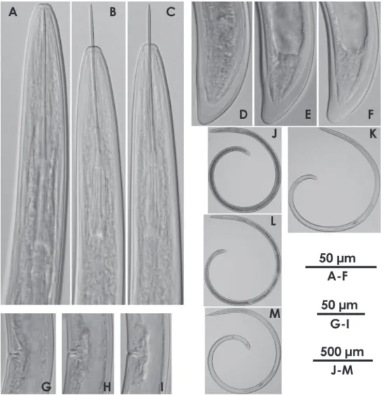
Xiphinema brevicolle Lordello & Costa, 1961
http://species-id.net/wiki/Xiphinema_brevicolle Figs 1A, 1C, 1E, 1G, 2, 4A, 5A, 6A
Measurements. See Tables 2, 3.
Remarks. Morphometrics of the specimens obtained here generally agree with
those of the type specimens and topotype specimens (Lamberti et al. 1992, Luc et al. 1998) of the species (Table 2). No male was detected.
Nomenclatorial note. he emended name of this species, Xiphinema brevicollum,
was proposed by Luc et al. (1998) and used in many works to date. Monteiro (2010) claimed that the correct species name is Xiphinema brevicolle Lordello et Costa, 1961, and should have been preserved unaltered. We support the claim by Monteiro (2010) and X. brevicolle is used here.
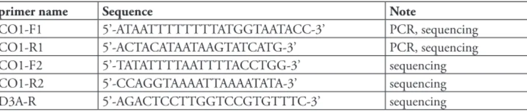
table 1. Primers designed and employed originally in this study for PCR ampliication and sequencing of mitochondrial COI region and rDNA D2/D3 region.
primer name Sequence Note
First report of Xiphinema brevicolle in Japan 25
Xiphinema sp.
Figs 1B, 1D, 1F, 1H, 3, 4B, 5B, 6B
Measurements. See Tables 2, 3.
Remarks. hese specimens morphometrically resemble Xiphinema paramonovi
Ro-manenko, 1981, except for the clearly diferent tail length (Table 2). he morphometrics of the specimens partly overlap those of X. brevicolle. General morphology and DNA information addressed below suggest that these specimens belong to some species related to X. brevicolle, though a speciic species accommodating them was not found. We i-nally regarded these specimens as an unidentiied X. americanum-group species. Further information is required to identify the specimens as a new species or determine if they represent intra-speciic variation of species previously described. No male was detected.
Molecular study. DNA sequences of 886 bp except for primer regions were
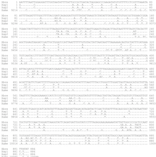
ob-tained for the mitochondrial COI region. he ive X. brevicolle specimens observed had identical sequences, whereas a single nucleotide in the sequence difered among the four specimens of Xiphinema sp. though this variation resulted in no diference between the translated amino acid sequences within the specimens. Sequence identity between these two species was 84.0-84.1%, whereas the sequences of X. brevicolle and Xiphinema sp. were 80.7 and 80.4-80.5% identical to that of X. americanum (He et al. 2005a) respec-tively, with no gap found among them (Fig. 7). Putative amino acid sequences were available without any stop codon when translations were made from the second base of the obtained sequences. DNA sequences of 1,566 bp except for primer regions were obtained for the 18S rDNA region from one specimen for each species. he diference between sequences of the two species studied here was only a single nucleotide, result-ing in 99.9% identity without any gap. DNA sequences of 788-791 bp except primer regions were obtained for the D2/D3 region from four specimens for each species. A single nucleotide variation of sequence among four specimens of X. brevicolle was ob-served, whereas no variation of sequence was observed among four specimens of Xiphin-ema sp. Sequence identity between these two species was 98.1-98.2%, with gaps found. ML trees inferred for the 18S rDNA and D2/D3 regions placed our specimens in similar clades, which include X. brevicolle and its junior synonyms by Luc et al. (1998) such as X. difusum Lamberti et Bleve-Zacheo, 1979, X. incognitum Lamberti and Bleve-Zacheo, 1979, X. taylori Lamberti et al., 1992, as well as other diferent species like X. inaequale (Khan et Ahmad, 1975) and X. lamberti Bajaj et Jairajpuri, 1977 (Figs 8, 9). On the other hand, the ML tree inferred for the COI region didn’t show strong support for such a clade because of a low bootstrap value though several subclades were strongly supported by high bootstrap values (Fig. 10).
Discussion
H
ir
omichi S
akai et al. / Z
ooK
eys 135: 21–40 (2011)
26
table 2. Morphometrics of Xiphinema brevicolle, Xiphinema sp., X. paramonovi and X. difusum (female). Mean ± standard deviation (range) in µm, except for L in mm, and ratios.
Xiphinema brevicolle Xiphinema sp.
(this study) Xiphinema paramonovi Paratypes (Romanenko 1981) Xiphinema difusum Paratypes (Lamberti and Bleve-Zacheo 1979) Population
in this study
Topotypes Types
(Lordello and Costa 1961) (Luc et al. 1998) (Lamberti et
al.1992)
n 22 25 17 - 13 27 10
L 1.93 ± 0.11 (1.71-2.10)
1.92 ± 0.122 (1.7-2.16)
2.1 ± 0.1 (1.8-2.2)
(1.82-2.20) 2.30 ± 0.12 (2.08-2.47)
2.1 (2.0-2.3)
1.7 (1.6-1.8) a 46.1 ± 2.6
(40.5-50.5)
46.1 ± 1.72 (44-51)
44.5 ± 2.3 (40.7-50.1)
(36.0-42.2) 48.8 ± 2.4 (45.0-52.3)
49.6 (44-54.2)
47 (46-51)
b 6.6 ± 0.6
(5.5-8.2) 5.92 ± 0.37(5.1-6.7) 6.4 ± 0.6(5.6-7.7) (7.0-10.5) 7.0 ± 0.4(6.5-8.1) (4.8-7.1)6.1 (5.3-8.9)6.9 c 69.8 ± 4.4
(60.5-79.1)
76.9 ± 5.64 (67.6-89.9)
77.8 ± 0.6 (60.3-94.0)
(62.5-93.0) 79.6 ± 8.5 (67.5-94.5)
60.5 (49.1-68.5)
72 (63-84) c’ 1.0 ± 0.1
(0.9-1.2)
0.96 ± 0.06 (0.89-1.10)
1.0 ± 0.06 (0.9-1.1)
- 0.9 ± 0.1 (0.8-1.2)
1.1 (0.9-1.2)
0.9 (0.8-1.1 V 50.5 ± 1.2
(48.1-53.6) 53 ± 1.9(50-55) (51.0-54.0)53 ± 0.9 (50.0-54.0) (50.9-54.0)52.4 ± 1.1 (50.8-55.0)52.1 (47-52)50 Total stylet 149.0 ± 4.4
(144-161)
159 ± 8.05 (144-173)
- (156.0-168.3) 166.5 ± 2.6 (161-171)
-
-Odontostyle 94.1 ± 3.3 (88-102)
101 ± 6.14 (89-110)
101.9 ± 7.2 (84.7-108.2)
- 107.3 ± 2.7 (103-111)
103.5 (88.5-120.0)
87 (84-89) Odontophore 54.9 ± 2.1
(51-59)
59 ± 3.43 (50-64)
57.0 ± 2.9 (48.8-60.0)
(61.2-62.7) 59.2 ± 1.6 (57-62) 56.7 (53.1-60.0) 50 (48-51) Oral aperture
to guide ring
76.5 ± 3.8 (67-83)
86.0 ± 4.23 (77-92)
86.3 ± 5.6 (72.3-92.3)
- 85.5 ± 5.0 (78-93) 79.6 (66.0-103.0) 62 (60-64) Pharyngeal bulb length
79.3 ± 4.0 (73-86; n = 10)
- - (61-79) 84.9 ± 2.7
(82-90; n = 7)
94.2 (65.0-117)
-Pharyngeal bulb width
21.2 ± 1.2 (18-23; n = 21)
- - (12-19) 22.9 ± 1.7
(20-26)
21.1 (18.0-24.0)
-F irst r epor t of Xiphinema br evicolle in J apan 27
Tail 27.8 ± 2.0 (25-32)
26.0 ± 1.71 (23-28)
26.8 ± 2.0 (24.1-31.2)
(24.5-29.0) 29.2 ± 2.8 (24-34) 36.1 (33.0-47.0) 24 (21-28) Hyaline portion
of tail (J)
10.4 ± 1.5 (8-13)
- 8.0 ±0.9 (5.9-9.4)
- 11.4 ± 1.5 (9-14) 9 (7-11) 12 (10-14) Body diam.
at lip region
12.5 ± 0.4 (12-13)
11.5 (11-13)
11.5 ± 0.5 (10.6-12.3)
- 12.4 ± 0.4 (11-13) 14.6 (13.5-15.0) 11 (10-12) Body diam.
at guide ring
30.4 ± 0.8 (29-32)
- 29.8 ± 1.5 (27.1-31.8)
- 32.6 ± 0.8 (31-34) 31.7 (30.0-36.0) 26 (26-27) Body diam.
at base of pharynx
37.9 ± 1.2 (35-40)
- 39.4 ± 2.9 (35.3-45.3)
- 41.3 ± 2.2 (38-45) 40.1 (36.0-42.0) 33 (31-35) Body diam. at vulva
42.0 ± 2.0 (38-46)
- 46.6 ± 3.4 (39.4-50.0)
(49.0-59.7) 47.2 ± 2.5 (44-52) 43.4 (39.0-47.2) 36 (33-38) Body diam. at anus
27.0 ± 1.6
(24-30) (22-29)26 (21.8-29.4)26.6 ± 1.7 (30.6-36.7) 29.7 ± 1.1(28-32) (27.0-41.0)32.4 (23-28)25 Body diam.
at beginning of J
16.2 ± 2.2 (12-20)
- 13.7 ± 1.1 (11.2-14.7)
- 18.4 ± 2.2 (15-22)
7 (6-8)
Hiromichi Sakai et al. / ZooKeys 135: 21–40 (2011)
28
incognitum Lamberti & Bleve-Zacheo, 1979, described as a new species for nematode specimens detected from soil of bonsai trees exported from Japan to England (Lam-berti and Bleve-Zacheo 1979).
he specimens of this study were obtained from mixed populations of the X. amer-icanum-group. Information on juveniles was not included because the coexistence of the two closely related nematodes requires special care to separate juvenile specimens of the diferent species. Situations like this will make matters worse if one is to iden-tify the species, since it is diicult enough to ideniden-tify even a single population of the group in many cases. Detection of mixed populations of X. americanum Cobb, 1913 and X. rivesi Dalmasso, 1969 were reported (Vrain et al. 1992, Vrain 1993), and such coexistence of multiple populations and/or species of the X. americanum-group may be common. herefore, it is strongly recommended to check the genetic uniformity of a given population to be identiied. he mitochondrial COI region has been recently used to examine variations in populations of Xiphinema species including members of the X. americanum-group (Lazarova et al. 2006, Kumari et al. 2010a, 2010b), where ca 400 bp sequences were examined. his genetic region has much more information to diferentiate between populations than 18S rDNA and D2/D3 regions. As shown above, we developed new primers and examined longer sequences of the COI region than previously used. Examination of longer sequences provides not only more reliable comparative results but also another option to develop a more speciic primer to the species to be tested since high variability of this region may contribute to some unit-ness of reported primers. Among options available at present, sequences of the COI region can be eiciently used to examine the diversity of Xiphinema specimens.
ML phylogenetic analyses of 18S rDNA and D2/D3 regions moderately sup-ported the clade including X. brevicolle, X. difusum, X. inaequale, X. incognitum, X. lamberti, and X. taylori with our materials (Figs 8, 9), whereas such a clade was not so highly supported in the ML tree of COI (Fig. 10). Member species of X. americanum-group harbor endosymbionts (Vandekerckhove et al. 2000). he table 3. Uterine and vaginal region lengths of Xiphinema brevicolle and Xiphinema sp. measured in this study (female). Mean ± standard deviation (range; number of specimens) in µm, except where indicated.
Xiphinema brevicolle Xiphinema sp.
Anterior uterus length 44.3 ± 4.8 (34-54; n = 14)
35.1 ± 2.9 (32-40; n = 9) Posterior uterus length 41.1 ± 2.9
(37-46; n = 9)
32.7 ± 2.7 (29-37; n = 9)
Pars proximalis vaginae 5.9 ± 0.7
(5-7; n = 22) 7.6 ± 1.5(5-10; n = 13)
Pars distalis vaginae 10.5 ± 0.7
(9-12; n = 22)
11.3 ± 1.0 (9-13; n = 13) Vagina length 16.4 ± 1.0
(14-18; n = 22)
18.9 ± 1.5 (17-22; n = 13) Vagina length /
body diam. at vulva (%)
39.2 ± 3.2 (31.3-44.6; n = 22)
First report of Xiphinema brevicolle in Japan 29
diference in phylogenetic inference between nuclear and mitochondrial genetic regions may result from the symbionts’ efect on mitochondrial DNA since unre-liable results of phylogenetic inference based on mitochondrial DNA due to the presence of a symbiont are known (Hurst and Jiggins 2005). Furthermore, the ML tree of COI brought another problem to us. It showed closer relationship of our X. brevicolle population to X. difusum than to other X. brevicolle populations. Our specimens identiied as X. brevicolle were reasonably larger than type specimens of X. difusum (Table 2), and our identiication was made considering more overlap-ping morphometrics of type specimens of X. brevicolle. If COI sequences can dif-ferentiate species of X. americanum-group, our specimens may be identiied as X. difusum rather than X. brevicolle. In such a situation, morphological features used to identify species should be reconsidered since a single species like X. difusum can have a wide range of morphometrics which may result in more diicult diagnoses without reducing the number of species by synonymizing them intensively. In any case, it should be desirable to collect much more COI sequences of other species and populations, such as the sequence data of topotype specimens of X. difusum, which is unavailable at present. Our results of phylogenetic analyses, however, may be helpful to reine the concept of a X. brevicolle-subgroup, which was previously discussed in some works (Romanenko and Stegaresku 1985, Lamberti and Ciancio 1993, He et al. 2005b). Taking our results of phylogenetic analyses into considera-tion, we suggest that the X. brevicolle-subgroup includes at least the ive species of X. brevicolle, X. difusum, X. inaequale, X. incognitum, and X. taylori, and our specimens also belong to the subgroup, though the validity of respective species is a diferent matter. Appropriate establishment of subgroups within the X. america-num-group will contribute to a more feasible identiication process of the member species and requires further research.
Conclusion
In this study, specimens from mixed populations of the X. americanum-group, pre-sent in the root zone of Japanese holly in Japan, were identiied as X. brevicolle and Xiphinema sp. his record of X. brevicolle is the irst for Japan. PCR primers to amplify longer sequences of the mitochondrial COI regions were originally designed and used to eiciently diferentiate the specimens. Phylogenetic analyses using 18S rDNA, D2/ D3, and COI regions supported a close relationship among our specimens and species related to X. brevicolle or the X. brevicolle-subgroup.
Acknowledgements
Hiromichi Sakai et al. / ZooKeys 135: 21–40 (2011)
30
T. Gaspard for improving the manuscript. his study was supported by Research and development projects for application in promoting new policy of Agriculture Forestry and Fisheries (21043) from the Ministry of Agriculture, Forestry and Fisheries of Japan.
References
De Grisse AT (1969) Redescription ou modiications de quelques techniques utilisées dans l’étude des nématodes phytoparasitaires. Mededelingen van de Rijksfaculteit Landbow-wetenschappen Gent 34: 351–369.
De Ley P, Félix M-A, Frisse LM, Nadler SA, Sternberg PW, homas K (1999) Molecular and morphological characterisation of two reproductively isolated species with mirror-image anatomy (Nematoda: Cephalobidae). Nematology 1: 591–612.
Edgar RC (2004) MUSCLE: multiple sequence alignment with high accuracy and high throughput. Nucleic Acids Research 32: 1792–1797. doi: 10.1093/nar/gkh340
Hall TA (1999) BioEdit: a user-friendly biological sequence alignment editor and analysis pro-gram for Windows 95/98/NT. Nucleic Acids Symposium Series 41: 95–98.
He Y, Jones J, Armstrong M, Lamberti F, Moens M (2005a) he mitochondrial genome of Xiphi-nema americanum sensu stricto (Nematoda: Enoplea): considerable economization in the length and structural features of encoded genes. Journal of Molecular Evolution 61: 819– 833. doi: 10.1007/s00239-005-0102-7
He Y, Subbotin SA, Rubtsova TV, Lamberti F, Brown DJF, Moens M (2005b) A molecular phy-logenetic approach to Longidoridae (Nematoda: Dorylaimida). Nematology 7: 111–124. doi: 10.1163/1568541054192108
Howe DK, Denver DR (2008) Muller’s Ratchet and compensatory mutation in Caenorhab-ditis briggsae mitochondrial genome evolution. BMC Evolutionary Biology 8: 62. doi: 10.1186/1471-2148-8-62
Hurst GDD, Jiggins FM (2005) Problems with mitochondrial DNA as a marker in population, phylogeographic and phylogenetic studies: the efects of inherited symbionts. Proceedings of the Royal Society B 272: 1525–1534. doi: 10.1098/rspb.2005.3056
Kumari S, Decraemer W, De Luca F (2010a) Molecular characterization of Xiphinema brevi-collum (Nematoda: Longidoridae) from the Czech Republic. European Journal of Plant Pathology 128: 243–250. doi: 10.1007/s10658-010-9651-8
Kumari S, Decraemer W, De Luca F, Tiefenbrunner W (2010b) Cytochrome c oxidase subunit 1 analysis of Xiphinema diversicaudatum, X. pachtaicum, X. simile and X. vuittenezi (Nema-toda, Dorylaimida). European Journal of Plant Pathology 127: 493–499. doi: 10.1007/ s10658-010-9614-0
Lamberti F, Bleve-Zacheo T (1979) Studies on Xiphinema americanum sensu lato with descriptions of ifteen new species (Nematoda, Longidoridae). Nematologia Mediterannea 7: 51–106. Lamberti F, Ciancio A (1993) Diversity of Xiphinema americanum-group species and
First report of Xiphinema brevicolle in Japan 31
Lamberti F, Ciancio A, Agostinelli A, Coiro MI (1992) Relationship between Xiphinema brevi-colle and X. difusum with a redescripiton of X. brevicolle and descriptions of three new species of Xiphinema (Nematoda: Dorylaimida). Nematologia Mediterranea 19 (1991): 311–326. Lazarova SS, Malloch G, Oliveira CMG, Hübschen J, Neilson R (2006) Ribosomal and
mito-chondrial DNA analyses of Xiphinema americanum-group populations. Journal of Nema-tology 38: 404–410.
Lordello LGE, Costa CP (1961) A new nematode parasite of cofee roots in Brazil. Revista Brasileira de Biologia, 21: 363–366.
Luc M, Coomans A, Loof PAA, Baujard P (1998) he Xiphinema americanum-group (Nema-toda: Longidoridae). 2. Observations on Xiphinema brevicollum Lordello & da Costa, 1961 and comments on the group. Fundamental and Applied Nematology 21: 475–490. Monteiro AR (2010) Nomenclatorial note on Xiphinema brevicolle Lordello & Costa, 1961
(Nematoda: Longidoridae). Nematologia Brasileira 34: 1–2.
Mullin PG, Harris TS, Powers TO (2005) Phylogenetic relationships of Nygolaimina and Do-rylaimina (Nematoda: Dorylaimida) inferred from small subunit ribosomal DNA sequenc-es. Nematology 7: 59–79. doi: 10.1163/1568541054192199
Netscher C, Seinhorst JW (1969) Propionic acid better than acetic acid for killing nematodes. Nematologica 15: 286.
Romanenko ND (1981) [Discovery of a new species of nematode Xiphinema paramonovi n. sp. (Nematoda: Longidoridae) in the territory of the soviet Union]. Tezisy Dokladov, Pervaya Konferentsiya po Nematodam Rastenii, Nasekomykh, Pochvy i Vod, 68–69.
Romanenko ND, Stegaresku OP (1985) [Taxonomic problem in nematodes of the Xiphinema americanum group (Nematoda, Dorylaimoidea)]. Parazitologiya 19: 434–442.
Sakai H (2010) A DNA extraction method with SDS from single nematodes for direct applica-tion to PCR ampliicaapplica-tion. Nematological Research 40: 13–14. doi: 10.3725/jjn.40.13 Seinhorst JM (1959) A rapid method for the transfer of nematodes from ixative to anhydrous
glycerin. Nematologica 4: 67–69. doi: 10.1163/187529259X00381
Tamura K, Peterson D, Peterson N, Stecher G, Nei M, Kumar S (2011) MEGA5: Molecu-lar Evolutionary Genetics Analysis using maximum likelihood, evolutionary distance, and maximum parsimony methods. Molecular Biology and Evolution. doi: 10.1093/molbev/ msr121
homas WK, Vida JT, Frisse LM, Mundo M, Baldwin JG (1997) DNA Sequences from for-malin-ixed nematodes: integrating molecular and morphological approaches to taxonomy. Journal of Nematology 29: 250–254.
Vandekerckhove TTM, Willems A, Gillis M, Coomans A (2000) Occurrence of novel ver-rucomicrobial species, endosymbiotic and associated with parthenogenesis in Xiphinema americanum-group species (Nematoda, Longidoridae). International Journal of Systematic and Evolutionary Microbiology 50: 2197–2205. doi: 10.1099/00207713-50-6-2197 Vrain TC (1993) Restriction fragment length polymorphism separates species of the Xiphinema
americanum group. Journal of Nematology 25: 361–364.
Hiromichi Sakai et al. / ZooKeys 135: 21–40 (2011)
32
Figures
First report of Xiphinema brevicolle in Japan 33
Hiromichi Sakai et al. / ZooKeys 135: 21–40 (2011)
34
First report of Xiphinema brevicolle in Japan 35
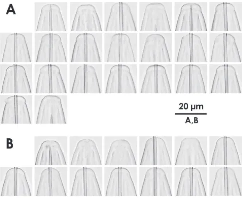
Figure 4. Intra-population variation of lip region in females AXiphinema brevicolleBXiphinema sp.
Hiromichi Sakai et al. / ZooKeys 135: 21–40 (2011)
36
First report of Xiphinema brevicolle in Japan 37
Hiromichi Sakai et al. / ZooKeys 135: 21–40 (2011)
38
Figure 8. Maximum likelihood tree for 18S rDNA region. Bootstrap values higher than 50 are shown. Arrows indicate specimens examined in this study: Xiphinema brevicolle GenBank accession AB604340;
First report of Xiphinema brevicolle in Japan 39
Hiromichi Sakai et al. / ZooKeys 135: 21–40 (2011)
40