Complex Function in Colorectal Cancer Cells
Dipak Barua1, William S. Hlavacek1,2,3*
1Theoretical Biology and Biophysics Group, Theoretical Division and Center for Nonlinear Studies, Los Alamos National Laboratory, Los Alamos, New Mexico, United States of America,2Department of Biology, University of New Mexico, Albuquerque, New Mexico, United States of America,3Clinical Translational Research Division, Translational Genomics Research Institute, Phoenix, Arizona, United States of America
Abstract
In colorectal cancer cells, APC, a tumor suppressor protein, is commonly expressed in truncated form. Truncation of APC is believed to disrupt degradation of b—catenin, which is regulated by a multiprotein complex called the destruction complex. The destruction complex comprises APC, Axin, b—catenin, serine/threonine kinases, and other proteins. The kinasesCK1a and GSK{3b, which are recruited by Axin, mediate phosphorylation of b—catenin, which initiates its ubiquitination and proteosomal degradation. The mechanism of regulation ofb—catenin degradation by the destruction complex and the role of truncation of APC in colorectal cancer are not entirely understood. Through formulation and analysis of a rule-based computational model, we investigated the regulation of b—catenin phosphorylation and degradation by APC and the effect of APC truncation on function of the destruction complex. The model integrates available mechanistic knowledge about site-specific interactions and phosphorylation of destruction complex components and is consistent with an array of published data. We find that the phosphorylated truncated form of APC can outcompete Axin for binding tob—catenin, provided that Axin is limiting, and thereby sequesterb—catenin away from Axin and the Axin-recruited kinasesCK1aandGSK{3b. Full-length APC also competes with Axin for binding tob—catenin; however, full-length APC is able, through its SAMP repeats, which bind Axin and which are missing in truncated oncogenic forms of APC, to bringb—catenin into indirect association with Axin and Axin-recruited kinases. Because our model indicates that the positive effects of truncated APC onb—catenin levels depend on phosphorylation of APC, at the first 20-amino acid repeat, and because phosphorylation of this site is mediated byCK1E, we suggest thatCK1E is a potential target for therapeutic intervention in colorectal cancer. Specific inhibition ofCK1Eis predicted to limit binding of b—catenin to truncated APC and thereby to reverse the effect of APC truncation.
Citation:Barua D, Hlavacek WS (2013) Modeling the Effect of APC Truncation on Destruction Complex Function in Colorectal Cancer Cells. PLoS Comput Biol 9(9): e1003217. doi:10.1371/journal.pcbi.1003217
Editor:Stanislav Shvartsman, Princeton University, United States of America
ReceivedFebruary 13, 2013;AcceptedJuly 10, 2013;PublishedSeptember 26, 2013
Copyright:ß2013 Barua, Hlavacek. This is an open-access article distributed under the terms of the Creative Commons Attribution License, which permits unrestricted use, distribution, and reproduction in any medium, provided the original author and source are credited.
Funding:This work was supported in part by NIH grants R01GM076570 and P50GM085273. DB acknowledges support from the Center for Nonlinear Studies at Los Alamos National Laboratory, which is operated for the US Department of Energy under contract DE-AC52-06NA25396. WSH acknowledges support from the Randy Pausch Scholars Program, which is sponsored by the TGen Foundation, Howard Young, and the Global Cure National Advisory Council. The funders had no role in study design, data collection and analysis, decision to publish, or preparation of the manuscript.
Competing Interests:The authors have declared that no competing interests exist.
* E-mail: [email protected]
Introduction
b{catenin (CTNNB1) is a key signaling protein in the Wnt=b{catenin pathway [1,2], a regulator of cadherin cell adhesion molecules [3], and a regulator of the Tcf and Lef family of transcription factors [4–7]. In mesenchymal cells,b{catenin levels increase when a Wnt ligand binds a cell-surface Frizzled (Fz)-family receptor. Activation of the Wnt/b{catenin pathway (transiently) inhibits proteosome-mediated degradation of b{catenin. Wnt binding also has other important effects on b{catenin, including regulation of phosphorylation state and redistribution ofb{cateninwithin subcellular compartments. In colorectal cancer cells, normal control ofb{catenindegradation is disrupted, resulting in elevated levels ofb{catenin.
Cellular degradation ofb{cateninis regulated by (in our view) oligomeric protein complexes, which have diverse compositions but common features; these complexes are often collectively referred to as the b{catenin destruction complex [8–10]. The destruction complex, which characteristically containsb{catenin
that alters regulation of b{catenin degradation, perhaps by destabilizing the destruction complex in a way similar to the destabilization brought about by Wnt signaling.
The interactions responsible for assembly of the destruction complex are complex and are mediated by multiple functional sites within the member proteins of the destruction complex. The characteristic core of the destruction complex can be viewed as a ternary complex that forms through interactions of APC, Axin, and b{catenin. b{catenin contains twelve ARM (Armadillo) repeats, allowing it to bind both APC and Axin. In particular, ARM repeats 3 and 4 constitutively bind a central region of Axin [22,23] as well as a phosphorylated 20-amino acid (20-aa) repeat region of APC [24,25]. There are total of seven 20-aa repeats in this region.b{cateninARM repeats 5–9 constitutively bind three 15-amino acid (15-aa) repeats in the N-terminal region of APC [26]. APC contains three SAMP (serine-alanine-methionine-proline) repeats, which bind the RGS (regulator of G protein signaling) domain of Axin [27]. These interactions connect the three core proteins of the destruction complex (APC, Axin, and b{catenin) and enable each protein to bind the other two core proteins, possibly within a closed/cyclic ternary complex. A cyclic complex would presumably be highly stable, because dissociation of such a complex would require the sequential break up of two protein-protein interactions.
Stability of the destruction complex may be important for its function as a platform for phosphorylation of b{catenin, and other proteins. The destruction complex mediates phosphorylation of b{catenin by allowing Axin to colocalize the kinases
GSK{3b and CK1a with their substrate b{catenin. Axin contains binding sites for both GSK{3b [11,28] and CK1a [12,29]. Interestingly, the destruction complex is also thought to mediate phosphorylation of APC by colocalizing another kinase,
CK1E(CSNK1E), with APC [30], although it is not known which protein in the destruction complex recruits CK1E. CK1E and
GSK{3btogether mediate phosphorylation at the 20-aa repeat region of APC [30]. Recall that this region in APC, when
phosphorylated, mediates interaction with a site in b{catenin that also interacts with Axin [22–25]. Thus, phosphorylated APC and Axin compete for binding tob{catenin. The outcome of this competition is perhaps dependent on stability of the destruction complex.
Much of what we know about the functional effects of APC truncation has come from studies of a human colon adenocarci-noma cell line (SW480). SW480 cells express a truncated form of APC termed APC1338, which contains only the first 1,338 amino acids of the full-length protein [9,31]. APC1338 contains all three 15-aa repeats and the first 20-aa repeat, but is devoid of the remaining six 20-aa repeats and the SAMP repeats, which bind Axin [9,31]. Therefore, a model can be conceptualized wherein assembly of the functional destruction complex cannot be completed in the absence of interaction between APC1338 and Axin, leading to decreased phosphorylation, ubiquitination, and degradation ofb{catenin [2]. However, an absence of SAMP repeats in APC does not prevent direct binding of Axin to b{catenin[22,23], and there are some uncertainties about the validity of this model [19] because reports from different laboratories have shown that expression of recombinant APC can either promote degradation ofb{cateninor have no or little effect, depending on cell type and whether APC is expressed transiently or stably [31–34].
As discussed above, APC plays an important role in destruction complex function. However, APC is a multifunctional protein, subject to numerous post-translational modifications. It is believed to play a role in regulating not only phosphorylation and ubiquitination ofb{cateninbut also localization ofb{catenin. There are several pools ofb{catenin: membrane-associated (e.g., complexed with E-cadherin), cytosolic (free, a{catenin bound and Tcf bound), and nuclear. Other components of the destruction complex are also multifunctional proteins, which can be found in distinct subcellular locations and states. For example, Axin, through self-polymerization mediated by its DIX (dishev-elled and axin) domain [35], localizes to cytoplasmic puncta. We will not consider these complexities, but they are mentioned at this point to caution the reader about the limitations of our study.
Here, our focus will be on APC regulation of b{catenin phosphorylation within an idealized destruction complex, taken to comprise a ternary complex of APC, Axin, andb{cateninwith 1:1:1 stoichiometry. We will consider the site-specific details of the interactions amongst these proteins, because these details are relevant for understanding how the interactions of APC, Axin, and b{catenin are perturbed by an absence of SAMP repeats in truncated APC (APC1338). We also consider, with less mechanis-tic resolution, proteins that mediate phosphorylation and dephos-phorylation of APC and b{catenin and degradation of b{catenin. The set of proteins of interest are considered in isolation. Thus, for example, we do not consider b{catenin interaction with E-cadherin, or the effects of Wnt. We also do not consider Axin puncta or the DIX domain in Axin. Axin puncta play a role in b{catenin degradation but are not required for phosphorylation ofb{catenin[36].
To investigate the roles of APC and its oncogenic truncated forms in destruction complex function, we formulated a compu-tational model for regulation ofb{cateninphosphorylation and degradation using local rules to represent the protein-protein interactions of interest [37–39]. This rule-based approach, ideal for modeling the chemical kinetics of biomolecular interaction networks, allowed us to consider the mechanistic details of protein-protein interactions at the resolution level of functional sites within the proteins of interest. These mechanistic details are complex, as summarized above, and arguably beyond our ability to compre-Author Summary
hend without reasoning aids, such the model considered here. Using this model, we interrogated system behavior, which emerges from the states and state changes of protein sites, with the goal of elucidating the distinctive mechanisms by which APC and APC1338 regulate the rate ofb{catenin destruction in normal and SW480 colorectal cancer cells. We also used our model to investigate the functional significance of intracomplex interactions among APC, Axin, andb{catenin, which have the potential to produce a highly stable cyclic ternary complex.
Although APC is a characteristic component of the destruction complex and thought to be important for degradation of b{catenin [31–34], our analyses suggest that APC does not promote degradation of b{catenin in a normal cell when overexpressed. However, we do predict that expression of recombinant full-length APC in SW480 cells promotes b{catenin degradation, as seen in several studies [31–34]. These results are obtained because, according to our model, phosphorylated APC1338 in SW480 cells competes with Axin for b{catenin. APC1338-mediated separation of b{catenin from Axin reduces phosphorylation of b{catenin by Axin-recruited kinases, and reduced phosphorylation of b{catenin decreases its rate of degradation. In contrast, in normal cells, binding of phosphorylated full-length APC to b{catenin, in competition with Axin, is not functionally equivalent because Axin can still colocalize with b{catenin through indirect association via the SAMP repeats in APC, which are missing in APC1338. Because of these results and because CK1E is responsible for phosphorylation of APC (but not b{catenin), we identify CK1E as a potential target for therapeutic intervention in colorectal cancer. Inhibition of CK1E is predicted to limit sequestration ofb{cateninaway from Axin and Axin-associated kinases and thereby to lower b{catenin levels in cancer cells expressing truncated APC.
Results
To investigate how the function of theb{catenin destruction complex changes when APC is mutated, especially as a result of a typical C-terminal truncation that removes the SAMP repeats and all but the first of the 20-aa repeats, we formulated a model (as described below) for full-length APC interactions with other components of the destruction complex. We then used this model and variants corresponding to different mutated forms of APC to predict how b{catenin levels and other readouts of system behavior depend on various parameters, such as the abundance of APC or truncated APC. Because APC contains multiple functional components or sites and we are interested in forms of APC containing different subsets of these sites, we formulated a model that tracks the chemical kinetics of the protein-protein interactions of interest with site-specific/structural resolution. This was accomplished by leveraging the rule-based modeling approach [37,40], in which local rules are used to represent biomolecular interactions and their consequences. Modeling with site-specific resolution is difficult with traditional modeling approaches, such as that of ordinary differential equations (ODEs), because of combinatorial complexity [41], which arises from multisite phosphorylation, multivalent binding, and other common aspects of biomolecular interactions involved in cellular regulation. Combinatorial complexity is a motivating factor for the use of rule-based modeling here.
Model
We developed a model for APC, Axin, and b{catenin
interactions and destruction complex function using the
rule-based modeling framework of BioNetGen [37–39] (see Materials and Methods). We considered a base model, corresponding to a normal cell with full-length APC, and several variant forms of the base model. The base model is illustrated in Figs. 1 and 2. The model is annotated in Text S1 (Supporting Information). Executable BioNetGen input files are provided in the Supporting Information for the base model (Text S2) and eight variant forms of the base model (Text S3 through Text S10).
In the base model, both explicit and implicit interactions are considered. We explicitly consider the interactions of five signaling proteins (and their isoforms presumed to be functionally equiva-lent): APC, Axin, b{catenin, CK1a, and GSK{3b. We implicitly consider the interactions of CK1E, PP2A, other phosphatases, and the proteins responsible for ubiquitination and proteosomal degradation ofb{catenin. In Fig. 2, proteins and their interactions are represented with site-specific/structural resolution using the conventions of Chylek et al. [42]. Briefly, proteins and their functional components are represented by nested boxes. Components excluded from consideration (e.g., the DIX domain of Axin) are not illustrated in Fig. 2. Arrows connecting boxes represent interactions. It should be noted that the visual elements of Fig. 2 correspond to the formal elements of our model [42]: boxes correspond to molecule types and arrows correspond to rules for interactions (Text S1). Each interaction included in the model is discussed below. The technical details of how these interactions are modeled/represented using rules are explained in Text S1. See also the Materials and Methods section. Arrow 1 in Fig. 2 represents reversible binding ofb{catenin ARM repeats 5–9 to the 15-aa repeats of APC [22,26]. In the model, ARM repeats 5–9 are considered to comprise a single binding site. Likewise, the three 15-aa repeats in APC are considered to comprise a single binding site.
Arrow 2 represents reversible binding of b{catenin ARM repeats 3 and 4 to phosphorylated APC 20-aa repeats [22]. In the model, ARM repeats 3 and 4 are considered to comprise a single binding site. The seven 20-aa repeats of APC are taken to function as two distinct binding sites, with binding activity of one site considered to be mutually exclusive with binding activity of the other site. The first site (labeled 1) corresponds to the first 20-aa repeat and the second site (labeled 3) corresponds to the third 20-aa repeat. We consider binding of APC to b{catenin to be mediated by the phosphorylated first repeat when the protein is APC1338 (or a comparable truncated form of APC), and predominantly (exclusively in the model as a simplification) by the phosphorylated third repeat if the protein is full-length APC. This distinction is made because APC1338 contains only the first 20-aa repeat, whereas full-length APC contains all seven 20-aa repeats. Binding of full-length APC to b{catenin is mediated primarily by the phosphorylated third 20-aa repeat [25] because the phosphorylated third repeat binds with 100- to 1000-fold higher affinity than that of any of the other phosphorylated 20-aa repeats [25]. We take the stoichiometry of a b{catenin-APC complex to be 1:1.
Arrow 3 represents reversible binding ofb{catenin to Axin. ARM repeats 3 and 4 ofb{cateninbind a central region of Axin [22,23]. As noted before, ARM repeats 3 and 4 also bind the phosphorylated 20-aa repeat region of APC (Arrow 2). Thus, ARM repeats 3 and 4 represent a b{catenin binding site recognized by both APC and Axin.
Arrows 5 and 6 represent reversible binding ofGSK{3band CK1ato Axin, respectively.GSK{3bbinds the GSK3 interac-tion domain (GID) of Axin [11,28,44]. CK1a binds a central region of Axin [29], which is distinct from the binding sites in Axin recognized by other binding partners. In the model, the binding of
CK1a, b{catenin and GSK{3b to Axin is taken to be non-competitive and non-cooperative.
Arrows 7 and 8 represent phosphorylation ofb{catenin by Axin-bound CK1a and GSK{3b, respectively. b{catenin phosphorylation takes place in a processive manner [12,13].
CK1afirst phosphorylates Ser-45 (labeled as S45 in Fig. 1), and
GSK{3bthen phosphorylates Ser-33, Ser-37, and Thr-41. In the model, as a simplification, the latter three sites are lumped together (labeled as S33/S37 in Fig. 2). We model the phosphorylation reactions as processes with first-order kinetics that occur only when kinases and substrates are colocalized within a complex. In the model, phosphorylation at S45 occurs when b{cateninis colocalized with Axin-associatedCK1a. Phosphor-ylation at S33/S37 occurs whenb{cateninis phosphorylated at S45 and colocalized with Axin-associatedGSK{3b[12,13]. We do not consider phosphorylation ofb{cateninoutside the context of the destruction complex.
Arrows 9 and 10 represent phosphorylation of APC 20-aa repeats by CK1E and GSK{3b [30,45]. Both CK1E and
GSK{3b are required for phosphorylation of APC [30]. In Fig. 2,CK1Eis shown for illustration purposes only. In the model,
we implicitly considerCK1Ebecause it is not known which protein is responsible for colocalizingCK1Ewith APC. Phosphorylation of APC is taken to occur through a process with first-order kinetics when APC and GSK{3b are colocalized via Axin. Thus, we assume thatCK1Eis colocalized with APC in proportion to the extent to whichGSK{3bis colocalized with APC via Axin. This assumption is equivalent to assuming thatCK1Eassociates non-competitively with Axin (or directly withGSK{3b).
We model dephosphorylation reactions as first-order processes (without explicit consideration of phosphatases). We allow dephosphorylation to occur if a site is exposed, i.e., not occupied and shielded by a binding partner. In the model, both phosphorylation sites ofb{catenin (i.e., S45 and S33/S37) are dephosphorylated according to the same rate law. In other words, the same first-order dephosphorylation rate constant is used for both sites. We allow the 20-aa repeats in APC to be dephosphor-ylated only if APC is in complex with Axin because Axin recruits PP2A, a phosphatase that mediates dephosphorylation of APC [46].
An important feature of the model is intracomplex binding of APC, Axin, andb{catenin. In Fig. 2, Arrows 1–4 each represents two distinct types of binding reactions: intermolecular binding, and intracomplex binding. The former type of binding reaction occurs when the reacting sites are freely diffusing, i.e., not tethered. The latter type of binding reaction occurs when the reacting sites are already in a complex together, i.e., tethered and Figure 1. Overview of the signaling proteins and interactions considered in the model.Panel A is a simplified version of Fig. 2, which follows and goes beyond the diagram shown here by illustrating the functional components of proteins responsible for interactions. Selected protein complexes considered in the model are illustrated in Panels B–D. (A) Proteins are represented by boxes. In the model, five proteins,b{catenin, APC,
Axin,GSK{3b, andCK1a, are considered explicitly, whereasCK1E, PP2A (not shown), and other proteins are considered implicitly.CK1E, which
mediates phosphorylation of APC, and PP2A, which mediates dephosphorylation of APC, are assumed to be constitutively associated with Axin. In the model, their activities are engaged when Axin is in complex with APC. Interactions included in the model are represented by arrows; numbering of arrows is the same as in Fig. 2. The arrows labeled 1–6 represent reversible direct binding interactions. The arrows labeled 7–10 represent catalytic (phosphorylation) interactions (and enzyme-substrate relationships). All phosphorylation events are taken to be reversed by phosphatases. The interaction represented by Arrow 1 is constitutive. The interaction represented by Arrow 2 depends on sequential phosphorylation of APC byCK1E
andGSK{3b(Arrows 9 and 10). The interactions represented by Arrows 2 and 3 are mutually exclusive, because they involve the same binding site inb{catenin, i.e., Axin and APC compete for binding to this site. The interaction between APC and Axin represented by Arrow 4 is missing for typical
truncated forms of APC (i.e., forms of APC, such as APC1338, missing SAMP repeats). Arrows 5 and 6 represent recruitment ofGSK{3bandCK1ato Axin. Arrows 7–10 represent phosphorylation reactions mediated by Axin-associated kinases. (B) A binary complex of APC andb{cateninconnected
through two distinct protein-protein interfaces. The interactions represented by Arrows 1 and 2 are allowed to occur simultaneously. (C) A complex whereinb{cateninis directly bound to Axin via the interaction represented by Arrow 3. Recall that this interaction cannot occur ifb{cateninis bound to APC via the interaction represented by Arrow 2. (D) A complex containing a linear (vs. cyclic) ternary complex of APC, Axin, andb{catenin.
co-confined to a small subvolume of the cytoplasm. An intracomplex reaction can be marked by a high apparent affinity because of the high local concentrations of the tethered binding partners [47]. In the model, these reactions lead to complex stabilization. We account for the high local concentration effect on an intracomplex reaction by multiplying the corresponding forward rate constant by an enhancement factorx. For instance, ifb{cateninand APC are already connected via Axin, then the effective forward rate constant for the reaction represented by Arrow 1 would be xkf, where kf is the intrinsic forward rate constant when the proteins are not tethered together.
It should be noted that in the modelb{cateninand APC can form a binary complex held together by two-point attachment i.e., b{cateninand APC can be held together through simultaneous
interaction betweenb{cateninARM repeats 3 and 4 and APC 20-aa repeats (Arrow 1) and interaction between b{catenin ARM repeats 5–9 and APC 15-aa repeats (Arrow 2). The intracomplex reactions between APC andb{cateninare allowed to occur outside the context of a completely assembled destruction complex.
In the model, except forb{catenin, the total concentrations of signaling proteins are taken to be conserved (i.e., constant). b{cateninis produced in a process with zeroth-order kinetics and degraded in either a slow or fast process with first-order kinetics. When S33/S37 is not phosphorylated,b{cateninis degraded at a slow rate, regardless of its bound state. When S33/S37 is phosphorylated, b{catenin is degraded at a fast rate, again regardless of its bound state. Thus, we allow b{catenin to be degraded, through a slow or fast process, independently of whether it is free or bound. We assume that b{catenin releases any binding partner(s) upon degradation.
The model has 27 independent parameters, including five protein concentrations and 14 binding constants (Table 1). Parameter values were specified as described in Materials and Methods. A local sensitivity analysis indicates that model behavior is not particularly sensitive to any individual parameter value (Table S1).
Effects of APC mutation onb–catenin expression Using the estimated parameter values summarized in Table 1 (see Materials and Methods), which were selected in part to allow the model to reproduce certain system behaviors (Figs. S1 and S2), we tested whether the model is able to predict the effects of transfection of SW480 cells with different truncated forms of APC. Munemitsu et al. [31] systematically transfected SW480 cells with various forms of APC. These experiments were designed to understand the effects of deletion of different functional compo-nents of APC onb{cateninlevels in SW480 cells, which almost exclusively express APC1338 instead of the full-length protein [31,48].
Munemitsu et al. [31] transfected SW480 cells with full-length APC or one of 11 different truncated forms of APC (illustrated in Fig. 3). In our model, full-length APC and the 11 truncated forms of the protein can be grouped into six distinctive classes, Classes A–F (Fig. 3). The proteins in each class are functionally equivalent based on the components and interactions of APC included in the model (Fig. 2). For example, Munemitsu et al. [31] considered three forms of APC each containing the following components: 1) a partial or complete set of the 15-aa repeats, 2) all of the 20-aa repeats, and 3) the SAMP repeats of APC. These are the functional sites that we consider to be included in full-length APC (Fig. 2). Therefore, we will use APC-A to represent all three proteins, as we take these forms of APC to be functionally equivalent. Similarly, we will use APC-B to represent two other proteins, which both contain the 15-aa repeats and only the first 20-aa repeat. We take these two forms to be equivalent to APC1338, the truncated protein in SW480 cells. Henceforth, we will use APC-A, APC-B, APC-C, APC-D, APC-E and APC-F to refer to the proteins in Classes A (e.g., full-length APC), B (e.g., APC1338), C, D, E and F (Fig. 3).
Using the model, we investigated the effects of transfection of SW480 cells with APC-A through APC-F. In the model, the endogeneous concentration of APC1338 in an SW480 cell is set at 100 nM. Similarly, the endogeneous concentration of full-length APC in a normal cell is set at 100 nM (Table 1). Because APC1338 does not contain the third 20-aa repeat, nor SAMP repeats, Axin interactions associated with these sites (Fig. 2) are absent in an SW480 cell. In contrast, in a normal cell, all Figure 2. Site-specific details of the proteins and interactions
considered in the model.Proteins, interactions, and the functional components that mediate interactions are represented according to the conventions of Chylek et al. [42]. The numbering of arrows is the same as in Fig. 1. The double-arrowed lines represent reversible binding interactions. The lines ending with an open circle represent enzyme-substrate relationships and point to sites of phosphorylation. In the model,b{cateninsites Ser-33, Ser-37, and Thr-41, which areGSK{3b substrates, are lumped together as a single site labeled S33/37. b{cateninsite Ser-45, which is aCK1asubstrate, is labeled S45. In the model, the seven 20-aa repeats of APC are lumped into two distinct sites labeled 1 and 3. For further information about the model, see Materials and Methods. A complete and executable specification of the model is provided in the Supporting Information as a plain-text BioNetGen input file (Text S2). Note that there is a correspondence between the arrows shown here and the rules of the model (Text S1). Model parameter values are summarized in Table 1.
interactions considered in the model are active, except for the low-affinity interaction between APC and b{catenin involving the phosphorylated first 20-aa repeat of APC and ARM repeats 3 and 4 of b{catenin. This low-affinity interaction is omitted when considering a normal cell as a simplification (see Materials and
Methods). In the model, when a representative of one of the six classes of APC is introduced into an SW480 cell, any novel interactions associated with the functional components of the transfected protein become active. For example, when APC-A is introduced, interactions associated with the third 20-aa repeat and Table 1.Model parametervalues1.
Parameters Comments
BCATtot= 35 nM (1.16104copies/cell) b–catenin concentration [53,59]
APCtot= 100 nM (3.26104copies/cell) APC concentration [53,59]
AXINtot= 10 nM (3.26103copies/cell) Axin concentration [59]
GSKtot= 100 nM (3.26104copies/cell) GSK–3bconcentration [53,59]
CKl1atot= 100 nM (3.26104copies/cell) CK1aconcentration (assumed)
b–catenin ARM repeats 5–9 binding to the APC 15-aa repeat region
KD1,bap= 273 nM Equilibrium dissociation constant [43]
kr1,bap~KD1,bap|kf1,bap~0:273 s{1 Dissociation rate constant2
b–catenin ARM repeats 3 and 4 binding to the phosphorylated APC 20-aa repeat
KD2,bap~1:5nM Equilibrium dissociation constant [25]
kr2,bap~KD2,bap|kf2,bap~0:0015 s{1 Dissociation rate constant2
b–catenin ARM repeats 3 and 4 binding to the phosphorylated APC1338 20-aa repeat
KD3,bap~85nM Equilibrium dissociation constant [25]
kr3,bap~KD3,bap|kf3,bap~0:085 s{1 Dissociation rate constant2
b–catenin ARM repeats 3 and 4 binding to Axin
KD,ba~227nM Equilibrium dissociation constant [43]
kr,ba~KD,ba|kf,ba~0:227 s{1 Dissociation rate constant2
Axin binding to the APC SAMP repeats
KD,apa~100nM Equilibrium dissociation constant (assumed)
kr,apa~KD,apa|kf,apa~0:1 s{1 Dissociation rate constant2
GSK–3bbinding to Axin
KD,ga~65nM Equilibrium dissociation constant [68]
kr,ga~KD,ga|kf,ga~0:065 s{1 Dissociation rate constant2
CK1abinding to Axin
KD,ca~100nM Equilibrium dissociation constant (assumed)
kr,ca~KD,ca|kf,ca~0:1 s{1 Dissociation rate constant2
b–catenin phosphorylation and dephosphorylation (at both S33/S37 and S45 sites)
kp,b~0:05 s{1 b–catenin phosphorylation rate constant
k{p,b~0:0012 s{1 b–catenin dephosphorylation rate constant3
APC/APC1338 phosphorylation and dephosphorylation at the 20-aa repeat region (site 1 or 3)
kp~0:05 s{1 APC phosphorylation rate constant4
k{p~0:05 s{1 APC dephosphorylation rate constant4
b–catenin synthesis and degradation
kd,b1~4:28|10{5s{1 Slow degradation rate constant4
kd,b2~4:28|10{3s{1 Fast degradation rate constant4
ks,b~0:013|10{3nM s{1(4.0 molecules/s) Synthesis rate constant4
Enhancement factor
x= 104nM Enhancement factor for intracomplex binding4
1Unit conversions are based on a cell cytoplasmic volume of10{12L [59]. 2For each binding reaction, the association rate constant (k
f) is assumed to be10{3nM{1s{1.
3The half-lifet
1=2ofb{cateninphosphorylation is approximately 10 min [74].
4The selected parameter values allow the model to reproduce a number of experimental observations, including 1) the steady-stateb
{cateninlevel, 2) the half-lives of
b{cateninand S33/S37-mutatedb{catenin(Fig. S1), and 3) the kinetics of dephosphorylation ofb{cateninat S33/S37 and S45 upon treatment with LiCl (Fig. S2). See Materials and Methods for more details.
the SAMP repeats (Fig. 2) become active. These interactions are normally missing in an SW480 cell. We assume that simulated transfections each introduce 100 nM of new protein into a cell. Thus, simulated transfection of SW480 with a particular form of APC implies that the cell contains 100 nM of a protein belonging to that form in addition to the 100 nM of the endogeneous form of APC (APC1338). (We systematically investigate how behavior depends on the amount of transfected protein below.)
In Fig. 4, we compare the model-predicted changes in b{catenin levels in SW480 cells after simulated transfection of different forms of APC against the findings of Munemitsu et al. [31] (Fig. 4). The model is able to recapitulate the qualitative increase or decrease in b{catenin level observed after transfec-tion of each class of protein. Consistent with the findings of Munemitsu et al. [31], the model predicts that only transfection of APC-A and APC-E leads to a decrease in b{catenin level, whereas the other four classes of APC have the opposite or no effect onb{cateninlevel (Fig. 4) [31]. It should be noted that the
results in Fig. 4 were obtained without adjustment or fitting of parameter values.
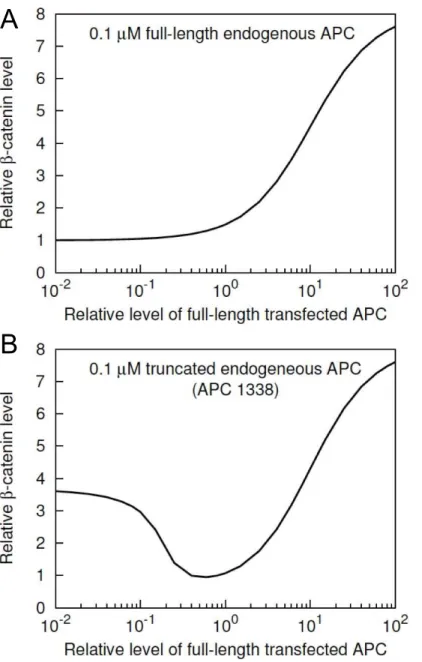
Role of full-length APC inb–catenin degradation The results of Munemitsu et al. [31] suggest that exogeneous full-length APC downregulates b{catenin by promoting b{catenin degradation in SW480 cells. Similar results for SW480 cells have been obtained in other studies [33,34]. However, transfection of different cell types have yielded different results [33]. Using our model, we investigated whether overex-pression of APC can generally be expected to increase the rate of b{catenindegradation in all cell types, or if the effect may be specific to SW480 cells only (Fig. 5).
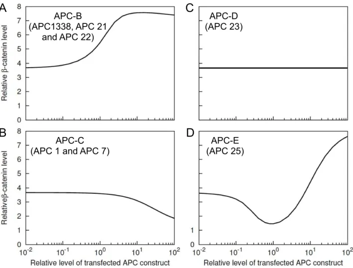
Fig. 5A shows the model-predictedb{cateninlevel in a normal cell as a function of APC level. A normal cell in the model is taken to have endogeneous full-length APC at a cytosolic concentration of 100 nM (Table 1). Fig. 5A illustrates the predicted effects of added APC. Fig. 5A shows that increased abundance of APC does not promoteb{catenindegradation, rather it has a concentra-Figure 3. Summary of APC constructs considered in simulated transfections and in the experimental study of Munemitsu et al.[31]. The 12 constructs used by Munemitsu et al. [31] are divided into six classes based on their structures. Proteins within the same class are functionally equivalent according to our model. A representative of Class A (APC-A) contains all three protein binding sites considered in the model for full-length APC. This class is regarded as equivalent to full-length APC. A representative of Class B (APC-B) contains 15-aa repeats and the first 20-aa repeat. This class is regarded as equivalent to APC1338, the truncated form of APC found in SW480 cells. A representative of Class C (APC-C) corresponds to a fragment that contains only the 15-aa repeats. A representative of Class D (APC-D) corresponds to a fragment that contains only the first 20-aa repeat. A representative of Class E (APC-E) corresponds to a fragment that contains the 20-aa and SAMP repeats. A representative of Class F (APC-F) corresponds to a nonfunctional fragment that contains none of the three APC sites included in the model.
tion-dependent positive effect onb{cateninlevel in normal cells, in contrast to the effect in SW480 cells (Fig. 4). The effects of exogenous full-length APC at different concentrations in SW480 cells are considered in Fig. 5B, which shows the model-predicted b{cateninlevel in SW480 cells as a function of full-length APC level. An SW480 cell is taken to have endogeneous APC1338 at a cytosolic concentration of 100 nM (Table 1). The predicted effect of added full-length APC is a significant decrease inb{catenin level in SW480 cells over a wide range of exogeneous full-length APC expression levels (Fig. 5B). This finding is consistent with the effects of transient expression of full-length APC in SW480 cells [31] and to some extent also with stable expression of full-length APC in SW480 cells [34].
Concentration-dependent effects of truncated forms of APC in SW480 cells
In Fig. 4, we assumed a fixed amount (100 nM) of exogeneous expression for all six classes of APC. However, the results in Fig. 4 could depend on APC concentration, as seen for APC-A (Fig. 5). Therefore, we investigated the predicted concentration-dependent effects of APC-B, -C, -D and -E onb{catenin levels in SW480 cells (Fig. 6). For APC-A, such effects have already been discussed (Fig. 5B). We do not consider APC-F because in our model it represents a non-functional form of APC with no binding sites.
The simulation results in Fig. 6 illustrate the concentration-dependent effects of APC-B, -C, -D and -E. As seen in Fig. 6A, added APC-B (e.g., APC1338) increasesb{cateninlevel over the entire concentration range considered. The level of b{catenin doubles as the amount of exogeneous APC1338 approaches a 10-fold higher amount of endogeneous APC1338 (Fig. 6A). Unlike APC-B, the other three proteins do not increaseb{cateninlevel over the entire concentration range. APC-C reducesb{catenin level at relatively high concentrations (Fig. 6B), APC-D does not alterb{cateninlevel at any concentration (Fig. 6C), and APC-E reducesb{cateninlevel over a range of intermediate concentra-tions in a manner similar to full-length APC (cf. Fig. 6D and Fig. 5B).
The only difference between APC-B and APC-C is that the former form of APC contains the first 20-aa repeat, whereas the latter form does not. This distinction leads to APC-B and APC-C having opposite effects onb{catenin level in SW480 cells (cf. Figs. 6A and 6B). APC-D contains the first 20-aa repeat but no other functional components of APC that are able to interact with b{catenin or Axin. Therefore, APC-D cannot interact with b{cateninbecause of the consequent absence of phosphorylation of the 20-aa repeat. The 20-aa repeat in APC-D is never phosphorylated because the unphosphorylated protein is unable to interact with Axin. Thus, APC-D has no effect on b{catenin level (Fig. 6C). APC-E entails all structural features of APC-B, but in addition it contains SAMP repeats, which mediate Axin binding (Fig. 2). Because of this distinctive feature, the model predicts that APC-E behaves differently from APC1338 and produces reduced b{cateninlevels at intermediate concentrations of APC-E similar to the predicted effects of full-length APC (Fig. 5B). These results indicate that the absence of SAMP repeats in APC1338 may have an important role in APC1338-mediated increases ofb{catenin levels in cancer cells.
Phosphorylation-dependent competition between APC1338 and Axin for binding tob–catenin
The analysis of Fig. 6 indicated that APC-B (e.g., APC1338) and APC-C have opposite effects onb{catenin level because APC-B contains a 20-aa repeat that APC-C does not. In Fig. 7, we analyze the effects of phosphorylation of the 20-aa repeat in APC-B on b{catenin levels in SW480 cells. Recall that phosphorylation of the 20-aa repeat in APC is mediated by
CK1E andGSK{3b[30,45] and that phosphorylation of this site is necessary for direct interaction of APC withb{catenin [22,25].
The simulation results shown in Fig. 7A indicate that phosphorylation of the 20-aa repeat is needed for APC-B/ APC1338-mediated stabilization ofb{catenin. In the figure, the solid line corresponds to default rate constants for phosphorylation and dephosphorylation of APC in the model (Table 1). For these Figure 4. Comparison of simulated and observed effects of transfection of SW480 cells with APC constructs.Relativeb{cateninlevels
in SW480 cells in response to transfection with the APC constructs of Fig. 3 are shown. The gray bars, which correspond to the lefty{axis, represent
experimental data from Munemitsu et al. [31]. The black bars, which correspond to the righty{axis, represent model predictions. The predicted concentrations (black bars) are each divided by the concentration ofb{cateninin a normal cell (35 nM, Table 1). For all APC constructs, the same
transfection efficiency is assumed. We take a transfected cell to contain 100 nM of added protein. The predicted results therefore represent the effects of 100 nM of a construct in addition to 100 nM of endogeneous APC1338. The simulation results shown here were obtained using BioNetGen input files provided in the Supporting Information: Text S3 was used for the APC-B and APC-F cases, Text S4 was used for the APC-A case, Text S5 was used for the APC-C case, Text S6 was used for the APC-D case, and Text S7 was used for the APC-E case.
parameter values, the 20-aa repeat is nearly always phosphorylat-ed. This case can be viewed as the extreme opposite of the case where the 20-aa repeat is deleted and therefore never present in phosphorylated form. When the 20-aa repeat is deleted, APC-B becomes equivalent to APC-C and downregulatesb{cateninin a similar manner (cf. Fig. 7A and Fig. 6C).
Phosphorylated APC1338 binds to ARM repeats 3 and 4 in b{catenin, which is also a binding site for Axin (Fig. 2). Thus, phosphorylated APC1338 competes with Axin for binding to b{catenin and can inhibit phosphorylation of b{catenin by sequestering b{catenin away from Axin-associated kinases. Fig. 7B illustrates the predicted effect of APC1338 phosphoryla-tion on associaphosphoryla-tion ofb{cateninand Axin. The simulation results
of Fig. 7B show that phosphorylation of APC1338 inhibits interaction ofb{cateninwith Axin.
Mechanism ofb–catenin upregulation by APC1338 The results of Fig. 7 suggest that competition between APC1338 and Axin forb{cateninbinding upregulatesb{cateninlevels in SW480 cells. These results however do not explain how APC1338 and APC regulateb{catenindifferentially. Differential regulation is somewhat paradoxical because both proteins have phosphory-lation sites in the 20-aa repeat region, which mediatesb{catenin binding. The distinction between APC and APC1338 can be attributed to the absence of SAMP repeats in APC1338, as explained fully below. In short, APC1338 sequestersb{catenin
Figure 5. Concentration-dependent effects of full-length APC onb{catenin.(A)b{cateninlevel in a normal cell is shown as a function of
APC concentration. Thex{axisrepresents the relative amount of APC introduced exogeneously with respect to the endogeneously present 100 nM of full-length APC in a normal cell. They{axisrepresents the level ofb{cateninrelative to its nominal level in a normal cell (Table 1). (B)b{catenin
level in an SW480 cell is shown as a function of APC concentration. Thex{axisrepresents the amount of APC introduced exogeneously relative to the endogeneously present 100 nM of APC1338 in an SW480 cell. They{axisrepresents the level ofb{cateninrelative to its nominal level in a
normal cell, as in panel A. The simulation results shown here were obtained using BioNetGen input files provided in the Supporting Information: Text S2 was used for panel A and Text S4 was used for panel B.
away from Axin, whereas APC fails to do so (Fig. 8). The sequestration effect arises because APC1338, lacking SAMP repeats, cannot mediate indirect association of b{catenin with Axin. We note that the bell-shaped curve in Fig. 8B represents a characteristic scaffold effect [49,50]. Here, the scaffold is APC and the scaffold ligands are Axin andb{catenin.
In normal cells, b{catenin can associate with Axin in two ways: 1) direct binding via ARM repeats 3 and 4 inb{catenin (Arrow 3; Fig. 2), and 2) indirect binding via APC, with APC acting as a linker betweenb{cateninand Axin (Arrows 2 and 4; Fig. 2). As in an SW480 cell, the direct interaction in a normal cell is also inhibited by phosphorylation of the 20-aa repeat region in APC because of competition between phosphorylated APC and Axin for binding to ARM repeats 3 and 4 in b{catenin. Nonetheless, in a normal cell, the indirect interaction still enables b{catenin to colocalize with Axin via APC [30], thus allowing phosphorylation of b{catenin to occur via Axin-associated kinases, which leads to degradation of b{catenin. In contrast, in SW480 cells,b{catenincan associate with Axin only through direct interaction. The indirect interaction does not occur because APC1338 lacks the SAMP repeats necessary for Axin binding. Thus, in SW480 cells, APC1338 phosphorylation effectively blocks
b{cateninassociation with Axin, leading to less degradation. A corollary of this finding is that increased expression of Axin would be expected to increase the degradation ofb{catenin, which has been observed [36,51,52].
Effect of stability of the core destruction complex onb– catenin level
The stability of the destruction complex can be perturbed (decreased) by preventing APC, Axin, and b{catenin from forming a closed/cyclic ternary complex. The cyclic complex, which we assume can form in normal cells, cannot form in SW480 cells as a result of APC truncation. Formation of the cyclic ternary complex can be prevented not only by truncation of APC but also by other mutations. Any mutation affecting one of the three protein-protein interfaces of the ternary complex would prevent closure of the cyclic structure. Using our model, we simulated inhibition of formation of the cyclic structure by systematically blocking each of the three protein-protein interfaces of the closed/ cyclic ternary complex, and we determined the resulting effect on b{cateninlevel. As seen in Fig. 9, blocking the contact between APC and b{catenin or b{catenin and Axin did not change Figure 6. Concentration-dependent effects of APC constructs in SW480 cells.Predicted b{catenin level is shown as a function of
expression level for APC-B, -C, -D, and -E. In each panel, thex{axisrepresents the amount of expression relative to the endogeneous level of APC1338 (100 nM). They{axisrepresents theb{cateninlevel relative to the nominal level in a normal cell (35 nM, Table 1). Thus, a value of 1 on
thex{axiscorresponds to a concentration of 100 nM of transfected protein, and a value of 1 on they{axiscorresponds to a concentration of 35 nM ofb{catenin. The simulation results shown here were obtained using BioNetGen input files provided in the Supporting Information: Text S3
b{cateninlevel, indicating that the cyclic structure is unimpor-tant for regulation of b{catenin level. Only blocking of the interface between APC and Axin (by removal of SAMP repeats) is predicted to upregulateb{cateninlevel. However, as established above, this behavior arises for reasons other than destablilization of the cyclic ternary complex of APC, Axin, and b{catenin. Thus, our model indicates that destabilization of this complex (through ablation of cyclization) is not an important effect of APC truncation.
Discussion
In this study, we have modeledb{catenin regulation by the destruction complex in normal and colorectal cancer cells, which express full-length and truncated APC, respectively. Our model,
illustrated in Figs. 1 and 2, incorporates site-specific mechanistic details about the destruction complex, which comprises a number of signaling proteins. In colorectal cancer cells (e.g., SW480 cells), the interactions of these proteins are altered by truncation of APC. We have used our model to study the function of APC and the effects of its truncation on b{catenin phosphorylation and phosphorylation-dependent degradation. We caution that our results pertain to only the function of APC within an idealized destruction complex and furthermore that we considered the interactions of particular (multifunctional) proteins in isolation from most of their binding partners. Thus, within the context of a cell, the functional effects of APC or truncated APC overexpres-sion could potentially be very different from what our model predicts. Nevertheless, the model is qualitatively consistent with the observed effects of transient expression of various recombinant Figure 7. APC1338 phosphorylation and its competition with Axin forb{catenin. (A)b{cateninlevel is shown at different levels of
APC1338 phosphorylation. Phosphorylation of APC1338 at the first 20-aa repeat is modulated by changing the values of the phosphorylation and dephosphorylation rate constantskpandk{p. In the figure, the ratio,kp=k{p~1corresponds to the default values ofkpandk{pin the model, which
are taken to be the same (Table 1). The case wherekp=k{p??represents an extreme, where the 20-aa repeat always remains phosphorylated. The case where kp=k{p~0 represents the opposite extreme, where APC1338 never becomes phosphorylated. (B) Competition effects on b{catenin{Axinbinding arising from APC1338 phosphorylation. They{axisrepresents the fraction of Axin in complex withb{catenin. The patterns of the lines represent different phosphorylation and dephosphorylation rate constants, as labeled in panel A. The simulation results shown here were obtained using Text S3, a BioNetGen input file provided in the Supporting Information.
forms of APC in SW480 cells [31] (Fig. 4). Stronger, less ambiguous tests of model predictions in the future would ideally be performed using anin vitroreconstituted or cell-free system [53] to eliminate the uncertainties and complexities of the cellular milieu.
Our analyses indicate that whilst the expression of full-length APC in SW480 cells can be expected to increase degradation of b{catenin, APC overexpression in normal cells may decrease b{catenin degradation or have no effect. We show that phosphorylation of the first 20-aa repeat in truncated APC, together with the absence of the SAMP repeats, is crucial for the effect of APC1338 on b{catenin levels in SW480 cells (Figs. 7 and 8). We suggest that phosphorylated APC1338 sequesters b{cateninfrom Axin, thus blockingb{cateninphosphorylation by Axin-bound kinases, viz. CK1a and GSK{3b. In contrast, phosphorylation of full-length APC, because of its SAMP repeats, which provide an indirect means for interaction between Axin and b{catenin, does not blockb{cateninassociation with Axin and
Axin-bound kinases, except at significantly higher levels of expression (cf. panels A and B in Fig. 8).
Several experimental studies have detected competition be-tween phosphorylated full-length APC and Axin forb{catenin binding [24,30,54,55], although the effect of such competition on b{cateninlevels has not been previously characterized.
Our results suggest that APC1338, similar to full-length APC, can efficiently mediate competition with Axin for b{catenin binding, even though it lacks the third 20-aa repeat (Fig. 3), the high-affinityb{catenin binding site in full-length APC. In the model, APC1338 associates with b{catenin with sufficient strength to displace Axin because of two-point attachment via its 15-aa and phosphorylated 20-aa repeat sites (i.e., because of the combined action of the interactions represented by Arrows 1 and 2 in Figs. 1 and 2). The single-siteKD’s for the interactions mediated by these sites are &273 and 80 nM, respectively [25,43]. The
affinities are comparable to the affinity of Axin for b{catenin
ARM repeats 3 and 4 (KD~227nM [43]). However, if two-point Figure 8. Sequestration ofb{cateninaway from Axin by APC1338.(A) Predicted amount ofb{cateninassociated either directly or indirectly
with Axin is shown as a function of APC1338 concentration in the background of an SW480 cell. The horizontal axis indicates the amount of APC1338 divided by the nominal amount of APC1338 in an SW480 cell (100 nM). The vertical axis indicates the amount of Axin-associatedb{catenindivided by the total amount ofb{cateninat steady state, which is a function of APC1338 concentration. (B) Predicted amount ofb{cateninassociated
either directly or indirectly with Axin is shown as a function of full-length APC concentration in the background of a normal cell. The horizontal axis indicates the amount of full-length APC divided by the nominal amount of full-length APC in a normal cell (100 nM). The vertical axis indicates the amount of Axin-associatedb{catenindivided by the total amount ofb{cateninat steady state, which is a function of full-length APC concentration. (C) Predicted amount ofb{cateninassociated directly with APC1338 as a function of relative APC1338 concentration. (D) Predicted amount of
b{cateninassociated directly with full-length APC as a function of relative full-length APC concentration. All results shown were obtained using the
parameter values of Table 1, except as indicated. The following BioNetGen input files were used to obtain simulation results: Text S3 was used for panels A and C and Text S2 was used for panels B and D.
attachment is possible, as we have postulated in our model, then there is an avidity effect. This effect has been studied in other systems [56–58] and may confer on phosphorylated APC1338 a competitive advantage, allowing it to outcompete Axin for b{catenin.
A critically important feature of our model is a greater abundance of APC than Axin (Table 1). According to our model, as discussed above, truncated APC in SW480 cells acts as a diversion sink that sequestersb{catenin away from Axin. This diversion-sink mechanism cannot be operative if Axin is more abundant than APC. Recent measurements of APC and Axin in SW480 cells indicate that the total amounts of Axin and APC are comparable [59]. At first, these results might seem to contradict the model presented here, which takes APC to be 10-fold more abundant than Axin. However, Axin is not homogeneously distributed in a cell. Much of the Axin in a cell is found in cytoplasmic puncta [35,36]. Thus, only a fraction of total Axin may be available in a form capable of joining a destruction complex having the composition and structure considered here. A more complicated model than that presented here would be required to account for subcellular compartmentalization of APC, Axin and b{catenin, which clearly play an important role in Wnt=b{catenin signaling [2,3]. Such an effort is beyond the intended scope of our study.
The role of colocalization of signaling proteins within the destruction complex is not completely understood. In our model, we assumed that the core of the destruction complex, formed by mutual interactions of APC, Axin, andb{catenin, has a closed/ cyclic structure (as depicted in the cartoon diagram at the far left of Fig. 9). Within this cyclic structure, there are three protein-protein interfaces, and each of the three interfaces involves interaction between two adjacent proteins, which are connected indirectly via the third protein. Therefore, the binding sites at each interface are confined together in a volume that is small relative to the total volume of the cytoplasm and the local concentrations of tethered
binding partners are high. Such high local concentrations can confer on a cyclic structure more stablity than a linear structure of the same composition [60]. It has been assumed that the destruction complex provides a stable platform for phosphoryla-tion ofb{catenin by the Axin-recruited kinasesGSK{3b and
CK1a. However, according to our analyses, stability of the core destruction complex (i.e., the cyclic ternary complex of APC, Axin and b{catenin) is not important for efficient degradation of b{catenin. By systematically simulating ablation of each possible contact betweenb{catenin, APC, and Axin, we demonstrate that stability of the complex has little if any influence onb{catenin degradation (Fig. 9). (Note that the bar at the far right of Fig. 9 is explained by the diversion-sink mechanism.) We caution that, in the model, stability of the cyclic structure is also determined by factors other than the local concentration effect. Degradation of b{cateninin a core complex can terminate its cyclic structure, leaving behind a complex of APC and Axin only. In addition, phosphorylated APC can disrupt the cyclic structure by breaking theb{catenin-Axin interface through competitive binding and sequestering ofb{cateninaway from the complex.
Questions may arise as to what other roles APC plays besides destruction complex-mediated regulation ofb{catenin because our model indicates that elevated expression of APC in a normal cell does not have a positive effect on b{catenin degradation (Fig. 5A), i.e., an increase in APC abundance is not predicted to cause a decrease inb{cateninlevel. A variety of other potential functions of APC have been suggested. Phosphorylated APC has been implicated in subcellular localization and nuclear shuttling of b{catenin [32,61–63], and high-affinity binding of phosphory-lated APC with b{catenin has been suggested to disrupt b{catenin interaction with other binding partners, such as E-cadherin and the Tcf and Lef family transcription factors [24]. It has been shown that APC competes with E-cadherin for binding to the ARM repeat region ofb{catenin [64]. Indeed, the main effect of stable expression of full-length APC in SW480 cells is not Figure 9. Effects of simulated perturbations of the putative closed/cyclic core destruction complex.Three cases are considered, as indicated in the descriptions of Text S8, S9, S10 (Supporting Information). Each of the three protein-protein interfaces,APC{b{catenin(Text S8),
b{catenin{Axin(Text S9), and APC-Axin (Text S10), is ablated such that a closed/cylic structure cannot form. The relativeb{cateninlevel in each
a reduction ofb{cateninlevel (although there is an approximate 2-fold reduction in the total amount ofb{catenin), but rather a redistribution of b{catenin from the nuclear and cytosolic compartments to the plasma membrane [34]. Transient expres-sion of APC (at higher levels) causes a more dramatic reduction in the level of b{catenin [31,34]. In future work, it would be interesting to investigate how E-cadherin may regulateb{catenin
andvice versa[3].
Our study identifiesCK1Eas a potential target for therapeutic intervention in colorectal cancer. InhibitingCK1Eis expected to reverse the effect of truncation of APC in SW480 cells. According to the model, phosphorylation of APC1338 at the first 20-aa repeat plays a key role in upregulatingb{cateninin cancer cells (Fig. 7). Therefore, inhibition of phosphorylation of APC at this site might be an effective way to normalizeb{cateninlevels in cancer cells. Phosphorylation of APC requires the combined action of two kinases, CK1E and GSK{3b [30]. Therefore, blocking of either kinase is predicted to reduce APC phosphor-ylation, as shown by Ha et al. [30]. Because GSK{3b is a common kinase for both b{catenin and APC (Fig. 1) and its inhibition would stabilizeb{catenin, only CK1E is a potential target. We note that targeting of CK1E should be feasible in preclinical studies, as pharamacological kinase inhibitors specific toCK1Eare available [65–67].
In this study, we used a detailed mechanistic modeling approach based on the principles of chemical kinetics to investigate regulation of b{catenin phosphorylation and degradation by full-length and truncated APC. In a previous study, Lee et al. [53] developed a related model to investigate regulation ofb{catenin by Wnt stimulation. However, this model does not consider truncated APC. Another notable difference is that the model of Lee et al. [53] is an ordinary differential equation (ODE)-based model, wherein molecules and complexes of signaling proteins are treated as reactive chemical species, which must be enumerated along with all possible reactions to obtain an executable model. In contrast, because of the goals of our study, we developed our model using the rule-based modeling approach [37–40]. With this approach, local rules are used to represent protein-protein interactions, which are assumed to be modular. Assumptions of modularity can greatly reduce the complexity of a model for protein-protein interactions, and as a result, enable explicit consideration of multiple functional components within proteins (e.g., the multiple sites of phosphorylation inb{catenin). In our model, the components of the proteins considered are the basic reactive elements, i.e., we consider biochemical reactions, such as reversible binding and phosphorylation, to occur at the level of protein sites. This approach was critical for the goals of our study, which included a characterization of the effects of loss of sites in APC. Such effects, and biomolecular site dynamics in general, are difficult to capture in an ODE model [40]. The study presented here provides an example of how rule-based modeling, a fairly new approach in biology, can be used to study biomolecular site dynamics.
We focused on a part of the Wnt/b{cateninsignaling pathway that controls b{catenin degradation and expression level. Our primary goal was to understand the differential regulation of b{cateninin normal and cancer cells at steady state and in the absence of Wnt signals, unlike in other modeling studies that have focused on the dynamics ofb{cateninregulation in response to a Wnt ligand [20,53]. In future work, it would be interesting to extend our model by connecting it to other components of the Wnt/b{cateninsignaling pathway and to further investigate the dynamics of regulation ofb{catenin.
Materials and Methods
Parameter values
Model parameter values are listed in Table 1. Most of the parameter values are based on previously reported estimates. However, some parameter values were set to allow the model to capture a set of observed system behaviors.
In the model, the concentration ofb{catenindepends in part on its rates of synthesis and degradation. As discussed below, we set parameters for these and other processes considered in the model such that the nominal, steady-state concentration of b{cateninis 35 nM [53], which corresponds to 11,000 copies/ cell assuming a cytoplasmic volume of 10{12
L [59]. This concentration is consistent with the concentration ofb{catenin measured inXenopusegg extract [53]. It is also consistent with the cytosolic (but not total) concentration ofb{cateninmeasured in HEK293T and MDCK cells, kidney epithelial cell lines, and in Caco-2 cells [59], an intestinal cell line. We take both APC and
GSK{3b concentration to be 100 nM (31,540 copies/cell). Concentrations of APC andGSK{3b have been measured to be 100 nM and 50 nM, respectively, inXenopusegg extract [53], and measured concentrations of these proteins in mammalian cells fall in the ranges of 4–34 nM and 10–120 nM, respectively [59]. We take CK1a concentration to be the same as GSK{3b, 100 nM. We assume Axin to be present at 10 nM (3,154 copy/ cell), which is consistent with recent measurements of Axin abundance in mammalian cells; the Axin concentration measured in mammalian cells ranges from 20 to 150 nM [59]. Lee et al. [53] reported that Axin is present inXenopusegg extract at a very low concentration, in the range of 10 to 20 pM [53]. We rejected this value, while accepting and using the qualitative observation of Lee et al. [53] that Axin is less abundant than APC, because a concentration of 20 pM corresponds to only six copies of Axin per cell for a human epithelial cell, which as stated above is taken to have a cytoplasmic volume of10{12
L [59].
For association of any two proteins that are not already in a complex together we assume the same forward rate constant (kf) for all interactions:10{3nM{1s{1
(Table 1). Each reverse rate constant (kr) is determined from the relationkr~kf|KD, where KD is the equilibrium dissociation constant for the reaction of interest. Equilibrium dissociation constants are set at values reported earlier in the literature, as indicated below.
Axin (Arrow 4 and 6, respectively) with the sameKD:KD,apa~100 nM (kr,apa~0:1 s{1) andKD,ca~100nM (kr,ca~0:1 s{1).
In the model, b{catenin level is determined not only by b{cateninsynthesis and degradation rate constants, but also by other parameters that affect the phosphorylation of b{catenin and APC. These parameters include phosphorylation and dephosphorylation rate constants and the enhancement factorx. All of these parameters directly or indirectly determine the total level of b{catenin. We selected values for these parameters, which are given in Table 1, such that the model captures a set of experimentally-observed system behaviors.
With the parameter values listed in Table 1, the model reproduces a steady-state concentration of (cytosolic)b{catenin of 35 nM, which as discussed above is consistent with some measurements [53,59]. Furthermore, the model predicts the effective half-life ofb{cateninto be&30 min, and the half-life
of b{catenin mutated at S33/S37 to be &4:5 h, which is
consistent with the observations of Rubinfeld et al. [14] (Fig. S1). The model also reproduces the experimentally-determined kinetics ofb{catenindephosphorylation at distinct phosphorylation sites in response to treatment with LiCl, an inhibitor ofGSK{3b[12] (Fig. S2). The consistency of the model with observed system behaviors is further discussed in the Results section.
We note that the model is not entirely consistent with recent measurements of APC, Axin, (cytosolic) b{catenin, and
GSK{3bconcentrations in SW480 cells [59]. The model cannot simultaneously reproduce these concentrations and observed system behaviors. This could be because the measured concen-trations do not reflect the subcellular concenconcen-trations relevant for destruction complex function. Alternatively, our mechanistic knowledge of destruction complex function could be incomplete. We formulated our model to probe the limits of understanding of destruction complex function. In setting parameter values, we put an emphasis on selecting values that allow the model to reproduce observed system behaviors, rather than measured system param-eters (viz., protein concentrations). This approach is guided by findings from computational analyses of model sloppiness, which indicate that fitting tends to improve the accuracy of model predictions more than parameter measurement [69].
Model specification
We will refer to the model illustrated in Figs. 1 and 2 as the base model. The base model corresponds to the case of a normal cell with full-length APC. Variants of the base model correspond to SW480 cells with APC1338, SW480 cells transfected with different forms of APC, and other cases considered in this study (see below for further details). The base model and each of its variants was formulated using BNGL, a model-specification language [39]. Executable model-specification files are provided in the Support-ing Information for the base model (Text S2) and its variants (Text S3, S4, S5, S6, S7, S8, S9, S10). BNGL is compatible with BioNetGen [39,70], a software tool for rule-based modeling, and a number of other tools, such as RuleBender [71], an interface for BioNetGen. In addition to parameter values and initial data, each model-specification included molecule type definitions and rules. Molecule type definitions delimit the functional components of proteins and their possible phosphorylation states. The rules characterize protein-protein interactions and other processes (viz., synthesis and degradation of b{catenin). The rules that characterize protein-protein interactions can be subdivided into 10 sets, with one set for each arrow in Fig. 1 or 2. An arrow corresponds to multiple rules if the interaction represented by the rule can take place in multiple contexts and the context influences the rate of reaction. For example, Arrow 3 in Fig. 1 or 2 represents
an interaction that can take place between either unconnected proteins or tethered proteins. Thus, there is a rule for each of these two cases.
Simulation
The base model (Figs. 1 and 2) and each variant model was simulated by submitting the corresponding model-specficiation file to BioNetGen [39,70] for processing. Model-specification files are provided in the Supporting Information (Text S2, S3, S4, S5, S6, S7, S8, S9, S10); the format of these files is plain text. Simulation protocols are included in each model-specification file. The captions of figures indicate which model-specification files were used in calculations. In all cases, the method used for simulation was an indirect method, meaning that the rules of the model being simulated were not used directly in the simulation procedure. Rather, the rules were expanded (i.e., used to exhaustively enumerate the distinct chemical species and individual chemical reactions implied by the rules) by invoking the generate_ network function of BioNetGen to obtain a reaction network. The corresponding system of ODEs describing the mass-action kinetics of this network were then numerically integrated by invoking thesimulate_odefunction of BioNetGen. BioNetGen uses CVODE [72,73] for numerical integration of ODEs. For the base model, the reaction network obtained by expansion of its rules comprises 410 distinct chemical species. The size of this network does not reflect the intrinsic complexity of the base model. Rather, the intrinsic complexity of this model is reflected by the number of its rules. The base model includes 29 rules. We used scripts to systematically vary default parameter values specified in BioNetGen input files to produce many of the figures shown in the Results section. Parameter scans are enabled by a function available within RuleBender [71].
Supporting Information
Figure S1 The model reproduces the half-lives of
b{cateninandb{cateninmutated at S33/S37.The model predicts the half-life ofb{cateninin a normal cell to be 30 min (solid line) and the half-life ofb{cateninmutated at the S33/S37 phosphorylation site to be 4.5 h (dashed line). These predicted values are consistent with the measured values reported by Rubinfeld et al. [14]. The figure illustrates model-predicted decay of cellularb{catenin or b{catenin mutated at S33/S37 in a simulated pulse-chase experiment [14]. The model was used to simulate the pulse-chase experiments of Rubenfield et al. [14], which were carried out to determine the half-lives of the proteins. (EPS)
Figure S2 The model reproduces the effects of LiCl treatment on phosphorylation of cellularb{catenin.The figure shows predicted and measured phosphorylation of b{catenin at S45 and S33/S37 in response to LiCl treatment of cells. The solid lines represent model predictions, and the points represent experimental data of Liu et al. [12]. The solid line/filled circles correspond to S45, and the dashed line/open circles correspond to S33/S37. In the model, LiCl addition is simulated by assuming a 20-fold decrease in kinase activity ofGSK{3b(i.e., 20-fold reduced rate of phosphorylation of the APC 20-aa repeats and S33/S37 inb{catenin). The kinase activities ofCK1aand