The role of autolysis loop in
determining the specificity of
coagulation proteases
Edward A. Doisy Department of Biochemistry and Molecular Biology, Saint Louis University School of Medicine, Saint Louis, MO, USA L. Yang,
C. Manithody and A.R. Rezaie
Abstract
We recently demonstrated that the substitution of the autolysis loop (residues 143 to 154 in the chymotrypsin numbering system) of activated protein C (APC) with the corresponding loop of factor Xa (fXa) renders the APC mutant (APC/fX143-154) susceptible to inhibi-tion by antithrombin (AT) in the presence of pentasaccharide. Our recent results further indicated, that in addition to an improvement in the reactivity of APC/fX143-154 with AT, both the amidolytic and anti-factor Va activities of the mutant APC have also been significantly increased. Since the autolysis loop of APC is five residues longer than the autolysis loop of fXa, it could not be ascertained whether this loop in the mutant APC specifically interacts with the activated conforma-tion of AT or if a shorter autolysis loop is responsible for a global improvement in the catalytic activity of the mutant protease. To answer this question, we prepared another APC mutant in which the autolysis loop of the protease was replaced with the corresponding loop of trypsin (APC/Tryp143-154). Unlike an ~500-fold improvement in the reactivity of APC/fX143-154 with AT in the presence of pentasac-charide, the reactivity of APC/Tryp143-154 with the serpin was im-proved ~10-fold. These results suggest that both the length and structure of residues of the autolysis loop are critical for the specificity of the coagulation protease interaction with AT. Further factor Va inactivation studies with the APC mutants revealed a similar role for the autolysis loop of APC in the interaction with its natural substrate. Correspondence
A.R. Rezaie
Department of Biochemistry and Molecular Biology
St. Louis University School of Medicine 1402 S. Grand Blvd. St. Louis, MO 63104 USA
Fax: +1-314-977-9205 E-mail: [email protected]
Research supported by the National Heart, Lung, and Blood Institute of the National Institutes of Health (HL 62565 and HL 68571 to A.R. Rezaie).
Received January 9, 2007 Accepted April 23, 2007
Key words
•Activated protein C •Factor Va
•Antithrombin •Factor Xa •Serpins
Introduction
The proteolytic activity of coagulation proteases of both the intrinsic and extrinsic clotting cascades is primarily regulated by the serpin inhibitor antithrombin (AT) in plasma (1-5). However, AT is a poor inhibi-tor of coagulation proteases unless it is bound to heparin-like glycosaminoglycans found on the surface of the endothelium (1,6). This
serpin, thereby facilitating its recognition by the coagulation enzymes (allosteric activa-tion mechanism) (9,10). Second, heparin can bind to a basic exosite on coagulation pro-teases, thus being capable of bridging AT and the target protease in one complex, there-by decreasing the dissociation constant for the initial interaction of two proteins (tem-plate mechanism) (3,5). The extent of the contribution of each mechanism to the rate-accelerating effect of heparin in protease inactivation by AT has been extensively stud-ied. For certain proteases such as thrombin, the co-factor effect of full-length heparin is mediated primarily through a template mech-anism with ~2-fold contribution from the activation mechanism (9). In the case of factors Xa (fXa) and IXa, however, the co-factor effect of full-length heparin is medi-ated by ~300- to 500-fold enhancement through activation and ~200- to 300-fold enhancement through the template mechan-ism in the presence of a physiological con-centration of Ca2+ (8,11-13). The
elucida-tion of these two distinct co-factor mechan-isms of heparin has been documented by the observation that a unique pentasaccharide fragment of high-affinity heparin which can bind with equal affinity to AT, but is not capable of bridging the serpin to the prote-ase, enhances the reactivity of fXa with AT ~300-fold by an allosteric mechanism with an ~2-fold co-factor effect on the AT inhibi-tion of thrombin (14,15).
We investigated the mechanism by which coagulation proteases differentially react with the activated conformation of AT and dem-onstrated that the autolysis loop of both fXa and fIXa (residues 143-154 in chymotrypsin numbering) (16) have several basic residues that specifically interact with the serpin in the presence of pentasaccharide (17-19). Thus, we hypothesized that thrombin does not react with the heparin-activated confor-mation of AT because the basic residues of fXa are not conserved in the autolysis loop of the protease (17). In support of this
hypo-thesis substitution of the autolysis loop of activated protein C (APC), a protease nonre-active with either the native or activated conformations of AT, with the correspond-ing sequence of fXa yielded a mutant (APC-fXa143-154) which reacted with AT with
~375-fold improved rate constant in the presence of pentasaccharide (17). A similar chimeric mutant of thrombin containing the autolysis loop of fXa from residues 143 to 154 exhib-ited dramatically improved reactivity with the activated conformation of AT (17). Nev-ertheless, further characterization of the APC-fXa143-154 mutant revealed that the catalytic
activity of the mutant protease toward small synthetic substrates and the natural substrate, factor Va (fVa), was also improved (17,20). Since the autolysis loop of APC also has several basic residues, but is five residues longer than the autolysis loop of other vita-min K-dependent coagulation proteases, the general improvement in the catalytic func-tion of the APC mutant in reacfunc-tion with AT, fVa and small chromogenic substrates raised the possibility that a shorter autolysis loop, but not specific interaction of the residues of this loop with the pentasaccharide-activated con-formation of AT, is responsible for the im-proved reactivity of the mutant with the serpin. This uncertainty was underscored by the ob-servation that thrombin also has an insertion of six residues in its autolysis loop (16).
To address this question, in the present study, we substituted the autolysis loop of protein C with the corresponding shorter and neutral loop of trypsin. Furthermore, we also substituted three basic residues of APC-fXa143-154 with Ala. The catalytic properties
the specificity of APC interaction with its natural substrate, fVa.
Material and Methods
Construction, expression and purification of recombinant proteins
The construction, expression and purifi-cation of wild-type protein C and the mutant in which the autolysis loop (143
Tyr-His-Ser- Ser-Arg-Glu-Lys-Glu-Ala-Lys-Arg-Asn-Arg-Thr-Phe-Val154) of the zymogen was
replaced with the corresponding sequence of fX (143
Arg-Thr-His-Glu-Lys-Gly-Arg-Gln-Ser-Thr-Arg154) (PC-fX143-154) has been
described previously (17). A PC-fX143-154
variant in which the three basic residues Arg-143, Lys-147 and Arg-150 of the au-tolysis loop were replaced with Ala residues (PC-fX143-154/3A) was constructed by the
poly-merase chain reaction mutagenesis approach and expressed in the same expression sys-tem as described (17). A protein C mutant in which the autolysis loop of the zymogen was replaced with the corresponding sequence of trypsin (143
Asn-Thr-Lys-Ser-Ser-Gly-Thr-Ser-Tyr-Pro-Asp-Val154) (PC-Tryp143-154)
was constructed by the polymerase chain reaction mutagenesis approach and expressed in the same vector system. The amino acid sequence of the autolysis loops (residues 143-154) of trypsin and coagulation pro-teases discussed in this manuscript is pre-sented in Table 1. Following confirmation of the accuracy of the mutagenesis, the mutant constructs were expressed in human
embry-onic kidney 293 cells as described (17). Both wild-type and mutant zymogens were purified to homogeneity by immunoaffinity chromatography using the Ca2+-dependent
monoclonal antibody HPC4 as described (17, 21). The thrombomodulin (TM) fragment containing epidermal growth factor-like do-mains 456 (TM456) (21) and protein C in-hibitor (PCI) (22) were expressed in mam-malian cells as described. All recombinant proteins were tested for homogeneity by sodium dodecyl sulfate-polyacrylamide gel electrophoresis.
Human plasma proteins including pro-thrombin, fVa, fXa, and AT were purchased from Haematologic Technologies Inc. (Essex Junction, VT, USA). Phospholipid vesicles containing 80% phosphatidylcholine and 20% phosphatidylserine (PC/PS) were pre-pared as described (23). The active AT-binding pentasaccharide fragment of hepa-rin (fondapahepa-rinux sodium) was purchased from Quintiles Clinical Supplies (Mt. Lau-rel, NJ, USA). The normal pooled plasma was purchased from George King Bio-Medi-cal, Inc. (Overland Park, KS, USA) and the activated partial prothrombin time (aPTT) reagent, Alexin, was purchased from Sigma (St. Louis, MO, USA). α1-Antitrypsin was
obtained from Athens Research and Technol-ogy, Inc. (Athens, GA, USA). The chromo-genic substrate, Spectrozyme PCa (SpPCa, H-D-Lys (γ-Cbo)-Pro-Arg-pNA.2AcOH) was
purchased from American Diagnostica (Greenwich, CT, USA) and S2238 was pur-chased from Kabi Pharmacia/Chromogenix (Franklin, OH, USA).
Table 1. Alignment of the sequence of amino acid residues of the autolysis loops of trypsin and coagulation proteases from residues 143 to 154. The chymotrypsin numbering system is used (16).
Inactivation by antithrombin
The rate of inactivation of wild-type and mutant proteases by AT in both the absence and presence of pentasaccharide was meas-ured under pseudo-first-order rate conditions by a discontinuous assay method as described (17). In the absence of the co-factor, both wild-type and mutant proteases (2-3 nM) were incubated with plasma AT (0.125-10 µM) in 0.1 M NaCl, 20 mM Tris-HCl, pH 7.4, containing 1 mg/mL bovine serum albu-min (BSA), 0.1% polyethylene glycol 8000 (PEG-8000) and 5 mM Ca2+ (TBS/Ca2+). In
the presence of pentasaccharide, the reac-tion condireac-tions were the same except that wild-type and mutant proteases were incu-bated with AT (0.025-10 µM) in the pres-ence of saturating concentrations of penta-saccharide (1-20 µM) in TBS/Ca2+. All
reac-tions were carried out in 50-mL volumes in 96-well polystyrene plates at room tempera-ture. After 30-300 min, depending on the rate of reactions, 50 µL of the chromogenic substrate (200 µM SpPCa) in TBS was added to each well and the remaining enzyme ac-tivity was measured with a Vmax kinetics
microplate reader (Molecular Devices, Menlo Park, CA, USA). The second-order inhibi-tion rate constants for both uncatalyzed and catalyzed reactions were obtained as de-scribed (17).
The PCI and α1-antitrypsin inhibition
rates of APC derivatives were measured un-der pseudo-first-orun-der rate conditions by a discontinuous assay method as described (20). Briefly, 3 nM APC was incubated at room temperature with 50-200 nM PCI or 10-20 µM α1-antitrypsin in 50-µL reactions
in TBS buffer containing 2.5 mM Ca2+, 1
mg/mL BSA and 0.1% PEG-8000.
Hydrolysis of chromogenic substrates
The steady-state kinetics of hydrolysis of SpPCa (15-2000 µM) by both wild-type and mutant APC (5 nM) was measured in TBS/
Ca2+ at 405 nm at room temperature in a V max
kinetic microplate reader as described above. The Km and kcat values for the substrate
hydrolysis were calculated from the Michae-lis-Menten equation as described (17).
Zymogen activation
Two milligrams of protein C derivatives was incubated with thrombin (25 µg) in 0.1 M NaCl, 20 mM Tris-HCl, pH 7.4 (TBS buffer), containing 5 mM EDTA for 2 h at 37ºC. The APC derivatives were separated from thrombin by an FPLC Mono-Q column developed with a 40-mL linear gradient from 0.1 to 1.0 M NaCl, 20 mM Tris-HCl, pH 7.4, as described (22,24). The concentrations of proteases were determined from the absorb-ance at 280 nm and extinction coefficient (E1%
1 cm) of 14.5 (assuming a molecular mass
of 56 kDa for APC), by an amidolytic activ-ity assay using SpPCa, and by stoichiomet-ric titration of enzymes with known concen-trations of PCI as described (22).
The initial rate of wild-type and mutant protein C activation by thrombin was meas-ured in both the absence and presence of TM456 and Ca2+ as described (21). In the
absence of TM456, the time course of pro-tein C (1 µM) activation by thrombin (5-50 nM) was studied at room temperature in 0.1 M NaCl, 20 mM Tris-HCl, pH 7.4 (TBS) containing 1 mg/mL BSA, 0.1% PEG-8000 and 2.5 mM Ca2+ in 96-well assay plates. At
different time intervals, thrombin activity was quenched by 1 µM AT and the rate of protein C activation was measured from the cleavage rate of SpPCa (200 µM) at 405 nm by a Vmax kinetic microplate reader as
prepared by the total activation of protein C derivatives with the excess thrombin at the time of the experiments.
Measurement of the anticoagulant activity
Anticoagulant activities of the APC de-rivatives were evaluated both in the purified and plasma-based assay systems. The APC concentration dependence of fVa inactiva-tion wasdeterminedby a three-stage assay in the purified system as described (20). Briefly, in the first stage, fVa (5 nM) was incubated with wild-type or mutant APC (1-5 nM) on 2(1-5 µM PC/PS vesicles in TBS/Ca2+
for 10 min. In the second stage, the remain-ing fVa activity was determined in a pro-thrombinase assay from the fVa-catalyzed prothrombin activation by fXa as described (20). The prothrombinase assay was carried out for 30 s with excess prothrombin (1 µM) and a saturating fXa concentration (10 nM) at room temperature. The remaining activity of fVa was determined from the decrease of the rate of thrombin generation as monitored by an amidolytic activity assay in the third stage using 100 µM S2238.
The anticoagulant activities in plasma were evaluated in an aPTT assay using a STart 4 fibrinometer (Diagnostica/Stago, Asnieres, France). Briefly, 0.050 mL TBS lacking or containing 1-20 nM final concen-trations of wild-type or mutant APC was incubated with a mixture of 0.05 mL normal pooled plasma plus 0.05 mL aPTT reagent (Alexin) for 5 min before the initiation of clotting by the addition of 0.05 mL of 35 mM CaCl2 at 37ºC as described (20).
Results
Protein C activation
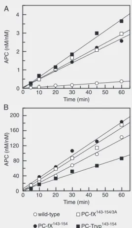
The initial rate of the activation of pro-tein C derivatives by thrombin both in the absence and presence of TM456 is presented in Figure 1. In the absence of TM, the
activa-tion of all protein C mutants by thrombin was improved 5- to 8-fold in the presence of Ca2+ (Figure 1A). The initial rate of
activa-tion of protein C mutants by thrombin in the presence of TM456 was not significantly changed, thus increasing ~50% for both PC-fX143-154 and PC-fX143-154/3A and decreasing
to a similar extent for the PC-Tryp143-154
mutant (Figure 1B). These results suggested that a longer autolysis loop in protein C plays a negative role in its recognition and subsequent activation by thrombin in the absence of TM.
Amidolytic activity
The capacity of autolysis loop mutants of APC to cleave the chromogenic substrate SpPCa was determined to assess the integ-rity of the catalytic domain active site. SpPCa (Lys-Pro-Arg-pNa) is a tripeptide sequence
Figure 1. Time course of the ini-tial rate of protein C activation by thrombin in the absence and presence of thrombomodulin 456 (TM456). A, Protein C de-rivatives (1 µM) were incubated with thrombin (10-50 nM) at room temperature in TBS con-taining 1 mg/mL BSA, 0.1% PEG-8000 and 2.5 mM Ca2+. At
that binds to the S1, S2, and S3 substrate-binding subsites of the catalytic groove, and distortion of these binding sites will alter SpPCa cleavage. Kinetic parameters for the SpPCa cleavage are shown in Table 2. All mutants exhibited improved activity toward SpPCa with an ~2- to 3-fold improvement in
Km for APC-Tryp143-154 and an ~2-fold
im-provement in kcat for the other two mutant
proteases. These results indicate that substi-tution of the autolysis loop of APC with either fXa or trypsin has no adverse effect on the conformation of the APC S1-S3 sub-strate-binding sites, but rather the mutagen-esis improves the catalytic efficiency of the mutants toward the chromogenic substrate.
Inactivation by antithrombin
The anticoagulant protease APC is es-sentially unreactive with AT in the absence of heparin co-factors (17). However, the incubation of APC with a very high concen-tration of AT (10 µM) in the presence of pentasaccharide for several hours leads to slow inactivation of the protease by the serpin with an estimated second-order association rate constant (k2) of 2.6 M-1 s-1 (Table 3).
Similar to the case with the wild type, nei-ther APC-fX143-154/3A nor APC-Tryp143-154
exhibited detectable reactivity with AT in the absence of pentasaccharide, though APC-fX143-154 was slowly inhibited by the serpin
under these conditions (Table 3). On the other hand, the APC-fX143-154 mutant reacted
efficiently with AT in the presence of penta-saccharide with a k2 value of 1.4 x 10³ M-1 s-1
(Table 3). Relative to wild-type APC, the reactivity of the other two mutants, APC-fX143-154/3A and APC-Tryp143-154 with AT in
the presence of pentasaccharide was also markedly improved (10- to 20-fold), thus yielding k2 values of 4.6 and 2.5 x 101 M-1 s-1,
respectively (Table 3). However, in contrast to a greater than 500-fold improvement in the reactivity of APC-fX143-154 with the
AT-pentasaccharide complex, a considerably lesser improvement of 10- to 20-fold was observed for the other two mutants of APC with the activated conformation of the serpin (Table 3). These results suggest that both the length and positively charged residues of the autolysis loop are critical for determining the specificity of the protease interaction with AT in the presence of pentasaccharide.
Anticoagulant activity
To understand the role of the autolysis loop in the anticoagulant activity of APC, the capacity of the mutants to inactivate fVa or to elevate the plasma clotting time was evaluated. As demonstrated in Figure 2A and consistent with our previous results (20),
Table 2. Kinetic constants for the hydrolysis of Spectrozyme PCa by the APC deriva-tives.
Km (µM) kcat (s-1) kcat/Km (s-1 µM-1)
APC wild-type 136.0 ± 25.1 31.1 ± 2.9 0.23 ± 0.06 APC-fXa143-154 115.5 ± 14.4 56.2 ± 4.9 0.49 ± 0.10
APC-fXa143-154/3A 111.2 ± 9.8 51.3 ± 3.0 0.46 ± 0.07
APC-Tryp143-154 43.5 ± 6.8 26.6 ± 3.9 0.61 ± 0.18
The kinetic constants were calculated from the hydrolysis rate of increasing concentra-tions of SpPCa (0.75 to 2000 µM) by wild-type and mutant activated protein C (APC) derivatives (3 nM) in TBS/Ca2+ as described in Material and Methods. The kinetic
values are reported as the mean ± SEM for three measurements. SpPCa = H-D-Lys (γ
-Cbo)-Pro-Arg-pNA.2AcOH.
Table 3. Second-order inhibition rate constants for the serpin inhibition of activated protein C (APC) derivatives.
k2 (AT) k2 (AT-H5) k2 (PCI) k2 (α1-AT)
(M-1 s-1) (M-1 s-1) (M-1 s-1) (M-1 s-1)
APC wild-type N/D 2.6 ± 0.6 (1.5 ± 0.3) x 103 2.6 ± 0.5
APC-fXa143-154 3.2 ± 1.2 (1.4 ± 0.1) x 103 (3.0 ± 0.8) x 103 3.0 ± 0.4
APC-fXa143-154/3A N/D (4.6 ± 0.4) x 101 (1.6 ± 0.2) x 103 N/D
APC-Tryp143-154 N/D (2.5 ± 0.2) x 101 (14.1 ± 0.3) x 103 26.8 ± 1.4
The APC derivatives (2 to 3 nM) were incubated with antithrombin (AT; 0.025 to 10 µM) in the absence and presence of saturating concentrations of pentasaccharide (H5; 1 to 20 µM), protein C inhibitor (PCI; 50 to 200 nM), and α1-antitrypsin (α1-AT; 10 to 20 µM)
at room temperature in TBS/Ca2+. The second-order rate constants (k2) were
the time course of fVa inactivation sug-gested that the activity of the APC-fX143-154
mutant was improved ~2-fold. The activity of APC-fX143-154/3A and APC-Tryp143-154
mutants was also slightly improved in this assay (Figure 2A). Comparisons of the anti-fVa activities of APC-fX143-154 and
APC-fX143-154/3A suggests that, similar to
interac-tion with AT, the basic charges of the autoly-sis loop contribute to the specificity of prote-ase interaction with the co-factor.
Unlike the improved anti-fVa activity of the APC mutant in the purified system, the plasma clotting activity of the APC-fX143-154
mutant was slightly impaired and the activ-ity of APC-fX143-154/3A and APC-Tryp143-154
mutants was nearly abolished (Figure 2B). To understand the basis for this paradoxical effect in the activity of the mutant in these two systems, the extent of the reactivity of APC derivatives with other plasma inhibi-tors was also studied. The time course of the incubation of each APC with normal plasma followed by monitoring the amidolytic ac-tivity yielded a half-life of ~30 min for wild-type APC at room temperature, which is consistent with the literature (25). Interest-ingly, the half-life of APC in plasma was decreased to less than 5 min for the mutants, suggesting that the APC mutants became susceptible to inhibition by the plasma in-hibitors. Thus, certain structural features in the autolysis loop of APC appear to be re-sponsible, at least partially, for the slow reactivity of the protease with plasma inhibi-tors. Further studies with other APC-specif-ic plasma serpin inhibitors, PCI and α1
-antitrypsin, in the purified system supported this conclusion since the reactivity of the APC mutants with these inhibitors was sig-nificantly improved. Thus, relative to wild-type APC (k2 = 1.5 ± 0.3 x 10³ M-1 s-1) the
reactivity of the APC-fX143-154 mutant with
the serpin was increased ~2-fold (k2 = 3.0 ±
0.8 x 10³ M-1 s-1). A much greater
improve-ment (~10-fold) in the reactivity of APC-Tryp143-154 (k
2 = 1.4 ± 0.3 x 10⁴ M-1 s-1)
Figure 2. Anticoagulant activity of activated protein C (APC) de-rivatives in factor Va (fVa) deg-radation and plasma-based clot-ting assays. A, The time course of fVa (5 nM) inactivation by in-creasing concentrations of the APC derivatives was carried out on phosphatidylcholine/phos-phatidylserine vesicles (25 µM) at room temperature in TBS con-taining 1 mg/mL BSA, 0.1% PEG-8000 and 5 mM Ca2+. At
each time, small aliquots of the inactivation reaction was trans-ferred to a 96-well assay plate and the remaining co-factor ac-tivity of fVa was determined by a prothrombinase assay as de-scribed in Material and Methods. B, The plasma clotting activity of the APC derivatives was deter-mined as a function of different concentration of APC at 37ºC as described in Material and Meth-ods. The plots are representa-tive experiments of 2-3 inde-pendent and reproducible meas-urements.
with PCI was observed (Table 3). Interest-ingly, a similar 10-fold improvement was also observed in the reactivity of the APC-Tryp143-154 mutant with
α1-antitrypsin (Table
3), possibly providing a basis for the poor anticoagulant activity of this mutant in the plasma-based clotting assay. Unlike an im-proved reactivity with AT, the reactivity of APC-fX143-154/3A with either PCI or
α1
-anti-trypsin was not improved. Whether the shorter half-life of this mutant is due to its susceptibility to other plasma serpins was not investigated.
Discussion
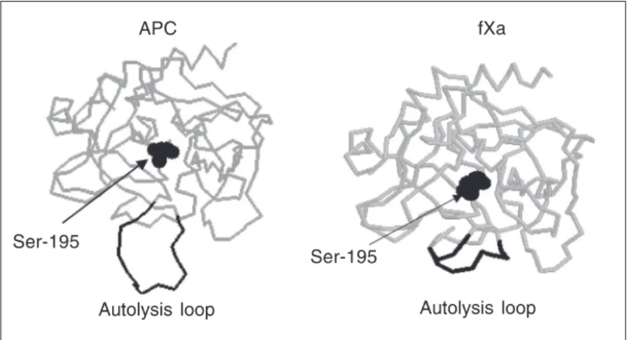
dem-onstrated that the substitution of the autoly-sis loop of APC with the corresponding loop of fXa renders the mutant protease suscep-tible to inactivation by AT specifically in the presence of pentasaccharide (17), suggest-ing that the autolysis loop of fXa carries recognition sites for interaction with the ac-tivated conformation of AT. The autolysis loop of fXa contains four basic residues at positions 143, 147, 150, and 154 (Table 1) and the Ala-scanning mutagenesis of these residues has identified Arg-150 as the criti-cal residue that specificriti-cally interacts with AT in the presence of pentasaccharide (18). The recent X-ray crystal structure determi-nation of a catalytically inactive mutant of fXa in complex with AT in the presence of pentasaccharide supported the mutagenesis results (26). Nevertheless, in a recent study we noted that, in addition to improvement in the reactivity of the APC-fX143-154 mutant
with AT, the activity of this mutant toward both small synthetic and the natural sub-strate fVa was also improved (20). The au-tolysis loop of APC is also basic but it is five residues longer than the corresponding loop of fXa (Figure 3). This raised the possibility that it is not only the specific interaction of charged residues of the autolysis loop that
determines the specificity, but the shorten-ing of this loop results in a global improve-ment in the reactivity of the catalytic groove of APC with macromolecular substrates and inhibitors in plasma. To test this possibility, in this study, we substituted the autolysis loop of APC with the corresponding loop of trypsin which has the same number of residues as in fXa, but the loop lacks any charged residue (Table 1). We also substi-tuted Arg-143, Lys-147 and Arg-150 of the APC-fX143-154 mutant with Ala. The
ob-servation that, relative to the APC-fX143-154
mutant, the reactivity of both APC-Tryp143-154
and APC-fX143-154/3A mutants with the
AT-pentasaccharide complex was markedly de-creased suggested that one or more of the charged residues of the autolysis loop in both fXa and in the APC-fX143-154
specifical-ly interact with the activated conformation of AT. Nevertheless, the observation that, rela-tive to wild-type APC, both APC-Tryp143-154
and APC-fX143-154/3A mutants exhibited
mark-edly improved inhibition rate constants with AT in the presence of pentasaccharide fur-ther suggests that the length of the autolysis loop is also important for the recognition of AT by these proteases. Thus, both the struc-ture of residues of the autolysis loop and its length play pivotal roles in determining the specificity of coagulation protease reactions with the native and activated conformation of AT. The other coagulation protease that exhibits similar reactivity with AT in both the absence and presence of pentasaccharide is thrombin which also contains an autolysis loop that has an insertion of 6 residues (Table 1). Based on data with APC, we hypothesize that a longer autolysis loop in thrombin con-tributes to the inability of this protease to react with an improved rate constant with the activated conformation of AT. We fur-ther hypothesize that a longer autolysis loop is responsible, at least partially, for the resis-tance of the anticoagulant APC to inactiva-tion by the serpin in the absence and pres-ence of the heparin co-factor.
fXa APC
Ser-195
Autolysis loop Autolysis loop
Ser-195
The autolysis loop of APC has five basic residues and previous mutagenesis of these residues resulted in impairments in the anti-coagulant activity of mutants in both fVa inactivation and plasma-based clotting as-says (29), suggesting that the basic residues of this loop are required for the recognition and subsequent proteolytic degradation of the substrate. The observation here that the activity of the APC-fX143-154 mutant
contain-ing the autolysis loop of fXa inactivated fVa with an improved activity compared to wild-type APC may seem somewhat contradic-tory. However, the observation that the ac-tivity of the APC-fX143-154/3A mutant toward
fVa was impaired compared to the APC-fX143-154 mutant suggests that the basic
resi-dues of the autolysis loop of fXa in the mutant APC functionally substitute for the basic residues of the APC autolysis loop in the mutant protease. Thus, similar to the case with AT, both the length and specific interaction of basic residues of the APC autolysis loop with the substrate fVa contri-bute to the anticoagulant function of APC.
Despite an improvement in the anti-fVa activity for APC-fX143-154 and normal
activi-ties for APC-fX143-154/3A and APC-Tryp143-154
mutants in the purified system, the antico-agulant activities of these mutants were ei-ther impaired or nearly abolished in the plas-ma-based aPTT assay. This observation raised the possibility that the mutant pro-teases react with other plasma serpin inhibi-tors. Indeed the evaluation of the kinetics of inhibition of these mutants after their incu-bation with normal plasma revealed that the half-life of mutant APC derivatives was
considerably shortened. Further studies in the purified system suggested that the reactivity of both APC-fX143-154/3A and APC-Tryp143-154
mutants with two specific serpin inhibitors of APC, PCI and α1-antitrypsin, was
im-proved by an order of magnitude. Taken together, these results suggest that a longer autolysis loop in APC plays a regulatory role in the function of APC: it renders the prote-ase resistant to inhibition by plasma serpins at the expense of lowering the catalytic effi-ciency of APC toward the procoagulant co-factor, fVa. In the latter case, however, we previously demonstrated that the inhibitory effect of a longer autolysis loop in APC can be overcome by the co-factor function of protein S, since both wild-type and APC-fX143-154 inactivated fVa with comparable
activities in the presence of protein S (20). Finally, the observation that all three pro-tein C mutants under study were activated by thrombin at a markedly improved rate in the absence of TM suggests that the unique structural features of the protein C autolysis loop also contribute to the regulation of zy-mogen activation by thrombin. Thrombin is a poor activator of protein C in the absence of TM under physiological concentrations of calcium (21). The results presented above suggest that the autolysis loop residues of protein C are involved in trapping the zy-mogen in a conformation that impedes its optimal recognition by thrombin in the ab-sence of TM. Thus, the residues of autolysis loop play critical roles in both zymogenic and enzymatic properties of the protein C anticoagulant pathway.
References
1. Damus PS, Hicks M, Rosenberg RD. Anticoagulant action of hepa-rin. Nature 1973; 246: 355-357.
2. Carrell RW, Stein PE, Fermi G, Wardell MR. Biological implications of a 3 A structure of dimeric antithrombin. Structure 1994; 2: 257-270.
3. Olson ST, Björk I. Regulation of thrombin by antithrombin and
hepa-rin co-factor II. In: Berliner LJ (Editor), Thrombin: structure and function. New York: Plenum Press; 1992. p 159-217.
4. Bock SC, Wion KL, Vehar GA, Lawn RM. Cloning and expression of the cDNA for human antithrombin III. Nucleic Acids Res 1982; 10: 8113-8125.
2002; 102: 4751-4804.
6. Jin L, Abrahams JP, Skinner R, Petitou M, Pike RN, Carrell RW. The anticoagulant activation of antithrombin by heparin. Proc Natl Acad Sci U S A 1997; 94: 14683-14688.
7. Weitz JI, Hirsh J, Samama MM. New anticoagulant drugs: the Sev-enth ACCP Conference on Antithrombotic and Thrombolytic Thera-py. Chest 2004; 126: 265S-286S.
8. Olson ST, Swanson R, Raub-Segall E, Bedsted T, Sadri M, Petitou M, et al. Accelerating ability of synthetic oligosaccharides on anti-thrombin inhibition of proteinases of the clotting and fibrinolytic systems. Comparison with heparin and low-molecular-weight hepa-rin. Thromb Haemost 2004; 92: 929-939.
9. Olson ST, Bjork I, Sheffer R, Craig PA, Shore JD, Choay J. Role of the antithrombin-binding pentasaccharide in heparin acceleration of antithrombin-proteinase reactions. Resolution of the antithrombin conformational change contribution to heparin rate enhancement. J Biol Chem 1992; 267: 12528-12538.
10. Huntington JA, McCoy A, Belzar KJ, Pei XY, Gettins PG, Carrell RW. The conformational activation of antithrombin. A 2.85-A struc-ture of a fluorescein derivative reveals an electrostatic link between the hinge and heparin binding regions. J Biol Chem 2000; 275: 15377-15383.
11. Rezaie AR. Calcium enhances heparin catalysis of the antithrom-bin-factor Xa reaction by a template mechanism. Evidence that calcium alleviates Gla domain antagonism of heparin binding to factor Xa. J Biol Chem 1998; 273: 16824-16827.
12. Rezaie AR, Olson ST. Calcium enhances heparin catalysis of the antithrombin-factor Xa reaction by promoting the assembly of an intermediate heparin-antithrombin-factor Xa bridging complex. Dem-onstration by rapid kinetics studies. Biochemistry 2000; 39: 12083-12090.
13. Wiebe EM, Stafford AR, Fredenburgh JC, Weitz JI. Mechanism of catalysis of inhibition of factor IXa by antithrombin in the presence of heparin or pentasaccharide. J Biol Chem 2003; 278: 35767-35774. 14. Lane DA, Denton J, Flynn AM, Thunberg L, Lindahl U. Anticoagulant activities of heparin oligosaccharides and their neutralization by platelet factor 4. Biochem J 1984; 218: 725-732.
15. Danielsson A, Raub E, Lindahl U, Bjork I. Role of ternary com-plexes, in which heparin binds both antithrombin and proteinase, in the acceleration of the reactions between antithrombin and thrombin or factor Xa. J Biol Chem 1986; 261: 15467-15473.
16. Bode W, Mayr I, Baumann U, Huber R, Stone SR, Hofsteenge J. The refined 1.9 Å crystal structure of human alpha-thrombin:
interac-tion with D-Phe-Pro-Arg chloromethylketone and significance of the Tyr-Pro-Pro-Trp insertion segment. EMBO J 1989; 8: 3467-3475. 17. Yang L, Manithody C, Rezaie AR. Heparin-activated antithrombin
interacts with the autolysis loop of target coagulation proteases. Blood 2004; 104: 1753-1759.
18. Manithody C, Yang L, Rezaie AR. Role of basic residues of the autolysis loop in the catalytic function of factor Xa. Biochemistry 2002; 41: 6780-6788.
19. Yang L, Manithody C, Olson ST, Rezaie AR. Contribution of basic residues of the autolysis loop to the substrate and inhibitor specific-ity of factor IXa. J Biol Chem 2003; 278: 25032-25038.
20. Yang L, Manithody C, Rezaie AR. The functional significance of the autolysis loop in protein C and activated protein C. Thromb Haemost 2005; 94: 60-68.
21. Rezaie AR, Esmon CT. The function of calcium in protein C activa-tion by thrombin and the thrombin-thrombomodulin complex can be distinguished by mutational analysis of protein C derivatives. J Biol Chem 1992; 267: 26104-26109.
22. Yang L, Manithody C, Rezaie AR. Contribution of basic residues of the 70-80-loop to heparin binding and anticoagulant function of activated protein C. Biochemistry 2002; 41: 6149-6157.
23. Smirnov MD, Esmon CT. Phosphatidylethanolamine incorporation into vesicles selectively enhances factor Va inactivation by acti-vated protein C. J Biol Chem 1994; 269: 816-819.
24. Chen L, Manithody C, Yang L, Rezaie AR. Zymogenic and enzy-matic properties of the 70-80 loop mutants of factor X/Xa. Protein Sci 2004; 13: 431-442.
25. Gruber A, Griffin JH. Direct detection of activated protein C in blood from human subjects. Blood 1992; 79: 2340-2348.
26. Johnson DJ, Li W, Adams TE, Huntington JA. Antithrombin-S195A factor Xa-heparin structure reveals the allosteric mechanism of anti-thrombin activation. EMBO J 2006; 25: 2029-2037.
27. Mather T, Oganessyan V, Hof P, Huber R, Foundling S, Esmon C, et al. The 2.8 Å crystal structure of Gla-domainless activated protein C. EMBO J 1996; 15: 6822-6831.
28. Adler M, Davey DD, Phillips GB, Kim SH, Jancarik J, Rumennik G, et al. Preparation, characterization, and the crystal structure of the inhibitor ZK-807834 (CI-1031) complexed with factor Xa. Biochem-istry 2000; 39: 12534-12542.